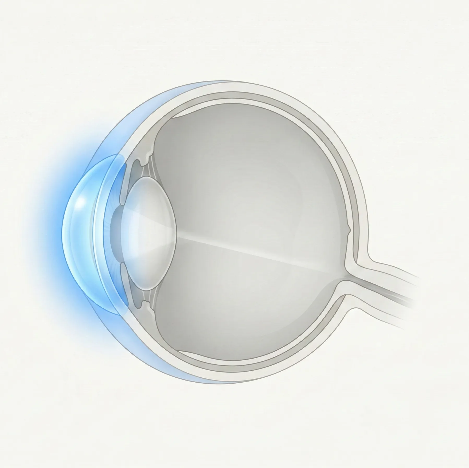
Cornea & External Eye
The cornea is the clear front window of the eye, and the conjunctiva covers the white of the eye and the inside of the eyelids. This category covers infections, inflammation, degeneration, shape abnormalities, and other conditions affecting the cornea and ocular surface.
239 English articles