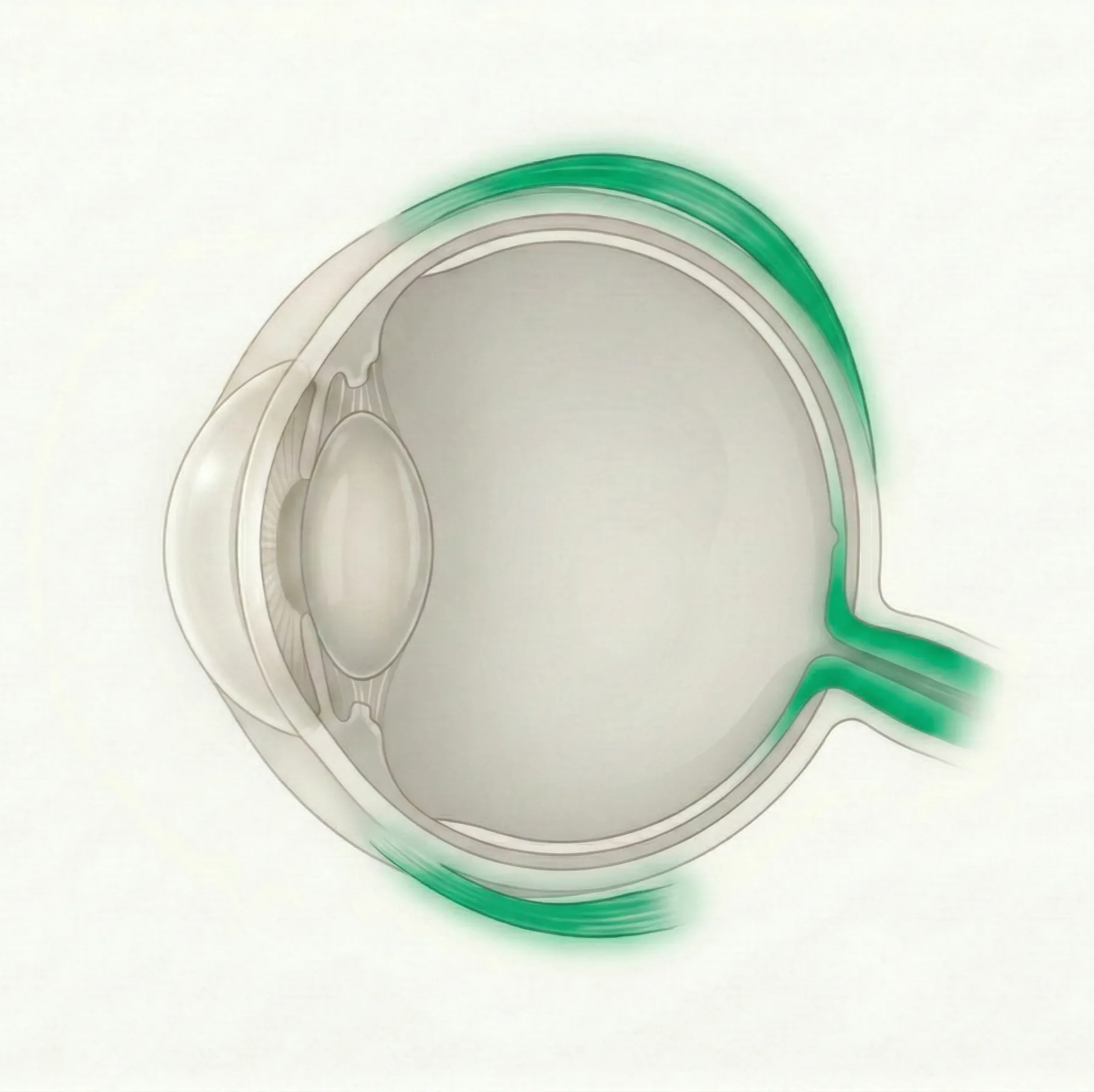
Pediatric Ophthalmology & Strabismus
Children's vision develops after birth and continues through early childhood. This category covers visual development problems, strabismus, amblyopia, and congenital eye diseases.
155 English articles
Children's vision develops after birth and continues through early childhood. This category covers visual development problems, strabismus, amblyopia, and congenital eye diseases.
155 English articles
A condition in which the lateral rectus muscle is paralyzed due to damage to the abducens nerve (sixth cranial nerve), resulting in limited abduction of the eye and incomitant esotropia. It is the most common ocular motor nerve palsy in adults, while in children, tumors and trauma are the main causes.
A rare ectodermal dysplasia caused by TWIST2 gene mutation. Characterized by eyelid hypoplasia, macrostomia, microtia, and loose skin. Treatment focuses on corneal protection and eyelid reconstruction.
A type of esotropia common in childhood, where one or both eyes turn inward due to accommodative effort caused by hyperopia or a high AC/A ratio. Spectacle correction is the mainstay of treatment, and early intervention is important for achieving binocular vision.
A technique to readjust the position of extraocular muscles after strabismus surgery to reduce overcorrection or undercorrection. Several methods exist, such as the bow-tie technique and the sliding noose technique.
An autosomal dominant genetic disorder caused by mutations in the JAG1 or NOTCH2 gene, affecting multiple organ systems including the liver, heart, eyes, skeleton, and kidneys. The most characteristic ocular finding is posterior embryotoxon.
Conjunctivitis caused by type I allergic reactions, common in children. Prevalence is about 20% and has been increasing in recent years with a trend toward younger age. It is classified into seasonal and perennial allergic conjunctivitis, vernal keratoconjunctivitis, and atopic keratoconjunctivitis. Treatment is based on anti-allergic eye drops, with immunosuppressive or steroid eye drops used in severe cases.
Alport syndrome is a hereditary basement membrane disease characterized by progressive renal dysfunction, sensorineural hearing loss, and ocular abnormalities, caused by mutations in type IV collagen genes (COL4A3, COL4A4, COL4A5). X-linked inheritance accounts for approximately 85% of cases, and males tend to have more severe disease.
A condition in which abnormal visual input occurs during the sensitive period of visual development, resulting in insufficient development of corrected visual acuity. It is classified into four types: refractive error, anisometropia, strabismus, and form deprivation. It is found in approximately 1-5% of children, and early detection and early treatment affect the visual prognosis.
Explains the indications, contraindications, and complications of general anesthesia, local block anesthesia, sub-Tenon's anesthesia, and topical anesthesia used in strabismus surgery. General anesthesia is standard for children, and for adults, the choice of anesthesia method according to the surgical procedure and patient background is important.
A neurodevelopmental disorder caused by loss of function of the maternally inherited copy of the UBE3A gene on chromosome 15q11-q13. It may be associated with ophthalmic complications such as strabismus, nystagmus, and hypopigmentation.
A rare congenital disease in which the iris is missing to varying degrees due to mutations in the PAX6 gene. It is accompanied by various ocular complications such as foveal hypoplasia, glaucoma, cataracts, and keratopathy, leading to progressive visual impairment.
A type of unilateral amblyopia that occurs in the eye with a higher refractive error when there is a difference in refractive error between the two eyes. It is the most common cause of amblyopia and is often detected during health checkups for 3-year-olds or preschool children. Early refractive correction and occlusion therapy can lead to good visual recovery.
A rare congenital eye malformation in which the eyeball is completely absent. Genetic and environmental factors are involved, and early orbital expansion treatment and multidisciplinary management are essential.
An extremely rare autosomal recessive disorder caused by loss-of-function mutations in the HOXA1 gene, leading to brainstem dysgenesis. Main features include congenital horizontal gaze palsy, sensorineural hearing loss, central hypoventilation, and developmental delay.
Autism spectrum disorder (ASD) is an early-onset neurodevelopmental disorder with a high risk of ocular conditions such as refractive errors, strabismus, and amblyopia. Due to communication difficulties, special approaches are needed for eye examinations.
A rare autosomal recessive disorder caused by biallelic mutations in the OPA1 gene. It presents with childhood-onset optic atrophy along with diverse neurological symptoms such as cerebellar ataxia, spastic paraplegia, and peripheral neuropathy.
A rare autosomal dominant disorder caused by mutations in the NR2F1 gene. It is characterized by optic atrophy, intellectual disability, developmental delay, and may involve cortical visual impairment.
Treatment of strabismus by injection of botulinum toxin type A into extraocular muscles. Approved in 2015 (Botox®), with insurance coverage for strabismus in patients aged 12 years and older. This article details the physician qualifications and administration protocols based on guidelines from the Japanese Association of Strabismus and Amblyopia and the Japanese Neuro-ophthalmology Society.
Treatment of strabismus by injection of botulinum toxin type A into the extraocular muscles. It can be an alternative to surgery for small to moderate angle esotropia.
A vertical strabismus characterized by limitation of elevation in adduction due to an abnormality of the superior oblique tendon-trochlea complex. It is classified into congenital and acquired types. Spontaneous resolution is common, but surgery is indicated when hypotropia or abnormal head posture is severe.
The most common benign orbital tumor in childhood. It grows within the first year of life and then spontaneously regresses over several years. If there is a risk of amblyopia, treatment with beta-blockers is the first-line therapy.
Visual impairment in children due to damage to the visual pathways beyond the lateral geniculate nucleus. It is the leading cause of visual impairment in children in developed countries, with hypoxic-ischemic encephalopathy being the most common etiology. Multidisciplinary rehabilitation is important.
An extremely rare autosomal recessive disorder caused by defects in the nucleotide excision repair pathway. It presents with microcephaly, congenital cataracts, microphthalmia, and multiple joint contractures, and the prognosis is very poor.
A multiple congenital anomaly syndrome characterized by coloboma, heart defects, choanal atresia, growth retardation, genital abnormalities, and ear anomalies due to CHD7 gene mutation. Multidisciplinary management is required, and ophthalmologically, attention to the risk of retinal detachment associated with coloboma is important.
A congenital eye disease in which tissue defects occur in various parts of the eye due to incomplete closure of the embryonic fissure. It is often accompanied by microphthalmia and causes visual impairment and complications depending on the location of the defect.
This article explains the incidence, diagnosis, and treatment of intraoperative and postoperative complications in strabismus surgery. It covers prevention and management of complications from scleral perforation, oculocardiac reflex, lost muscle to anterior segment ischemia and postoperative infection.
Lens opacity that develops at birth or during infancy, causing form deprivation amblyopia. The timing of surgery, appropriateness of IOL implantation, and postoperative amblyopia treatment determine visual prognosis.
A condition in which color discrimination ability differs from normal due to congenital deficiency or functional abnormality of cone photopigments. Congenital red-green color vision deficiency is found in about 5% of Japanese males and shows X-linked recessive inheritance. Visual functions other than color vision are normal and do not progress.
Congenital cranial dysinnervation disorders (CCDDs) are a group of congenital, non-progressive eye movement disorders caused by abnormal development of cranial nerves. They include Duane syndrome and CFEOM, presenting with paralytic strabismus due to aberrant innervation of extraocular muscles.
Congenital cystic eye (CCE) is an extremely rare congenital ocular malformation in which a cyst forms in the orbit instead of the eyeball due to impaired invagination of the primary optic vesicle during early embryonic development. Standard treatment involves cyst removal and prosthetic eye fitting.
A heterogeneous group of disorders caused by genetic abnormalities of the neuromuscular junction. It presents at birth or in childhood with easy fatigability, ptosis, ophthalmoplegia, and limb muscle weakness, and treatment is based on the genetic subtype.
Congenital nasolacrimal duct obstruction is a congenital blockage of the tear drainage system primarily caused by membranous obstruction at the lower end of the nasolacrimal duct, occurring in 6–20% of newborns. Approximately 90% resolve spontaneously by 12 months of age, but persistent cases are treated with conservative therapy such as lacrimal sac massage (Crigler method), probing, or dacryoendoscopic surgery.
Congenital nasolacrimal duct obstruction is an obstruction of the lacrimal drainage system occurring in 6–20% of newborns, primarily due to membranous obstruction of the valve of Hasner. Most cases resolve spontaneously within the first year of life, but persistent cases may require surgical treatments such as probing or stent placement.
Congenital palsy of the oculomotor nerve (third cranial nerve) is a pediatric ophthalmologic condition presenting with ptosis, exotropia, and limited eye movement. It is primarily caused by peripheral nerve injury during the perinatal period, and early intervention is important to prevent amblyopia.
Congenital rubella syndrome (CRS) is a congenital anomaly caused by maternal rubella infection during early pregnancy, characterized by the triad of cataracts, congenital heart disease, and hearing loss. Ocular symptoms are the most frequent, including pigmentary retinopathy, glaucoma, and microphthalmia.
A non-progressive congenital retinal dysfunction with a nearly normal fundus, diagnosed by ERG. It is classified into two types: complete (cCSNB) and incomplete (iCSNB), with X-linked recessive inheritance being the most common.
This article explains the evaluation of convergence ability using the near point of convergence (NPC) test and jump convergence test, as well as the diagnosis and treatment of convergence insufficiency, convergence paralysis, and convergence spasm.
A binocular vision disorder in which the eyes' ability to converge during near vision is insufficient, causing eye strain, double vision, and headaches. Main treatments include convergence exercises and prism glasses. It occurs across a wide age range, from children to adults.
The cover test is the gold standard objective examination for evaluating the presence, type, and degree of ocular misalignment (strabismus). This article explains the procedures and interpretation of the cover test, cover-uncover test, alternate cover test, and prism cover test.
A group of disorders in which premature fusion of cranial sutures leads to skull deformity and neurological complications. Includes Crouzon syndrome, Apert syndrome, Pfeiffer syndrome, etc., requiring multidisciplinary management.
Cryptophthalmos is a rare congenital anomaly in which fused eyelids cover the eyeball and the palpebral fissure is absent. There are three types: complete, incomplete, and abortive, and it is closely associated with Fraser syndrome.
Cyclopia is the most severe form of alobar holoprosencephaly, a lethal congenital anomaly characterized by a single orbit in the center of the face. The main pathogenesis is disruption of the Sonic Hedgehog pathway.
Cystic fibrosis (CF) is a systemic disease caused by mutations in the CFTR gene, and is associated with various ocular complications such as xerophthalmia due to vitamin A deficiency, CFRD-related retinopathy, and CFTR modulator-related cataracts.
Dacryocystocele is a neonatal condition in which the lacrimal sac becomes cystically dilated due to congenital nasolacrimal duct obstruction. This article explains its diagnosis, treatment, and complications.
A dermoid cyst is a congenital orbital choristoma that arises from ectodermal rests along suture lines. This article discusses its pathology, diagnosis, and surgical treatment.
Dissociated vertical deviation (DVD) is an abnormal eye movement that does not follow Hering's law, in which the non-fixing eye slowly elevates. It is frequently associated with infantile esotropia, and treatment options include anterior transposition of the inferior oblique muscle and recession of the superior rectus muscle.
Duane retraction syndrome is a non-progressive strabismus syndrome caused by a congenital abnormality of the abducens nerve. This article explains its clinical classification, pathophysiology, and treatment.
Duane retraction syndrome (DRS) is a non-progressive strabismus syndrome characterized by congenital abnormality of the abducens nerve and aberrant innervation of the lateral rectus muscle by the oculomotor nerve. This article explains its classification, pathophysiology, diagnosis, and surgical indications.
Dyslexia is a learning disability caused by neurological abnormalities in phonological processing. This article explains its relationship with vision, diagnosis, and treatment.
Dyslexia is a learning disability caused by neurological abnormalities in phonological processing. This article explains its relationship with vision, diagnosis, and treatment.
Esotropia is a misalignment of the eyes in which one eye deviates inward (toward the nose). The main types are infantile esotropia and accommodative esotropia. For infantile esotropia, very early surgery (≤8 months) is advantageous for achieving binocular vision, while for accommodative esotropia, full refractive correction with glasses is the basic treatment.
This article explains the indications, preoperative evaluation, selection of sedatives, and procedure for examination under anesthesia (EUA) in children. Main indications include retinoblastoma screening, diagnosis of developmental glaucoma, and electrophysiological tests such as ERG and VEP. It comprehensively covers doses of outpatient sedatives such as triclofos sodium and chloral hydrate, fasting guidelines, management of the oculocardiac reflex, and discharge criteria after sedation.
Exotropia is a misalignment of the eyes in which one eye deviates outward. Intermittent exotropia is the most common type. This article explains classification, diagnosis, and treatment including surgery.
The extraocular muscles consist of four rectus muscles, two oblique muscles, and the levator palpebrae superioris, and are responsible for eye movement and eyelid elevation under the control of three cranial nerves.
This article explains the causes, symptoms, diagnosis, and treatment of eye fatigue (asthenopia), including the latest meta-analysis data.
A treatment that occludes the healthy eye with an eye patch to improve visual acuity in the amblyopic eye. Evidence for dosing has been established by large-scale RCTs from PEDIG.
Fetal alcohol syndrome is an irreversible congenital condition caused by alcohol consumption during pregnancy, characterized by distinctive facial features, growth retardation, neurobehavioral disorders, and various ocular complications.
A progressive, large-angle esotropia and hypotropia associated with high myopia, accompanied by restricted eye movement. Surgical treatment using the Yokoyama procedure (loop myopexy) is the first choice.
Galactokinase deficiency, also called type II galactosemia, is a congenital metabolic disorder caused by mutations in the GALK1 gene, leading to bilateral cataracts. Early dietary intervention can reverse the cataracts.
A group of autosomal recessive disorders caused by congenital deficiency of enzymes involved in galactose metabolism, leading to accumulation of metabolites. Cataract is the main ophthalmic complication, and early lactose restriction is the basis of treatment.
A rare benign condition in which the optic disc is congenitally enlarged. It resembles glaucomatous optic neuropathy, but the disc rim area and retinal nerve fiber layer thickness are preserved, and it is not associated with progressive visual dysfunction.
A congenital craniofacial syndrome resulting from abnormal development of the first and second branchial arches, characterized by the triad of epibulbar dermoid, preauricular tags, and preauricular fistulas. It may also involve abnormalities of the vertebrae, heart, kidneys, and central nervous system.
A very rare congenital syndrome characterized by distinctive craniofacial features, congenital cataracts, microphthalmia, hypotrichosis, skin atrophy, and proportionate short stature. Approximately 90% of cases have ocular findings, requiring multidisciplinary management.
An autosomal dominant genetic disorder characterized by hyperferritinemia without iron overload and early-onset bilateral cataracts, caused by an IRE mutation in the FTL gene leading to overproduction of L-ferritin. Differentiation from hereditary hemochromatosis is important.
A clinical test to record dysfunction or overaction of the extraocular muscles and evaluate incomitant strabismus. This article explains the Hess chart, which uses red-green goggles to diagrammatically record ocular deviation.
Hypertropia is a vertical strabismus in which one eye is deviated upward relative to the other. The most common cause is superior oblique muscle palsy, which can be congenital or acquired. Diagnosis involves the Parks three-step test and the Bielschowsky head tilt test. Treatment options include prism correction and strabismus surgery, depending on the cause.
This article explains the pathology, diagnosis, and treatment of hypotropia associated with thyroid eye disease (Graves' ophthalmopathy). It covers the mechanism of restrictive strabismus due to fibrosis of the inferior rectus muscle, indications and surgical techniques for prism therapy and strabismus surgery, and the latest findings on teprotumumab.
A large-angle constant esotropia that develops within the first 6 months of life. Early surgical correction is important for achieving binocular vision.
Intermittent exotropia is a type of strabismus in which the eye alignment is normal most of the time but one eye drifts outward when tired or looking at distant objects. It is the most common type of strabismus in children. This article explains its classification, diagnosis, and treatment.
Non-congenital cataract that can develop in children as a side effect of CFTR modulators including ivacaftor, used to treat cystic fibrosis (CF). Regular ophthalmic screening is recommended.
A ciliopathy caused by dysfunction of primary cilia, characterized by the triad of molar tooth sign, hypotonia, and developmental delay. It is associated with various ophthalmic findings such as ocular motor apraxia and retinal dystrophy.
Juvenile xanthogranuloma (JXG) is the most common form of non-Langerhans cell histiocytosis, predominantly affecting infants and young children, characterized by yellow skin nodules. The eye is the most common site of extracutaneous involvement in JXG, and iris lesions can cause hyphema and secondary glaucoma.
A rare congenital genetic disorder characterized by distinctive facial features, intellectual disability, postnatal growth deficiency, skeletal abnormalities, and dermatoglyphic anomalies. Caused by mutations in KMT2D or KDM6A genes, it can be associated with various ophthalmic abnormalities.
Kasabach-Merritt phenomenon (KMP) is a life-threatening clinical syndrome characterized by thrombocytopenia and consumptive coagulopathy associated with kaposiform hemangioendothelioma (KHE) or tufted angioma (TA).
Strabismus surgery to correct abnormal head posture (face turn) in patients with nystagmus. It improves visual function by moving the null point to the primary position.
Langerhans cell histiocytosis (LCH) is a rare disease characterized by clonal proliferation of Langerhans cells, with proptosis due to osteolytic lesions of the orbital bone as the main symptom. It commonly occurs in children, with an incidence of 2 to 9 per million people.
The most severe form of congenital retinal dystrophy causing severe visual impairment from birth to infancy. Mainly autosomal recessive inheritance, with over 27 causative genes identified.
A congenital neurogenic ptosis in which the drooping eyelid elevates with jaw movement. Caused by abnormal connections between the trigeminal and oculomotor nerves, accounting for 2–13% of congenital ptosis.
Marfan syndrome (MFS) is an autosomal dominant connective tissue disorder caused by mutations in the FBN1 gene, with approximately 60% of patients developing ectopia lentis. It is frequently associated with high myopia, retinal detachment, glaucoma, and cataract. Regular dilated eye examinations and early intervention are crucial for visual prognosis.
Marfan syndrome (MFS) is an autosomal dominant connective tissue disorder caused by mutations in the FBN1 gene, with approximately 60% of patients developing ectopia lentis. It is frequently associated with high myopia, retinal detachment, glaucoma, and cataract. Regular examinations under dilated pupils and early intervention are crucial for visual prognosis.
Mucopolysaccharidosis type VI is a lysosomal storage disease caused by deficiency of arylsulfatase B, leading to accumulation of dermatan sulfate. It is characterized by progressive corneal opacification, glaucoma, and optic neuropathy, without intellectual disability.
Microphthalmia is a congenital ocular developmental anomaly in which the axial length of the eye is at least two standard deviations shorter than the age-adjusted mean. It is often associated with complications such as coloboma, cataract, and glaucoma, requiring early intervention and long-term management through multidisciplinary collaboration.
Moebius syndrome is a rare congenital disorder characterized by horizontal gaze palsy and facial paralysis due to congenital dysfunction of the sixth and seventh cranial nerves. It is classified under congenital cranial dysinnervation disorders (CCDDs).
An eye movement disorder in which one eye is limited in upward gaze in both adduction and abduction. It is often congenital and presents with hypotropia, ptosis, and abnormal head posture.
A sensory adaptation state in which foveal fusion is absent due to a macular scotoma in one eye, but peripheral fusion is maintained. Externally normal and asymptomatic, but associated with lack of fine stereopsis and mild amblyopia. Often recognized as a favorable outcome after strabismus surgery.
A congenital optic disc anomaly characterized by funnel-shaped excavation of the optic disc, white glial tissue, and radial blood vessels. It is frequently associated with retinal detachment and is known to be related to transsphenoidal encephalocele.
A mucocutaneous disease associated with Mycoplasma pneumoniae infection. It is a distinct disease entity separate from SJS/TEN, predominantly affecting young individuals, with mucositis of the oral cavity, eyes, and genitalia as the main feature. The ocular prognosis is better than that of SJS/TEN.
A congenital anomaly in which myelin sheaths are focally formed on retinal nerve fibers. Often discovered incidentally on fundus examination as brush-like white opacities, mostly asymptomatic and requiring no treatment.
Childhood myopia is a refractive error that develops during school age, primarily caused by axial elongation. Multiple progression control therapies, including low-dose atropine eye drops, myopia management spectacles, multifocal contact lenses, and orthokeratology, have accumulated evidence.
A specific type of myopia occurring in preterm infants, primarily caused by abnormal development of the anterior segment (cornea and lens) rather than axial elongation. It is closely associated with retinopathy of prematurity (ROP) and its treatment, and the choice of treatment significantly affects refractive prognosis.
Nanophthalmos is a rare developmental anomaly in which both the anterior and posterior segments of the eye are shortened due to arrested ocular growth during the embryonic period. It is characterized by high hyperopia, shallow anterior chamber, and thickened sclera, and is frequently complicated by glaucoma and uveal effusion.
Neonatal conjunctivitis is inflammation of the conjunctiva occurring within the first 28 days of life. Chlamydia and gonorrhea are the main causes. Early diagnosis and treatment lead to a good prognosis, but delayed treatment can result in serious complications.
A comprehensive explanation of the definition, classification, diagnosis, and treatment of congenital nystagmus (jerky nystagmus, pendular nystagmus), nystagmus blockage syndrome, latent nystagmus, spasmus nutans, and periodic alternating nystagmus. Includes indications for refractive correction, prism therapy, and surgical procedures such as the Kestenbaum procedure.
This article explains ocular complications associated with neurofibromatosis type 1 (NF1 / von Recklinghausen disease). Major findings include iris Lisch nodules, optic glioma, glaucoma, and eyelid/orbital neurofibromas. Regular ophthalmic examinations from childhood and long-term management are necessary.
This article explains ocular complications associated with neurofibromatosis type 1 (NF1/von Recklinghausen disease). Major findings include iris Lisch nodules, optic pathway glioma, glaucoma, and eyelid/orbital neurofibromas. Regular ophthalmic examinations from childhood and long-term management are necessary.
This article explains the types, frequency, diagnosis, and treatment of ophthalmic complications associated with Down syndrome (trisomy 21). Patients present with various ocular findings such as strabismus, refractive errors, keratoconus, and cataracts, making regular pediatric ophthalmologic screening important.
Mucopolysaccharidosis (MPS) is a group of diseases caused by genetic deficiencies of lysosomal enzymes, leading to accumulation of glycosaminoglycans in ocular tissues. Main ocular findings include corneal opacity, glaucoma, retinopathy, and optic disc abnormalities, with different characteristics depending on the type.
Classification, clinical findings, diagnosis, and treatment of ocular manifestations associated with HLH. Retinal hemorrhage is the most common, and various ocular complications have been reported.
This article explains the causes, diagnosis, and treatment of abnormal head posture (ocular torticollis) that occurs as a compensatory mechanism for ocular abnormalities.
The oculocardiac reflex (OCR) is a trigeminal-vagal reflex in which heart rate decreases by 20% or more due to stimulation of the extraocular muscles or eyeball. It occurs frequently during strabismus surgery and orbital trauma. This article explains its pathophysiology, diagnosis, management, and prevention.
A group of genetic disorders characterized by reduced melanin production, leading to pigment deficiency in the eyes and skin. The three main symptoms are nystagmus, photophobia, and reduced visual acuity, accompanied by foveal hypoplasia. There is no curative treatment; management focuses on refractive correction, tinted lenses, and low vision care.
A group of hereditary disorders characterized by reduced melanin production, leading to pigment deficiency in the eyes and skin. The three main symptoms are nystagmus, photophobia, and reduced visual acuity, accompanied by foveal hypoplasia. There is no curative treatment; management focuses on refractive correction, tinted lenses, and low vision care.
This article explains the various ophthalmological abnormalities associated with lissencephaly (a disorder of neuronal migration in the brain), categorized by type 1 (classic) and type 2 (cobblestone) lissencephaly.
A congenital condition characterized by a depression or defect in the optic disc due to incomplete closure of the posterior part of the embryonic fissure (optic cup fissure). Visual acuity depends on the degree of involvement of the papillomacular bundle. It can be complicated by serous retinal detachment or rhegmatogenous retinal detachment. It is important to screen for systemic associations such as CHARGE syndrome and renal coloboma syndrome.
Optic nerve hypoplasia (ONH) is the most common congenital optic nerve anomaly, characterized by a reduced number of optic nerve axons. This article explains its association with septo-optic dysplasia (SOD), diagnostic imaging, endocrine screening, and multidisciplinary management.
Optic neuropathy caused by fibrous dysplasia of the skull compressing the optic canal in McCune-Albright syndrome (MAS). Early diagnosis and appropriate timing of surgical intervention determine visual prognosis.
An infection of the soft tissues within the orbit posterior to the orbital septum. Most commonly spreads from sinusitis and frequently occurs in children. It presents with proptosis, ophthalmoplegia, and vision loss, requiring prompt antibiotic therapy and, if necessary, surgical drainage.
Orbital dermolipoma is a type of congenital choristoma containing adipose tissue, typically a benign mass occurring in the subconjunctival space of the superotemporal quadrant. This article explains its diagnosis, treatment, and associated syndromes.
An orthoptist (ORT) is a nationally licensed medical professional who performs orthoptic training for binocular vision and ophthalmic examinations under the direction of a physician. This article explains their scope of practice, legal status, education system, and work settings.
Pattern strabismus (A-V pattern strabismus) is a condition in which the horizontal deviation differs between upward and downward gaze. This article explains the classification into V, A, Y, X, and λ patterns, etiology, diagnosis, and surgical treatment.
A disease in children characterized by chronic inflammation of the eyelid margin accompanied by corneal and conjunctival lesions. It is based on meibomian gland dysfunction and bacterial blepharitis, and can lead to permanent visual impairment due to corneal scarring and amblyopia.
Contact lens prescription for children covers a wide range of purposes including amblyopia treatment, refractive correction, and myopia progression control. This article explains key points for determining indications, lens selection, and safety management.
Administering eye drops to children presents challenges different from adults, such as lack of cooperation and risk of systemic side effects. This article explains age-specific instillation techniques, guidance on cycloplegic agents like atropine, nasolacrimal occlusion, and key points for instructing caregivers.
Acute purulent inflammation (hordeolum) and chronic granulomatous inflammation (chalazion) of the eyelid, commonly occurring in children. Hordeolum is primarily treated with antibiotic eye drops, and most cases spontaneously drain; however, in infants and young children, rapid progression to eyelid abscess or orbital cellulitis requires caution. Chalazion is managed conservatively with warm compresses and local steroid injection; if ineffective, surgical removal under general anesthesia is considered.
Pediatric low vision is irreversible visual impairment in individuals under 21 years of age that cannot be improved by refractive correction, medical treatment, or surgical intervention. This article explains causes, age-specific assessment methods, and multidisciplinary management.
A demyelinating disease of the central nervous system in children characterized by MOG antibody positivity. Main phenotypes include ADEM and optic neuritis, with clinical features varying by age.
Pediatric vision screening aimed at early detection of amblyopia, strabismus, and refractive errors. This article explains the selection of examination methods according to age and the screening system centered on Japan's 3-year-old health checkup.
Explains visual acuity testing methods for infants to preschool children by age. Introduces the characteristics and procedures of each test, such as preferential looking, Teller acuity cards, LEA symbols, and Landolt C rings.
A hyperreflective ovoid structure observed around the optic disc on EDI-OCT, a nonspecific OCT finding associated with various optic nerve diseases as a marker of axoplasmic flow stasis.
Persistent fetal vasculature (PFV) is a congenital eye disease caused by incomplete regression of the fetal hyaloid vascular system. It is usually unilateral and non-hereditary, often presenting as leukocoria with microphthalmia. It is classified into anterior, posterior, and mixed types. In cases limited to the anterior type, visual improvement can be expected with lensectomy and amblyopia treatment, but the posterior type has a poor prognosis.
An anterior segment dysgenesis characterized by congenital corneal opacity due to central Descemet membrane defect and posterior corneal defect, often associated with iridocorneal adhesions and lens abnormalities. Glaucoma occurs in 50-70% of cases, and visual prognosis is poor.
PHACES syndrome is a rare neurocutaneous syndrome characterized by a large facial infantile hemangioma associated with posterior fossa malformations, arterial anomalies, cardiac anomalies, eye anomalies, and sternal defects.
Phakomatous choristoma is an extremely rare benign congenital tumor composed of ectopic lens tissue. It commonly occurs on the nasal side of the lower eyelid and can be cured by surgical excision, with no reports of recurrence.
Photoscreening is a vision screening method that uses a camera to capture and analyze the red reflex in children, detecting refractive errors and strabismus that are risk factors for amblyopia.
A genetic disorder caused by lack of expression of paternally inherited genes in the 15q11.2-q13 region of chromosome 15. It involves various ophthalmic abnormalities such as strabismus (40%), refractive errors, and hypopigmentation, requiring multidisciplinary management.
A rare congenital glaucoma in which intraocular pressure rises due to impaired aqueous humor outflow caused by developmental abnormalities of the anterior chamber angle. Surgery is the first-line treatment, and early diagnosis and treatment determine visual prognosis.
This article explains the types, indications, prescription methods, prism adaptation test, and weaning therapy of prism glasses used as optical treatment for strabismus and diplopia. It covers the characteristics and limitations of Fresnel membrane prisms, as well as usage scenarios for both children and adults.
A condition in which the eyes appear misaligned despite the absence of true deviation of the visual axes. It is most commonly pseudoesotropia due to facial morphological features such as epicanthal folds or an abnormal angle kappa.
A congenital absence of the lacrimal puncta. The main symptom is epiphora, and approximately 43% of cases are associated with systemic syndromes such as ectodermal dysplasia or Down syndrome. Surgical treatment may be necessary depending on the level of obstruction.
A proliferative vascular disease affecting the immature retinal vessels of preterm infants. The lower the gestational age and birth weight, the more severe the disease tends to be, accounting for approximately 30% of childhood blindness. It is classified by zone, stage, and plus disease based on ICROP3, and laser photocoagulation or anti-VEGF therapy is performed for type 1 ROP according to ETROP criteria.
An objective refraction method that uses a retinoscope to measure refractive errors of the eye. It is essential for evaluating patients who cannot undergo subjective testing, such as infants and individuals with developmental delays.
Saethre-Chotzen syndrome (acrocephalosyndactyly type III) is an autosomal dominant craniofacial syndrome caused by mutations in the TWIST1 gene. It is frequently associated with ophthalmic abnormalities such as ptosis and strabismus.
A common acquired strabismus in older adults caused by age-related degeneration of connective tissue between the extraocular muscle pulleys, leading to downward displacement of the lateral rectus muscle, resulting in distance esotropia and cyclovertical strabismus. Managed with prism correction or strabismus surgery.
Congenital hypoparathyroidism, growth retardation, and dysmorphism syndrome (HRD syndrome) caused by TBCE gene mutation. Frequently associated with ocular findings such as microphthalmia, corneal opacity, and tortuous retinal vessels.
A mysterious destructive panuveitis localized to Nepal. Epidemiological association with white moths (Gazalina species) is established; it predominantly affects children, and two-thirds are already blind at presentation.
A collective term for ophthalmic examination methods to evaluate strabismus and binocular vision abnormalities. Includes eye movement tests, ocular alignment tests, cover tests, and stereopsis tests, widely applicable from children to adults.
Abusive head trauma (AHT) is a form of inflicted head injury in infants and young children, caused by shaking or impact, leading to characteristic fundus findings such as retinal hemorrhage, retinoschisis, and retinal folds. The sensitivity of retinal hemorrhage is 85% and specificity is 94%, making fundus examination highly contributory to the diagnosis of AHT.
This article explains the effects of prolonged digital device use on children's visual acuity and refraction, the risk of myopia progression, and practical guidelines for screen time management.
A condition characterized by the triad of accommodative spasm, acute esotropia, and miosis. Often psychogenic and self-limiting, but it is important to rule out organic causes such as head trauma or neurological disease.
A type of acquired nystagmus that develops within the first two years of life, characterized by the triad of nystagmus, head nodding, and torticollis. Most cases are idiopathic and resolve spontaneously by age 3–4, but optic pathway glioma and retinal dystrophy must be ruled out.
Restrictive strabismus caused by inflammation and fibrosis of extraocular muscles associated with thyroid eye disease (Graves' ophthalmopathy). The main symptom is diplopia, and the inferior rectus and medial rectus muscles are commonly affected. Treatment includes prism therapy and strabismus surgery, provided thyroid function is normalized.
This article provides an overview of strabismus surgery, including indications, surgical techniques (recession, resection, muscle transposition, adjustable sutures), timing, complications, and postoperative management. It covers everything from early surgery for infantile esotropia to surgical planning for adult strabismus.
The synoptophore (major amblyoscope) is an instrument with independent optical systems for each eye that examines and trains binocular vision functions (simultaneous perception, fusion, and stereopsis). It enables quantitative measurement of subjective squint angle in nine directions, measurement of fusion range, and measurement of cyclodeviation. It is also used for vision training in children aged 3 years and older with strabismus or amblyopia.
An overview of basic genetics terminology in ophthalmology, types and selection strategies of genetic testing, and resources useful for diagnosis and management of hereditary eye diseases.
Parks-Bielschowsky three-step test for identifying the paretic muscle in hypertropia (e.g., superior oblique palsy). The three steps (primary gaze, lateral gaze, head tilt) narrow down the candidate muscles.
An autosomal dominant genetic disorder caused by mutations in the SALL1 gene, characterized by the triad of imperforate anus, ear malformations, and thumb anomalies. It may be associated with renal abnormalities, congenital heart disease, hearing loss, and ophthalmic abnormalities.
This article explains the indications, surgical techniques, and complications of muscle transposition surgery for dysfunction of the extraocular muscles, such as paralytic strabismus and Duane syndrome. It covers major procedures including full tendon transposition, Hummelsheim procedure, Jensen procedure, and Nishida procedure.
A chromosomal abnormality in which there are three copies of chromosome 13, frequently associated with severe ocular malformations such as microphthalmia, anophthalmia, and coloboma. The prognosis is poor, but survival rates have been improving due to advances in intensive care.
Vertical strabismus is a general term for vertical misalignment of the eyes. Congenital superior oblique palsy is the most common cause, and also includes inferior oblique overaction, Brown syndrome, A-V pattern strabismus, and double elevator palsy. Diagnosis is made using the Parks 3-step test, and surgery such as inferior oblique weakening, superior oblique tuck, or Harada-Ito procedure is performed depending on the angle and type of strabismus.
For children who are deaf or hard of hearing, conventional visual acuity tests that rely on verbal responses are difficult, so it is important to select non-verbal methods appropriate for their age and developmental stage. This article explains the characteristics and appropriate ages for various testing methods, as well as how to use the JEI/JEI test chart specifically designed for deaf and hard-of-hearing children.
Weill-Marchesani syndrome (WMS) is a rare hereditary connective tissue disorder characterized by microspherophakia, lens dislocation, secondary glaucoma, short stature, and brachydactyly. It occurs in 1 in 100,000 individuals, and early ophthalmologic management determines visual prognosis.
White pupil (Leukocoria) is a condition in which the pupil appears white, and is a sign of many eye diseases including retinoblastoma. It is detected as an abnormality of the red reflex in children, and prompt differential diagnosis is required.
The Worth 4-Light Test (W4LT) is a clinical examination that uses red-green glasses to evaluate binocular vision (fusion, suppression, anomalous retinal correspondence, diplopia). It can be performed from around 3 years of age and is tested at both near and far distances.