Exotropia is a condition in which, when one eye is fixated on a target, the other eye deviates outward (toward the ear). Most cases of exotropia without vertical or torsional deviation are comitant exotropia.
In Western countries, the ratio of esotropia to exotropia is 1.8:1, with esotropia being more common, whereas in Japan the ratio is 1:1.2–2.8, with exotropia being more frequent. A similar trend is observed in Southeast Asia. Intermittent exotropia is the most common type of strabismus in Japanese children, found in about 0.14% of elementary school students. The frequency in the United States is about 0.06%, lower than in Japan. Onset occurs from early childhood to around 8 years of age, most commonly at 3–4 years. Exotropia is estimated to be present in about 1% of the total population. The estimated prevalence of adult strabismus is about 4%, and the IRIS Registry reports 2.7% 5).
Initially, fusional convergence can maintain orthophoria, but exotropia appears during fatigue, poor physical condition, or immediately after waking.
Constant, Sensory, and Special Types of Exotropia
Constant exotropia: Constant outward deviation. Often transitions from intermittent exotropia.
Infantile exotropia: Ocular alignment does not normalize by age 1. Binocular vision development is severely impaired. Since 60% of infantile-onset cases are associated with systemic diseases or central nervous system abnormalities, CT/MRI is essential.
Sensory exotropia: Caused by long-term blurred retinal image in one eye. If the primary disease occurs before age 4, esotropia tends to develop; after age 4, exotropia is more common.
Consecutive exotropia: Exotropia occurring after esotropia surgery. Frequency 2–29%. Incidence increases with longer postoperative follow-up.
Long-term observation shows spontaneous resolution in about 10%, no change in about 40%, and progression to constant exotropia in about 50%. A North American report following 138 cases for 20 years found that 74.0% eventually required surgery. Von Noorden followed 51 untreated patients for an average of 3.5 years and reported progression in 75%, no change in 9%, and improvement in 16%.
QDoes intermittent exotropia worsen if left untreated?
A
Long-term observation indicates that about 50% progress to constant exotropia, while spontaneous resolution occurs in only about 10%. In a 20-year follow-up of 138 cases, 74% eventually required surgery. However, the rate of progression varies greatly among individuals, and some cases remain unchanged for several years. Surgery is considered when control scores worsen or stereopsis declines.
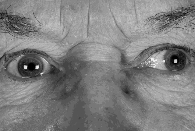
Breton S An Interesting Case of Pupillary Changes During the Testing of Ocular Movements and its Influence on the Diagnosis?. Br Ir Orthopt J. 2018. Figure 3. PMCID: PMC7510384. License: CC BY.
Frontal view clinical photograph showing outward deviation of one eye and asymmetry of corneal light reflexes. This demonstrates a typical ocular alignment abnormality in exotropia and is suitable for explaining the main symptoms and clinical findings.
Diplopia: Noticed if suppression is absent during exodeviation. Small-angle deviations may be perceived as blurred vision.
Asthenopia: Excessive accommodative effort to maintain fusion can lead to headache and vomiting.
Pseudo-myopia: Accommodation associated with convergence effort induces myopia. Uncorrected visual acuity decreases only when both eyes are open.
Monocular eye closure: In bright outdoor or strong light, fusion becomes difficult, exodeviation becomes manifest, and one eye is closed to avoid diplopia.
Asymptomatic: In children up to 10 years old, sensory adaptation (suppression) often prevents awareness of diplopia.
In intermittent exotropia, binocular vision develops well even during exodeviation, and most cases show normal retinal correspondence (NRC) and normal stereoacuity (<60 arcseconds). The binocular visual field (BVF) during exodeviation is 20–30 degrees, narrower than the normal 40 degrees. Infantile-onset cases may be complicated by monofixation syndrome and mild amblyopia (approximately 5%).
Assesses severity of control of eye alignment (maximum 9 points; higher scores indicate poorer control). Calculated by summing home assessment (0–3 points) and clinic assessment (0–3 points):
Score
Home Assessment
Clinic Assessment
3
Strabismus or monocular eye closure ≥50% of time at both distance and near
Exotropia without occlusion
2
Strabismus or eye closure at distance ≥50% of the time
Exotropia after cover test, does not return to phoria after cover removal
1
Strabismus or eye closure at distance <50% of the time
Exotropia after cover test, returns to phoria with blink after cover removal
0
No strabismus or eye closure at all
Exotropia after cover test, immediately returns to phoria after cover removal
QWhy do people with exotropia close one eye?
A
In bright outdoor environments, glare makes fusion difficult, and the outward deviation becomes apparent. When exotropia occurs, double vision results, and it is thought that one eye is closed to avoid this, but the exact mechanism is unknown. This “eye closure” is an important sign suggestive of intermittent exotropia.
Risk factors: Maternal smoking during pregnancy and low birth weight are significant and independent risk factors for the development of horizontal strabismus
Craniofacial abnormalities and neurodevelopmental disorders: Prone to exotropia
Causes of sensory exotropia: Fusion disturbance due to poor vision in one eye
Causes of postoperative exotropia: Overcorrection after esotropia surgery, or deterioration of binocular vision over the long term. In adults, adduction deficit is considered the main cause5)
In recent years, there have been reports of an association between prolonged near work such as smartphone use and convergence insufficiency-type exotropia.
Alternate prism cover test (APCT): Quantitative measurement of maximum strabismus angle. In intermittent exotropia, the angle may vary between measurements due to wide fusional range. It is important to detect the maximum angle
Comparison of distance and near deviation angle: Essential for classification into basic type, divergence excess type, and convergence insufficiency type
Classification and differential diagnosis of intermittent exotropia
Changes in near deviation after monocular occlusion/prism adaptation/+3D lens wear
Methods to differentiate pseudo divergence excess type (3 methods):
Monocular occlusion for 30–60 minutes (elimination of fusion)
Prism adaptation test under binocular open condition for 30 minutes to 1 hour
Wearing +3.0D lenses (elimination of accommodative convergence)
If any of these methods brings the near deviation angle close to the distance deviation (difference within 10Δ), it is diagnosed as basic type (pseudo divergence excess type).
Based on distance-near disparity, it is classified into three types: basic type (difference ≤10Δ), divergence excess type (distance > near by 10Δ or more), and convergence insufficiency type (near > distance by 10Δ or more). It is also important to differentiate from the “pseudo-divergence excess type (latent basic type)” where the near deviation angle increases with occlusion or +3D lens loading even if it appears as divergence excess type.
Observation: If control is good and asymptomatic, observation is the first step.
Part-time occlusion: Occlude the healthy eye for 3 hours per day. Effective for IXT with distance control score ≥2 in children aged 3–10 years 1). At 3 months, distance control score improves by 0.4 points (95% CI 0.1–0.7) and distance deviation improves by 2.1 PD 1).
Optical treatment: Fresnel membrane prisms, or overcorrecting glasses with +2 to +3 D minus lenses added to the cycloplegic refraction. Indicated when the deviation angle is small and the patient complains of asthenopia or diplopia.
Refractive correction: Myopia may improve control with corrective lenses.
Vision Therapy / Convergence Training
Indications for vision therapy: Corrected visual acuity equal in both eyes, deviation angle <25Δ, intermittent, near stereopsis present, and patient motivation essential. Optimal age is 8–12 years.
Sequence of training: Suppression elimination training → fusion training → convergence training. Combined therapy (surgery + vision therapy/occlusion) is more effective than either alone.
Convergence training: Particularly useful for convergence insufficiency type. Office-based training is more effective than home-based in children 5). Results are inconsistent in adults 5).
Base-in prisms: Promote fusion but reduce fusional convergence reserve, so they are rarely used for long-term management.
Surgery is generally considered after age 4. To achieve normal stereoacuity (60” or better), surgery should be performed by age 7 and within 5 years of onset. Surgery during the intermittent exotropia stage results in 93% achieving normal stereoacuity, compared to only 39% after progression to constant exotropia.
Postoperative Drift and Intentional Overcorrection
In pediatric surgery, a drift of 10–25Δ is observed compared to the immediate postoperative alignment. It is ideal to achieve an immediate postoperative esotropia within 10 PD (intentional overcorrection). Drift in adults is less than in children.
Postoperative outcomes of BLR for divergence excess type IXT: distance 38.1±8.0PD → −1.5±7.6PD, near 26.3±9.1PD → −0.9±6.2PD, distance-near disparity (NDD) improved from 15.4 to 0.62).
QWhen should surgery for intermittent exotropia be performed?
A
In principle, after age 4. To achieve normal stereopsis (60” or better), surgery is recommended by age 7 and within 5 years of onset. Surgery during the intermittent stage allows 93% to achieve normal stereopsis, but only 39% after it becomes constant. Deterioration of control score, decreased stereopsis, asthenopia, and cosmetic concerns are criteria for surgical indication.
QWhich is better: bilateral lateral rectus recession or unilateral recession-resection?
A
For divergence excess type intermittent exotropia, bilateral lateral rectus recession (BLR) and unilateral recession-resection (R&R) have been reported to have equal success rates of 83.3%2). The choice of procedure is determined by considering the amount of deviation, ocular alignment characteristics, and surgeon’s experience. For convergence insufficiency type, bilateral medial rectus resection may be effective.
The fusion maintenance mechanism in intermittent exotropia is primarily mediated by fusional convergence. Near fixation can relatively easily maintain orthophoria, but control breaks down at distance, leading to manifest exotropia.
Sensory adaptation (suppression): In early-onset cases, suppression develops instead of diplopia during exotropic deviation. The binocular visual field (BVF) is limited to 20–30 degrees (narrower than the normal 40 degrees), but stereopsis during orthophoria is preserved.
Relationship with refractive error (Donders theory): In uncorrected myopia, near vision is clear without accommodation, so accommodative convergence decreases, making exophoria more likely.
Involvement of AC/A ratio: The AC/A ratio (accommodative convergence to accommodation ratio) can be measured using the heterophoria method and the gradient method. A difference of 10 PD or more between distance and near indicates a high AC/A ratio. A high AC/A ratio is characteristic of the divergence excess type, and its involvement is assessed by adding +3.0 D lenses.
Mechanism of progression: It progresses through stages: exophoria → intermittent exotropia → constant exotropia.
Mechanism of sensory exotropia: Visual impairment in one eye → inability to maintain fusion → deviation occurs. Age 4 is considered the boundary between esotropia and exotropia (PPP also reports age 2 as a boundary: 69% exotropia vs 31% esotropia) 5).
Mechanism of consecutive exotropia: After esotropia surgery, if binocular vision is poor, eye position control becomes difficult, leading to exotropia. In adults, adduction deficit is considered the main cause 5).
Neuroplasticity in infantile-onset exotropia: Cases have been reported where stereopsis (55 arc seconds) is restored even after surgery in adulthood 3), suggesting the presence of residual visual plasticity.
In the PEDIG randomized controlled trial by Hatt et al. (2023), the part-time occlusion group (3 hours/day) showed a significant improvement of 0.4 points in distance control score at 3 months compared to the observation group (95% CI 0.1 to 0.7) 1). At 6 months, a significant difference of 0.3 points persisted (95% CI 0.02 to 0.6), but verification of long-term effects remains a challenge.
Expanded indications for R&R procedure in divergence excess type IXT
Han et al. (2023) demonstrated the potential for expanded indications of the R&R procedure in divergence excess type IXT 2). It was confirmed that R&R has a comparable success rate (83.3%) even in divergence excess type, for which BLR was standard.
Surgery and neuroplasticity in adult infantile-onset exotropia
Littlewood et al. (2021) reported that an adult patient who developed exotropia in infancy and underwent four surgeries over 15 years achieved recovery of binocular single vision (BSV) and stereopsis (55 arc seconds) postoperatively 3). This suggests that surgery for adult infantile-onset exotropia may utilize residual neuroplasticity.
Case report of transient high myopia after surgery
Yoshimura et al. (2022) reported a case of transient high myopia in a 6-year-old girl after exotropia surgery (lateral rectus recession 6.0mm + medial rectus resection 6.5mm), where the operated eye changed from +0.25D to −9.00D 4). AS-OCT showed abnormal anterior segment findings (anterior chamber depth 1.955mm vs. contralateral 3.007mm, lens thickness 4.216mm vs. contralateral 3.528mm). Spontaneous recovery occurred within 8 weeks. The mechanism was speculated to be anterior segment ischemia → ciliary body detachment → zonular relaxation → lens deformation.
Parameter
Operated eye
Contralateral eye
Refractive value (postop)
−9.00D
+0.25D
Anterior chamber depth
1.955mm
3.007mm
Lens thickness
4.216mm
3.528mm
Use of adjustable sutures and botulinum toxin
Adjustable sutures are considered useful for reoperations and unpredictable cases in adults 5). Botulinum toxin injection can be used for new-onset cases, reinforcement of large-angle deviations, postoperative residuals, and small-angle cases 5), and is attracting attention as one of the options.
Hatt SR, Kraker RT, Leske DA, Chandler DL, Fallaha N, Mohney BG, et al. Improved control of intermittent exotropia with part-time patching. Journal of AAPOS : the official publication of the American Association for Pediatric Ophthalmology and Strabismus. 2023;27(3):160-163. doi:10.1016/j.jaapos.2023.02.011. PMID:37187406; PMCID:PMC10330853.
Han M, Shen T, Wang X, Yu X, Zhu B, Wen Y, et al. Surgical outcomes of bilateral lateral rectus recession versus unilateral recession and resection for the divergence excess type of intermittent exotropia. Indian journal of ophthalmology. 2023;71(11):3558-3562. doi:10.4103/IJO.IJO_2977_22. PMID:37870024; PMCID:PMC10752302.
Littlewood RA, Rhodes M, Burke J.. A Post-Surgical Stereovision Surprise in an Adult With an Exotropia Since Infancy Previously Managed, at Two Years With Surgery. Br Ir Orthopt J. 2021;17(1):97-103. doi:10.22599/bioj.174. PMID:34278225; PMCID:PMC8269783.
Yoshimura A, Miyata M, Muraoka Y, et al. Unilateral transient high myopization after pediatric strabismus surgery: Observation by anterior segment optical coherence tomography. Am J Ophthalmol Case Rep. 2022;25:101421. doi:10.1016/j.ajoc.2022.101421. PMID:35198829; PMCID:PMC8850326.
American Academy of Ophthalmology Pediatric Ophthalmology/Strabismus Panel. Adult Strabismus Preferred Practice Pattern®. San Francisco, CA: American Academy of Ophthalmology; 2023.
Copy the article text and paste it into your preferred AI assistant.
Article copied to clipboard
Open an AI assistant below and paste the copied text into the chat box.