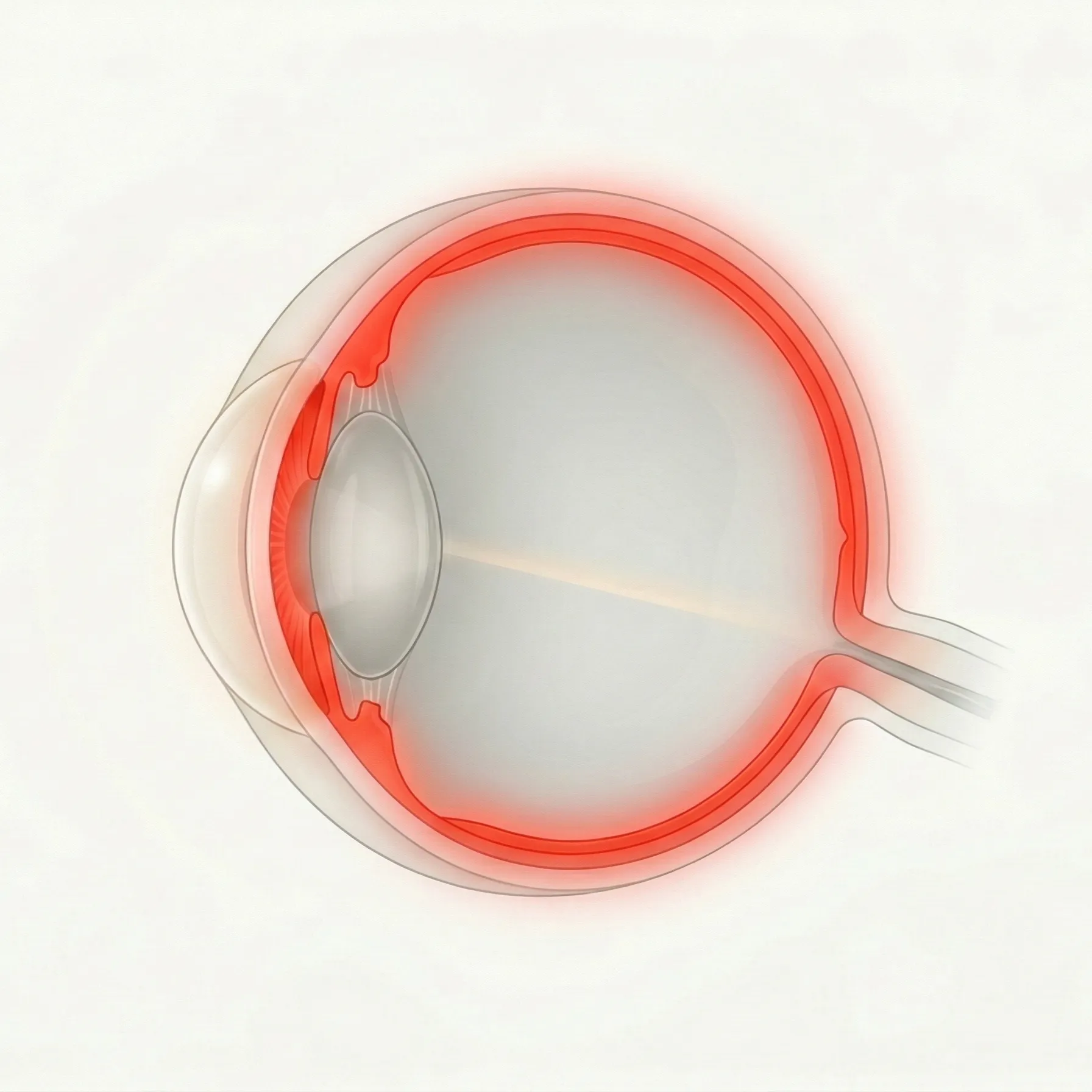
Uveitis
The uvea is the vascular middle layer of the eye, including the iris, ciliary body, and choroid. This category covers infectious, autoimmune, and systemic inflammatory causes of uveitis.
102 English articles
The uvea is the vascular middle layer of the eye, including the iris, ciliary body, and choroid. This category covers infectious, autoimmune, and systemic inflammatory causes of uveitis.
102 English articles
Acute anterior uveitis (AAU) is the most common form of uveitis, characterized by acute eye pain, redness, and photophobia. It is strongly associated with HLA-B27, and local treatment with steroid eye drops and mydriatics is the mainstay.
An acute inflammatory disease characterized by multiple disc-shaped white spots at the level of the retinal pigment epithelium in the posterior pole of both eyes. It predominantly affects young adults in their 20s to 30s, tends to resolve spontaneously, but attention should be paid to the complication of central nervous system vasculitis.
Rapidly progressive necrotizing herpetic retinitis caused by herpesviruses (HSV, VZV). First reported in Japan in 1971 by Urayama et al. as "Kirisawa-type uveitis," this is an ophthalmic emergency requiring early treatment based on the ASAP principle (antiviral therapy, anti-inflammatory therapy, antithrombotic therapy, and prevention of retinal detachment).
An acute outer retinal disorder of unknown cause. Despite minimal fundus findings, it presents acutely with photopsia and visual field defects. It predominantly affects young myopic women. The key diagnostic findings are loss of the ellipsoid zone on OCT and reduced multifocal ERG amplitudes.
This article explains the mechanism of action, indications, administration, side effects, and treatment evidence of the biologic agent adalimumab (Humira) for non-infectious uveitis.
An invasive procedure to collect aqueous humor for diagnostic purposes in uveitis. Used for viral DNA detection (HSV, VZV, CMV, Toxoplasma) by PCR and cytokine analysis (IL-10/IL-6 ratio for differentiating intraocular lymphoma). Lower complication risk than vitreous sampling and can be performed on an outpatient basis.
An active immunosuppressive (immune tolerance) phenomenon that confers immune privilege to the anterior chamber of the eye. Antibody production is maintained against antigens that enter the anterior chamber, but cell-mediated immunity such as delayed-type hypersensitivity is antigen-specifically suppressed. This is considered the main reason why the rejection rate of corneal transplantation is only about 20%, compared to about 100% for other organ transplants.
A rare autosomal dominant hereditary ocular inflammatory disease caused by CAPN5 gene mutation. It is characterized by progressive uveitis, retinal degeneration, neovascularization, and tractional retinal detachment, ultimately leading to blindness.
An OCT finding characterized by separation at the level of the inner segment myoid of photoreceptors and intraretinal fluid accumulation. It is observed in many uveitis and retinal diseases and was first reported as an independent concept in 2018.
This article explains the concept, epidemiology, symptoms, diagnostic criteria, and treatment (colchicine, cyclosporine, infliximab, adalimumab) of Behçet's disease based on the uveitis treatment guidelines and TNF inhibitor usage guidelines.
A chronic bilateral posterior uveitis characterized by fundus lesions resembling shotgun pellet scars, distributed from the posterior pole to the equator in both eyes. A strong association with HLA-A29 has been reported in Caucasians, and long-term immunomodulatory therapy centered on mycophenolate mofetil and adalimumab is required.
Ocular inflammation caused by bisphosphonates, drugs used for osteoporosis and bone metastases. It mainly presents as acute anterior uveitis, scleritis, and orbital inflammation, often occurring within one week after intravenous zoledronic acid infusion.
A rare monogenic autoinflammatory disease caused by a gain-of-function mutation in the NOD2 gene. It is characterized by the triad of granulomatous dermatitis, arthritis, and uveitis, and typically presents in childhood.
Inflammatory choroidal neovascularization (I-CNV) is a serious complication of chorioretinitis and posterior uveitis, and is the third most common cause of CNV after age-related macular degeneration and pathologic myopia. This article explains treatment strategies combining control of underlying inflammation with intravitreal anti-VEGF injections, multimodal imaging diagnosis using OCTA and ICGA, and characteristic findings such as the pitchfork sign.
A comprehensive explanation of eye drops used in the management of uveitis. Indicates the selection and precautions for steroids, mydriatics, and intraocular pressure-lowering drugs.
This article explains the diagnostic performance, technique, and indications of conjunctival biopsy for histological diagnosis of ocular sarcoidosis. It also presents the IWOS diagnostic criteria and the latest treatment strategies.
Infectious chorioretinitis caused by Cryptococcus neoformans. It commonly occurs in immunocompromised patients such as those with AIDS and may present as an initial ocular symptom of meningitis. Diagnosis and treatment are explained.
This article explains the use, efficacy, side effects, and drug interactions of the calcineurin inhibitor cyclosporine in ophthalmology for non-infectious uveitis.
Diagnosis and treatment of cytomegalovirus (CMV) anterior uveitis and corneal endotheliitis in immunocompetent individuals. Characterized by high intraocular pressure, coin-shaped keratic precipitates, linear keratic precipitates, and corneal endothelial cell loss. Treatment mainly involves ganciclovir gel eye drops and oral valganciclovir.
Necrotizing retinitis of the full thickness of the retina caused by cytomegalovirus (CMV). It is an opportunistic infection that occurs in immunocompromised individuals such as those with AIDS, post-organ transplant, or on immunosuppressive therapy. The mainstays of treatment are anti-CMV therapy centered on ganciclovir and prevention of retinal detachment.
Diagnosis and treatment of ocular complications associated with dengue virus infection. Covers a wide range of ocular findings including subconjunctival hemorrhage, dengue maculopathy, serous retinal detachment, and anterior uveitis to panuveitis.
Diagnosis and treatment of uveitis caused by systemic and topical medications. Explains the characteristics and management of causative agents such as rifabutin, bisphosphonates, immune checkpoint inhibitors, anti-VEGF drugs, brimonidine, and vancomycin.
Ophthalmic complications in survivors of Ebola virus disease (EVD). Various ocular symptoms, including uveitis, appear during the recovery phase and can lead to long-term visual impairment.
A severe infection where bacteria spread hematogenously into the eye from sepsis, liver abscess, etc. Klebsiella pneumoniae is the main causative organism, and progression is rapid. Early triple-route antibiotic administration and vitrectomy determine the prognosis.
Fuchs heterochromic iridocyclitis (FHI) is a unilateral uveitis characterized by the triad of iris heterochromia, chronic iridocyclitis, and cataract. Stellate keratic precipitates, iris atrophy, and Amsler sign are characteristic. Steroids are ineffective, so observation is generally recommended. An association with rubella virus has been suggested.
Endophthalmitis caused by various fungi migrating into the eye. Most cases are endogenous (hematogenous metastasis), with IVH patients and candidemia as major risk factors. Standard treatment includes systemic administration of antifungal agents such as fluconazole and voriconazole, along with vitrectomy.
A rare subtype of sarcoidosis characterized by four main symptoms: anterior uveitis, parotid gland swelling, facial nerve palsy, and fever. Also known as uveoparotid fever, it occurs in 4–6% of sarcoidosis patients.
Anterior uveitis caused by intraocular reactivation of herpes simplex virus (HSV). It is a representative cause of unilateral anterior uveitis with elevated intraocular pressure, accounting for 5–10% of all uveitis cases.
Anterior or posterior uveitis caused by reactivation of varicella-zoster virus (VZV). It occurs in 40–60% of herpes zoster ophthalmicus (HZO) cases and is characterized by elevated intraocular pressure, chronicity, and sectoral iris atrophy.
Acute, recurrent non-granulomatous anterior uveitis that frequently occurs in HLA-B27-positive individuals. It is commonly associated with spondyloarthropathies such as ankylosing spondylitis and presents with acute eye pain, photophobia, and redness. This article explains diagnosis, treatment, and indications for biologic agents based on the uveitis clinical practice guidelines.
Granulomatous or non-granulomatous uveitis occurring in carriers of human T-cell leukemia virus type 1 (HTLV-1). Common in Kyushu, Okinawa, and southern Shikoku, characterized by distinctive veil-like vitreous opacities and retinal vasculitis. Responds well to steroids but recurs in about 60% of cases.
Hyperreflective foci (HRF) observed on optical coherence tomography (OCT) are biomarkers of inflammation and degeneration found in various ocular diseases such as uveitis, age-related macular degeneration, and diabetic retinopathy.
Idiopathic multifocal choroiditis (IMFC) is a bilateral autoimmune disease characterized by multiple inflammatory lesions in the retina and choroid, predominantly affecting young myopic women, with choroidal neovascularization as a serious complication.
Ocular and orbital immune-related adverse events caused by immune checkpoint inhibitors (ICIs) used in cancer immunotherapy. They present with various conditions such as dry eye, uveitis, orbital myositis, and retinal vasculitis.
Immunomodulatory therapy (IMT) for ocular inflammation is an important treatment strategy to preserve vision in steroid-resistant or steroid-dependent non-infectious uveitis, with a wide range of options from conventional drugs to biologics.
General overview of uveitis caused by viruses, bacteria, fungi, and parasites. A comprehensive hub article explaining classification, diagnostic strategies, indications for intraocular fluid PCR, and the principle of steroid monotherapy contraindication.
This section explains ocular complications such as uveitis, scleritis, and chorioretinopathy associated with Crohn's disease and ulcerative colitis. It focuses on HLA-B27-related acute anterior uveitis, and TNF-α inhibitors can simultaneously control inflammation in the gut and eyes.
Infliximab is a mouse/human chimeric anti-TNF-α monoclonal antibody that plays an important role as a steroid-sparing treatment for refractory non-infectious ocular inflammation, particularly uveitis associated with Behçet's disease and JIA.
Intermediate uveitis is a chronic recurrent intraocular inflammation primarily involving the vitreous and peripheral retina, and includes the entity of pars planitis characterized by snowballs and snowbanks. It is common in young individuals, and macular edema is the main cause of vision loss.
Iris synechiae is a condition in which the iris adheres to adjacent structures due to intraocular inflammation, and is broadly classified into posterior synechiae and peripheral anterior synechiae. It is an important complication of uveitis and can cause secondary glaucoma and visual impairment.
Chronic granulomatous infection caused by Mycobacterium leprae. The eye is frequently affected, and chronic iridocyclitis, iris pearls, corneal lesions, and lagophthalmos are major causes of visual impairment.
Leukemic ocular infiltration can occur in the retina, anterior segment, optic nerve, and orbit. Retinal lesions are found in approximately 70% of all leukemia patients. This article explains Roth spots, pseudohypopyon, optic nerve infiltration, and GVHD-related ocular complications, and summarizes treatment options including radiotherapy, leukapheresis, and systemic chemotherapy.
A multi-organ infectious disease caused by Borrelia spirochetes transmitted through ticks. It has three stages, and ocular symptoms range from conjunctivitis in stage 1 to uveitis, keratitis, and cranial nerve palsy in stages 2 and 3. In Japan, Ixodes persulcatus and Ixodes ovatus are vectors, with northern Japan (mainly Hokkaido) being endemic. It is a Class IV infectious disease under the Infectious Diseases Control Law.
The most widely used immunomodulatory drug for non-infectious uveitis. It is an antimetabolite with antifolate activity, used globally as a first-line steroid-sparing agent.
A chronic bilateral disease presenting with multiple inflammatory lesions at the level of the retinal pigment epithelium and choriocapillaris. It is distinguished from punctate inner choroidopathy by the presence of anterior chamber and vitreous inflammation.
An acute inflammatory disease typically affecting one eye in young myopic women. Transient dysfunction of the outer retina and ellipsoid zone leads to gray-white spots that resolve spontaneously within weeks.
An antimetabolite immunosuppressant used for non-infectious uveitis. It selectively inhibits IMPDH to suppress lymphocyte proliferation and is positioned as a steroid-sparing agent with a favorable side effect profile.
Rapidly progressive necrotizing retinitis caused by herpes viruses (HSV, VZV, CMV). It is an ophthalmic emergency that presents as acute retinal necrosis (ARN) in immunocompetent individuals and progressive outer retinal necrosis (PORN) in immunocompromised individuals.
A rare autoimmune retinal disease in which anti-retinal antibodies destroy photoreceptors without associated malignancy. Characterized by bilateral progressive vision loss, night blindness, and photopsia, with diagnosis of exclusion and immunosuppressive therapy as mainstays.
An ophthalmology specialist explains the symptoms, diagnosis, and treatment of ocular complications (neuroretinitis, Parinaud syndrome, etc.) caused by cat scratch disease (Bartonella henselae infection).
An ophthalmology specialist explains the symptoms, diagnosis, and treatment of ocular complications (such as uveitis, keratitis, and optic neuropathy) caused by Chikungunya virus (CHIKV) infection.
Ocular corticosteroid therapy includes five routes: eye drops, subconjunctival, sub-Tenon's, intracameral, and intravitreal. It is the first-line treatment for uveitis and postoperative inflammation, but caution is needed for steroid-induced glaucoma and cataracts, and it should be avoided in infectious inflammation. This article systematically explains the drugs, doses, indications, and side effects for each route.
Leptospirosis is a zoonotic infection caused by a Gram-negative bacterium of the spirochete group, presenting various ocular findings such as non-granulomatous uveitis with hypopyon and panuveitis.
Comprehensive overview of various ocular complications associated with HIV infection. Covers the pathology, diagnosis, and treatment of HIV retinopathy, cytomegalovirus retinitis, opportunistic infections, malignancies, and immune recovery uveitis (IRU) that appear according to CD4-positive T lymphocyte count.
Coccidioidomycosis (Valley fever) is a systemic fungal infection caused by the dimorphic fungus Coccidioides. Ocular manifestations are rare but can cause severe intraocular inflammation when disseminated. It is endemic in the southwestern United States. Evaluation of extraocular symptoms and antifungal therapy are the mainstays of treatment.
Overview of ocular complications associated with familial Mediterranean fever (FMF). This article describes the various ocular symptoms reported in FMF, including episcleritis, uveitis, retinal vasculitis, and amyloid-related eye diseases, as well as their management.
Ocular involvement in an autoimmune disease characterized by recurrent inflammation of cartilage. Mainly scleritis, episcleritis, anterior uveitis, and peripheral corneal ulcers. Differentiation from Behçet's disease is important.
Systemic lupus erythematosus (SLE) is an autoimmune disease that causes chronic inflammation in multiple organs, with ocular symptoms occurring in approximately 30–50% of patients. It presents with various ocular lesions such as dry keratoconjunctivitis and lupus retinopathy, and in severe cases can lead to visual impairment.
An autoimmune disease in which autoantibodies against the conjunctival basement membrane cause chronic conjunctivitis and progressive scarring. Without treatment, it leads to symblepharon, corneal opacity, and blindness.
A rare zoonotic infection caused by larvae of the Pentastomida class parasitizing the eye. Infection occurs through ingestion of snake meat in endemic areas of Africa and Southeast Asia, and the larvae invade the anterior chamber, vitreous body, or subretinal space, causing severe visual impairment.
A parasitic uveitis caused by the invasion of larvae of the dog roundworm (Toxocara canis) or cat roundworm (Toxocara cati) into the eye. It primarily occurs in children and is characterized by unilateral vision loss and retinal granulomas.
A filarial infection caused by the nematode Onchocerca volvulus. Transmitted by black flies, it can cause keratitis, uveitis, and chorioretinitis, leading to blindness. It is the second leading cause of blindness due to infection worldwide.
Explanation of ocular complications caused by Rift Valley fever virus (RVFV). Posterior segment lesions, primarily macular retinitis, are characteristic, with ocular symptoms occurring in 0.5–15% of infected individuals, and severe cases leading to permanent vision loss.
A severe purulent infection that spreads to all structures of the eye and surrounding orbital tissues. It is the most advanced form of endophthalmitis, leading to blindness or loss of the eye without prompt treatment.
Severe uveitis involving inflammation of the entire uveal tract (iris, ciliary body, and choroid). It can be caused by various conditions such as sarcoidosis, Behçet's disease, Vogt-Koyanagi-Harada disease, syphilis, and infections. Without appropriate treatment, it can lead to severe visual impairment.
A condition characterized by acute, unilateral, recurrent elevation of intraocular pressure with mild anterior chamber inflammation. First reported by Posner and Schlossman in 1948. Strongly associated with CMV infection, and recurrent attacks increase the risk of secondary glaucoma.
A condition in which anterior chamber inflammation recurs after cataract surgery upon tapering or discontinuing steroid eye drops. Proper postoperative anti-inflammatory management and medication adherence are key to prevention and treatment.
A rare uveitis that develops via immune-mediated mechanisms after group A beta-hemolytic streptococcal infection. It primarily affects children and presents with bilateral non-granulomatous anterior uveitis.
Primary intraocular lymphoma (PIOL) is a primary intraocular lymphoma that forms lesions in the vitreous and retina, and is almost always diffuse large B-cell lymphoma. It is suspected in cases of uveitis resistant to steroid therapy, and diagnosis is made by measuring the IL-10/IL-6 ratio and vitreous biopsy. Intravitreal methotrexate injection and local ocular radiation are standard treatments.
A necrotizing herpetic retinopathy caused by varicella-zoster virus (VZV) that occurs in severely immunocompromised individuals (e.g., AIDS, post-organ transplant, malignant lymphoma). It is characterized by rapidly spreading white lesions from the outer retina and minimal anterior inflammation. It requires combination therapy with ganciclovir and foscarnet and has an extremely poor prognosis.
This article describes the clinical features, diagnosis, and treatment of uveitis associated with psoriasis and psoriatic arthritis. Anterior uveitis is predominant, and caution is needed regarding new onset or exacerbation risk when using IL-17 inhibitors.
An idiopathic inflammatory choroidal disease that predominantly affects young myopic women. It presents with small yellowish-white lesions in the posterior pole and is frequently complicated by choroidal neovascularization (CNV).
Rituximab, an anti-CD20 monoclonal antibody, is a biologic agent targeting B cells and is used for refractory non-infectious uveitis. It is internationally positioned as a third-line option after adalimumab and infliximab.
Uveitis associated with rubella virus infection. This article discusses acquired uveitis occurring during the course of adult rubella, ocular complications of congenital rubella syndrome (cataract, salt-and-pepper retinopathy), and its association with Fuchs heterochromic iridocyclitis.
Sarcoidosis is a systemic granulomatous disease and is the leading cause of uveitis. Non-caseating granulomas form within the eye, presenting as anterior, intermediate, posterior, or panuveitis. This article provides a comprehensive overview of diagnosis, treatment, and complication management.
A rare and severe eye disease in which scleral necrosis and thinning progress in a quiet eye without hyperemia or pain, associated with autoimmune diseases such as rheumatoid arthritis. It is classified as necrotizing scleritis without inflammation.
A chronic progressive posterior uveitis of unknown etiology affecting the retinal pigment epithelium, choriocapillaris, and choroid. It is characterized by serpiginous atrophic lesions extending from the peripapillary area, leading to irreversible vision loss when the fovea is involved.
Systematic classification criteria for the 25 most common types of uveitis published by the SUN Working Group in 2021. Developed and validated using machine learning, it aims to homogenize patient populations in research.
A novel drug delivery technique that directly administers medication into the suprachoroidal space (the space between the sclera and choroid). This article focuses on triamcinolone acetonide suprachoroidal injection, the only FDA-approved treatment for macular edema associated with non-infectious uveitis, and explains the procedure, efficacy, and safety.
Sweet syndrome is an autoinflammatory disease characterized by fever, neutrophilia, and painful erythematous plaques. Ocular manifestations include conjunctivitis, uveitis, and retinal vasculitis. Systemic corticosteroids are first-line therapy.
A rare autoimmune disease that causes bilateral granulomatous uveitis following penetrating trauma or intraocular surgery in one eye. Rapid systemic corticosteroid therapy combined with immunosuppressive agents is the mainstay of treatment.
Intraocular inflammation caused by Treponema pallidum. Known as the "great imitator," it presents with diverse ocular findings and has been increasing as a re-emerging infection. Cases with HIV co-infection tend to be more severe. High-dose penicillin therapy, as for neurosyphilis, is standard.
This article explains ocular side effects associated with systemic chemotherapy, including molecular targeted drugs and immune checkpoint inhibitors, categorized by drug class. It covers the mechanisms and management of various ocular toxicities such as uveitis, serous retinopathy, and corneal disorders.
Uveitis that develops after tattooing, accompanied by granulomatous inflammation at the tattoo site. It is a rare disease whose etiology is thought to be related to sarcoidosis or a delayed-type hypersensitivity reaction to tattoo ink.
A rare systemic inflammatory disease characterized by acute tubulointerstitial nephritis and bilateral anterior uveitis. It predominantly affects adolescent females and is thought to be immune-mediated. Renal prognosis is generally good, but uveitis tends to become chronic and recurrent.
Tocilizumab, an IL-6 receptor inhibitor, is a biologic agent reported to be effective for refractory non-infectious uveitis and uveitic cystoid macular edema that are resistant to TNF-α inhibitors. In juvenile idiopathic arthritis-associated uveitis, a phase II study reported some response.
Retinochoroiditis caused by intraocular infection with Toxoplasma gondii. It is the most common cause of infectious uveitis and can occur both as a recurrence of congenital infection and as acquired infection.
Anterior uveitis caused by blunt ocular trauma, resulting in inflammation of the iris and ciliary body. Main symptoms include eye pain, photophobia, and decreased vision. Treatment involves mydriatic agents and steroid eye drops. Usually resolves within 1 to 2 weeks.
Comprehensive overview of treatments for uveitis. Includes mydriatics, corticosteroids (eyedrops, local injection, systemic administration), immunomodulatory therapy (antimetabolites, biologics), and surgical interventions, along with evidence from major clinical trials.
Uveitis caused by cercariae of freshwater trematodes invading the eye and forming granulomas in various parts. Common in children and adolescents in developing countries; ciliary body granulomas can cause severe visual impairment.
Uveitis caused by intraocular infection or immune reaction to Mycobacterium tuberculosis. Presents with three major lesions: occlusive retinal phlebitis, choroidal miliary tuberculosis, and tuberculoma. Standard treatment is multidrug antitubercular therapy.
Uveitis caused by intraocular infection with Mycobacterium tuberculosis or an immune reaction to the bacterium. It presents with a wide variety of clinical manifestations and is difficult to diagnose and treat.
This article explains the mechanism of action, indications, administration, side effects, and monitoring of TNF inhibitors (infliximab, adalimumab, etanercept), which are biologic agents for refractory non-infectious uveitis.
A rare disease causing idiopathic exudative detachment of the choroid, ciliary body, and retina. It is thought to be primarily caused by impaired drainage of intraocular fluid due to scleral abnormalities, and treatments include scleral window surgery and steroid therapy.
Macular edema secondary to uveitis, a major cause of visual impairment. Steroid administration is the mainstay of treatment, but new therapies such as suprachoroidal injection and dexamethasone implant have emerged in recent years.
Chronic uveitis complicating juvenile idiopathic arthritis (JIA). It accounts for up to 47% of pediatric uveitis cases and is a refractory ocular disease that often progresses asymptomatically, leading to visual impairment.
A group of diseases that present with intraocular inflammation similar to uveitis but are not immune-mediated or infectious. They are broadly divided into neoplastic and non-neoplastic types, with intraocular lymphoma being the most common. Early differential diagnosis is crucial for visual function and life prognosis.
A postoperative complication of cataract surgery characterized by the triad of uveitis, glaucoma, and hyphema, caused by mechanical chafing of intraocular tissues by an intraocular lens (IOL). Early diagnosis and surgical intervention are key to preserving visual function.