Oculoplastic
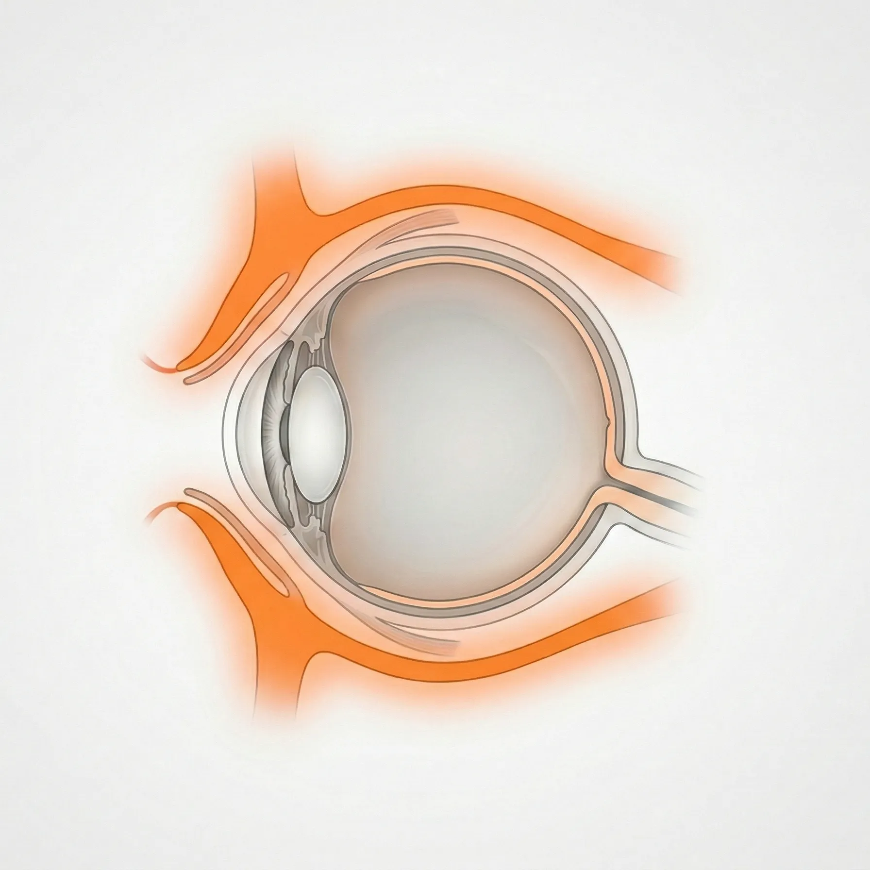
The eyelids, tear drainage system, orbit, and surrounding tissues support and protect the eye. This category covers structural, functional, and inflammatory diseases of these periocular tissues.
43 English articles
The eyelids, tear drainage system, orbit, and surrounding tissues support and protect the eye. This category covers structural, functional, and inflammatory diseases of these periocular tissues.
43 English articles
Comprehensive explanation of acute and chronic dacryocystitis including definition, classification, epidemiology, diagnosis, causative bacteria, treatment (DCR, probing), and dacryolithiasis. Also details management of neonatal dacryocystitis and the risk of chronic dacryocystitis before intraocular surgery.
Anophthalmia and microphthalmia are congenital conditions in which the eyeball is absent or small. They occur in 1 to 3 per 10,000 people. Starting expander use within the first 6 months after birth is important for orbital growth, and long-term plastic surgical management, including prosthetic eye fitting and orbital reconstruction surgery, is required.
Anophthalmic socket atrophy is a condition in which the socket shrinks and becomes sunken after eye removal, making it difficult to keep an artificial eye in place and causing cosmetic problems. It is classified as conjunctival sac contracture type, orbital depression type, or mixed type, and treated with enlargement of the conjunctival sac, dermis-fat grafting, bone grafting, or augmentation with artificial materials.
The first-line treatment for blepharospasm is botulinum toxin type A injections (Botox injections). The effectiveness rate is 90%, the effect starts after 2 to 3 days, and it lasts for 3 to 4 months. The orbicularis oculi muscle and corrugator muscle are targeted, with 2.5 units injected at each site in a spread-out pattern. Indications include essential blepharospasm, Meige syndrome, and hemifacial spasm.
This article explains the definition, classification (paralytic, age-related, symptomatic), diagnosis, and treatment of brow ptosis, including subbrow skin excision and frontalis suspension.
A chronic infection of the lacrimal canaliculus, most commonly caused by Actinomyces israelii. It forms concretions (sulfur granules) within the canaliculus and presents as unilateral refractory conjunctivitis. First-line treatment is canaliculotomy and curettage under local anesthesia, followed by topical new quinolone and systemic penicillin.
Comprehensive explanation of chalazion (non-infectious chronic granulomatous inflammation of the meibomian gland): definition, symptoms, classification, diagnosis (differentiation from sebaceous gland carcinoma), treatment (warm compresses, steroid injection, transconjunctival/transcutaneous excision), pathophysiology, and prognosis.
Complications after cosmetic eyelid surgery (double-eyelid surgery, eyelid fat removal, etc.) are classified into early (infection, hematoma, overcorrection) and late (ptosis, incomplete eyelid closure, sunken eye). Incomplete eyelid closure carries a risk of exposure keratitis, so ophthalmic care is important. Revision surgery may include levator refixation, skin grafting, and fat injection.
Hyaluronic acid fillers and autologous fat injections around the orbit carry a risk of irreversible vision loss due to vascular occlusion. The glabella and nasal root are the most dangerous areas because of dense anastomoses between the supratrochlear artery and the ophthalmic artery. For hyaluronic acid, emergency hyaluronidase injection is available as a treatment, but there is no specific treatment for autologous fat.
Conjunctival prolapse is a condition in which the bulbar conjunctiva protrudes beyond the eyelid margin. It can occur after eye trauma, as a postoperative complication, or in severe conjunctival laxity. Mild cases are managed with manual repositioning and a pressure bandage; severe or recurrent cases are treated with conjunctival excision and suturing, or conjunctival fixation surgery.
An inflammatory disease of the lacrimal gland, broadly classified into acute (viral or bacterial) and chronic (associated with systemic diseases or IgG4-related). Acute cases present with redness, swelling, and tenderness of the lateral upper eyelid; chronic cases show painless bilateral lacrimal gland enlargement. IgG4-related dacryoadenitis responds well to steroid therapy.
Dacryocystorhinostomy (DCR) is a definitive surgery for epiphora and dacryocystitis caused by nasolacrimal duct obstruction. A bony window is created between the lacrimal sac and the nasal cavity to form a new tear drainage pathway. In the external approach, a bone window of approximately 1×1 cm is made, and the reocclusion rate is less than 10%, indicating a high success rate. This article describes the procedural steps under general anesthesia, preoperative nasal management, mucosal flap suturing, and stent placement.
This article explains the definition, symptoms, diagnosis (evaluation based on MRD-1), differential diagnosis (distinction from ptosis), treatment (upper eyelid skin excision, subbrow skin excision), pathophysiology, and prognosis of dermatochalasis.
Explanation of the causes, classification, symptoms, diagnosis, and treatment of ectropion (a condition where the eyelid turns outward). It is classified into four types: involutional, paralytic, cicatricial, and mechanical. Surgical treatments such as the lateral tarsal strip procedure and Kuhnt-Szymanowski procedure are mainly discussed.
Comprehensive explanation of the definition, classification, symptoms, diagnosis, and surgical treatment of entropion (congenital, involutional, cicatricial, spastic, mechanical). Detailed description of key points in surgical technique selection such as the Hotz procedure, Jones modification, and lateral tarsal strip procedure.
Entropion is a condition in which the eyelid margin turns toward the eye and the eyelashes touch the cornea. The main types are congenital (trichiasis entropion) and age-related (involutional). For congenital cases, the suture method or Hotz procedure is used; for age-related cases, procedures that shorten the supporting tissues, such as the Jones modification or the lateral tarsal strip method, are selected. Ending with slight overcorrection is the key to reducing recurrence.
Trichiasis is a condition in which eyelashes grow abnormally toward the eye, while epiblepharon is a congenital condition in which excess skin causes the eyelashes to contact the cornea. Treatment options include eyelash removal, electrolysis, hair root resection, and modified Hotz procedure, selected based on severity and cause.
Explains the definition, classification, diagnosis, and treatment of epicanthus (Mongolian fold). Covers the distinction between epicanthus and epicanthus inversus, its relationship to blepharophimosis syndrome, pseudoesotropia, and the indications and techniques of epicanthoplasty.
Enucleation is a surgery that removes the entire eyeball and cuts the optic nerve, while evisceration is a procedure that removes only the intraocular contents while preserving the sclera and extraocular muscles. Main indications include intraocular malignant tumors, ocular trauma with no hope of vision recovery, and painful blind eyes. After surgery, cosmetic and functional maintenance is achieved with an orbital implant and prosthetic eye.
This article explains the definition and causes of eyelid retraction, its relation to thyroid eye disease, diagnosis, MRD evaluation, and surgical treatment including Müller muscle resection and levator recession.
An ocular prosthesis is an artificial eye worn for cosmetic and functional purposes after enucleation or evisceration. Today, custom-made acrylic (PMMA) ocular prostheses are the main standard, and they are made and adjusted in cooperation between an ocularist and an ophthalmologist. Daily care, socket management, and support that matches a child’s growth are important.
Comprehensive explanation of the definition, epidemiology, pathophysiology, diagnosis, and treatment of floppy eyelid syndrome (FES). Details the association with obstructive sleep apnea, clinical evaluation of tarsal laxity, and from conservative therapy to surgery.
A nonspecific inflammatory disease of unknown cause occurring in the orbit, formerly called "orbital inflammatory pseudotumor." It is defined pathologically by three conditions: nonspecific inflammation, no response to antibiotics, and marked response to steroids. Oral prednisolone is the first-line treatment, tapered over 3 to 6 months. For refractory cases, radiation therapy or methotrexate may be tried.
A systemic disease in which fibroinflammatory lesions rich in IgG4-positive plasma cells occur in the orbit. Painless swelling of the lacrimal gland is the most common symptom (86%), and the mainstay of treatment is steroid tapering therapy or immunosuppressive therapy with rituximab. The 2023 revised diagnostic criteria added a warning about optic neuropathy.
A disease causing epiphora due to obstruction of the lacrimal canaliculus or common canaliculus. Causes include inflammatory scarring, drug-induced (S-1), trauma, and post-infection. Dacryoendoscopic tube insertion is the first choice; for cases that cannot be recanalized, CDCR or dacryocystorhinostomy with canalicular bypass is indicated.
Lacrimal tube intubation is a surgery to recanalize the lacrimal pathway by placing a silicone tube in cases of obstruction or stenosis of the punctum, canaliculus, or nasolacrimal duct. Tube insertion using DEP/SEP perforation under dacryoendoscopy and SGI has become widespread, with a long-term survival rate of 94% for grade 1 canalicular obstruction. Complications include cheese-wiring, submucosal misinsertion, and granulation formation.
Lagophthalmos is a condition in which the eyeball is exposed due to incomplete eyelid closure, caused by facial nerve palsy, scarring, proptosis, etc. There is a risk of progression from corneal epithelial damage to perforation, and management is staged from conservative treatment to surgical intervention.
Ptosis surgery is selected based on levator function. When levator function is 10 mm or more, levator advancement (aponeurosis advancement) is standard; when it is less than 4 mm, frontalis suspension is standard. Preoperative MRD-1 measurement, levator function testing, and confirmation of Hering's law are important. Watch for complications such as hematoma, overcorrection, and undercorrection.
Orbital blowout fracture is a condition in which the orbital floor or medial wall is fractured due to blunt trauma to the eye, with main symptoms of diplopia, enophthalmos, and restricted eye movement. In closed fractures with entrapment of extraocular muscles, emergency surgery within 24 hours is required.
This article explains the definition, imaging diagnosis, and surgical treatment of cavernous hemangioma, a representative benign orbital tumor in adults. It is an encapsulated vascular mass commonly found within the muscle cone, with characteristic delayed enhancement on dynamic MRI. The standard surgical procedure is complete en bloc excision via a lateral orbitotomy, and the prognosis after complete resection is favorable.
An infection of the soft tissues within the orbit posterior to the orbital septum. Most commonly spreads from sinusitis and frequently occurs in children. It presents with proptosis, ophthalmoplegia, and vision loss, requiring prompt antibiotic therapy and, if necessary, surgical drainage.
An orbital dermoid cyst (dermoid cyst) is a congenital choristoma that develops along a bony suture because embryonic ectoderm becomes trapped. It accounts for 46% of pediatric orbital neoplasms and most often occurs just outside the eyebrow. Imaging with CT/MRI and complete removal without rupturing the cyst wall are key to treatment.
Orbital decompression is a surgery that removes orbital walls to expand orbital volume for proptosis and compressive optic neuropathy associated with thyroid eye disease. Techniques range from 1-wall to 3-wall plus fat decompression, with greater reduction in proptosis as the number of walls increases. During the inflammatory phase, steroid pulse therapy is prioritized; decompression is performed in cases unresponsive to medication or in emergencies.
Orbital fracture repair is a surgery to reduce incarcerated tissue and reconstruct the bony wall for fractures of the orbital floor and medial wall caused by blunt ocular trauma. Closed (trapdoor) fractures are common in children and require emergency surgery due to extraocular muscle entrapment. Selection of reconstruction materials such as titanium mesh, absorbable plates, and autologous bone is important.
A vascular malformation of the orbit (lymphatic malformation) that is common in childhood. It can present suddenly with eye bulging and eye pain because of bleeding within the mass (chocolate cyst). On MRI, a multilocular mass with fluid-fluid levels is a diagnostic finding. Conservative observation is the basic approach, but in severe cases, debulking surgery and sclerotherapy may be considered. The rebleeding rate is about 70%, so long-term follow-up is necessary.
This article explains the pathology, diagnosis, and treatment of lymphoma occurring in the orbit. It covers the characteristics and treatment strategies for each histological type, from the most common MALT lymphoma to high-grade DLBCL.
A rapidly progressive, fatal fungal infection caused by Mucorales fungi that spreads from the paranasal sinuses to the orbit and brain. It commonly occurs in patients with diabetes or immunodeficiency, and without treatment, the mortality rate reaches 79%. The mainstay of treatment is a combination of liposomal amphotericin B antifungal therapy and surgical debridement.
Acute infectious inflammation of the eyelids and periorbital soft tissues anterior to the orbital septum. Unlike orbital cellulitis, it does not involve proptosis or ophthalmoplegia. Main causes include sinusitis, trauma, and insect bites, and it commonly occurs in children. Mild cases can be managed with oral antibiotics on an outpatient basis, but progression to orbital cellulitis must be monitored.
A comprehensive explanation of the definition, classification by cause (congenital, aponeurotic, neurogenic, myogenic, pseudoptosis), diagnosis, surgical technique selection, and conservative treatment (oxymetazoline eye drops) of blepharoptosis.
A condition that causes epiphora due to narrowing or occlusion of the punctum, the tear drainage opening. It is classified into congenital punctal agenesis and acquired types (inflammatory, drug-induced, age-related, traumatic). Main acquired causes include Stevens-Johnson syndrome, ocular cicatricial pemphigoid, anticancer drug S-1, and glaucoma eye drops. First-line treatment is punctal dilation or incision; for recurrent occlusion, silicone tube intubation is performed.