Neuro-ophthalmology
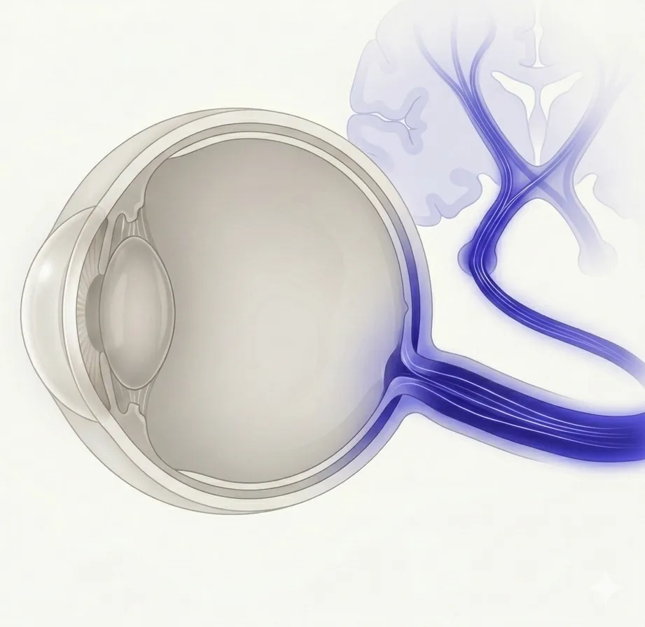
The eye is closely connected to the brain and nervous system. This category covers optic nerve inflammation, eye movement disorders, pupil abnormalities, and other conditions caused by neurologic disease.
282 English articles
The eye is closely connected to the brain and nervous system. This category covers optic nerve inflammation, eye movement disorders, pupil abnormalities, and other conditions caused by neurologic disease.
282 English articles
Paralysis of the sixth cranial nerve (abducens nerve) causes dysfunction of the lateral rectus muscle, leading to paralytic esotropia and ipsilateral diplopia. Ischemia due to diabetes and hypertension is the most common cause, but the frequency of tumors is higher than in other ocular motor nerve palsies.
Abnormal retinal correspondence (ARC) is a sensory adaptation phenomenon associated with strabismus, where the fovea of one eye corresponds to an extrafoveal retinal point of the other eye. This article explains the definition, classification, diagnostic methods, and treatment.
A condition caused by damage to the third cranial nerve (oculomotor nerve), leading to ptosis, impaired eye movement, and pupillary dilation. Common causes include aneurysm, ischemia, trauma, and tumors. When accompanied by pupillary dilation, it is a medical emergency.
An acute autoimmune demyelinating disease triggered by infection or vaccination. It causes multifocal lesions in the brain and spinal cord, presenting with encephalopathy, optic neuritis, motor paralysis, etc. Common in children but also occurs in adults.
First reported by Fletcher et al. in 1988, this is a self-limiting subclinical peripapillary retinopathy. It is characterized by acute blind spot enlargement and photopsia, and because fundus findings are nearly normal, it is a rare disease often misdiagnosed as optic neuritis.
Adie's pupil (tonic pupil) is a condition in which the light reflex is absent or diminished due to a lesion of the ciliary ganglion, but the near response is preserved. It is common in young women and is characterized by vermiform movements of the pupil and light-near dissociation.
An extremely rare and fatal malignant glioma (MOGA/MONG) arising in the anterior visual pathway and optic chiasm in adults. This article covers epidemiology, symptoms, imaging diagnosis, pathological findings, treatment, and prognosis.
Ageism is widespread in neuro-ophthalmology, posing social and ethical challenges that lead to delayed diagnosis, undertreatment, and worsened mental health in older patients.
An acquired reading disorder in which only reading ability is lost while writing ability is preserved. The most common cause is stroke due to occlusion of the left posterior cerebral artery, and it is a representative example of disconnection syndrome.
A neurological syndrome characterized by distortions in visual perception, body image, and time sense. Migraine is the most common cause, and it is more frequent in children and young adults. Most cases resolve spontaneously, but diagnosis and treatment of underlying conditions are important.
A condition in which vision in one eye transiently decreases and recovers within seconds to minutes. The most common cause is thromboembolism from internal carotid artery disease, and it can be a precursor to stroke, requiring urgent evaluation.
Optic neuropathy occurring as an ocular side effect of the antiarrhythmic drug amiodarone. It is characterized by insidious vision loss and persistent optic disc edema, and differentiation from nonarteritic anterior ischemic optic neuropathy (NAION) is important.
Anisocoria is a condition in which the pupil sizes differ between the two eyes, with causes ranging from physiological to life-threatening diseases. It is important to differentiate between sympathetic and parasympathetic nerve disorders and drug-induced causes, and to perform appropriate evaluation and management.
Anoxic brain injury (ABI) causes permanent damage to the visual system due to reduced oxygen supply to the brain, leading to ophthalmic symptoms such as cortical blindness, homonymous hemianopia, and ocular motility disorders. This article explains the pathophysiology, diagnosis, and rehabilitation.
This article explains the mechanisms of Wallerian (anterograde) degeneration and retrograde degeneration associated with neural damage in the visual pathway, diagnostic methods, and their relationship with optic atrophy.
Myasthenia gravis in which both anti-acetylcholine receptor antibodies and anti-MuSK antibodies are negative (double seronegative MG). Ocular symptoms are predominant, often occurring in children and young adults, and presents specific challenges in testing and diagnosis.
Anton syndrome is a rare neuro-ophthalmic disorder in which patients with cortical blindness due to bilateral occipital lobe damage deny vision loss and confabulate.
Aphantasia is a condition characterized by the lack or significant reduction of the ability to visualize mental images. It is found in 2-4% of the general population and can be congenital or acquired.
A pupillary abnormality characterized by the triad of miosis, loss of light reflex, and preserved near reflex. Classically caused by neurosyphilis (tabes dorsalis), but in modern times it is often associated with diabetes, cerebrovascular disease, and demyelinating diseases. The lack of response to low-concentration pilocarpine is an important distinguishing feature from Adie's pupil.
An ischemic disease of the optic nerve associated with giant cell arteritis (GCA). It causes sudden severe vision loss, and emergency treatment with high-dose steroids is essential to prevent progression to the fellow eye.
Autoimmune-related retinopathy and optic neuropathy (ARRON) is a rare disease in which autoimmune retinopathy and optic neuropathy occur together without malignancy. Because its clinical presentation is similar to cancer-associated retinopathy (CAR), it requires exclusion diagnosis.
Autosomal dominant optic atrophy (ADOA) is the most common hereditary optic neuropathy, primarily caused by OPA1 gene mutations, characterized by slowly progressive bilateral visual loss and optic atrophy starting in childhood.
Balint syndrome is a rare visuospatial coordination disorder caused by bilateral parieto-occipital lesions, characterized by the triad of simultanagnosia, optic ataxia, and oculomotor apraxia.
This article explains barriers to neuro-ophthalmology care, including specialist shortages, pre-referral misdiagnosis, and social determinants of health, as well as solutions such as telemedicine and educational improvements.
Diplopia (double vision) is a common complaint in neuro-ophthalmology and is broadly classified into monocular and binocular diplopia. Causes range from local ocular issues to intracranial diseases, and it is important to identify the causative site through systematic history taking, examination, and imaging.
Benign episodic mydriasis (BEM) is a rare condition characterized by transient, self-limiting unilateral pupillary dilation, and has been reported to be associated with migraine. Diagnosis is made by excluding serious causes.
Benign paroxysmal positional vertigo (BPPV) is the most common peripheral vertigo induced by head position changes. Ophthalmologists can contribute to diagnosis and differential diagnosis through evaluation of nystagmus. This article comprehensively explains pathophysiology, diagnosis, and treatment including canalith repositioning maneuvers.
Bickerstaff brainstem encephalitis (BBE) is a rare autoimmune brainstem encephalitis characterized by the triad of ophthalmoplegia, ataxia, and impaired consciousness following a preceding infection. As a subtype of anti-GQ1b antibody syndrome, it forms a continuous spectrum with Guillain-Barré syndrome and Fisher syndrome.
This article explains the classification, diagnosis, and treatments such as botulinum toxin therapy and microvascular decompression for blepharospasm (essential blepharospasm and Meige syndrome) and hemifacial spasm.
CAPOS syndrome is a rare neurological disease caused by mutations in the ATP1A3 gene. Its five main signs are cerebellar ataxia, areflexia, pes cavus, optic atrophy, and sensorineural hearing loss. It is characterized by paroxysmal neurological symptoms triggered by fever, as well as progressive vision loss and hearing loss.
Carotid-cavernous fistula (CCF) is an abnormal vascular connection between the internal carotid artery or external carotid artery and the cavernous sinus. The triad includes pulsatile exophthalmos, conjunctival chemosis, and vascular bruit. Endovascular treatment is the first-line therapy.
Carotidynia (TIPIC syndrome) is a self-limiting disease characterized by unilateral neck pain due to perivascular inflammation at the carotid bifurcation. This article explains diagnostic criteria, imaging findings, treatment, and ophthalmic signs.
A syndrome presenting with ophthalmoplegia, facial sensory disturbance, and Horner syndrome due to lesions of the cavernous sinus. The most common cause is tumor, but vascular, inflammatory, and infectious lesions are also important differential diagnoses.
A rare benign neuronal tumor (WHO grade II) that commonly occurs in the lateral ventricle. It predominantly affects young adults aged 20-40 and presents with symptoms of increased intracranial pressure due to obstructive hydrocephalus. Total resection is the standard treatment, and the prognosis is favorable.
A rare visual perseveration phenomenon in which multiple images are perceived from a single visual stimulus due to brain injury. Associated with lesions in the occipital lobe or posterior parietal cortex.
Cerebral venous and dural sinus thrombosis (CVST) is a blood clot in the cerebral venous drainage system, causing papilledema and visual impairment due to increased intracranial pressure. It is a rare disease accounting for 0.5–3% of all strokes.
This article explains the symptoms, diagnosis, and treatment of cerebral venous sinus thrombosis (CVST) caused by coagulopathy due to snakebite.
This article explains the mechanism of action, efficacy, safety, and neuro-ophthalmological significance of CGRP-targeted monoclonal antibodies (erenumab, fremanezumab, galcanezumab, eptinezumab) for migraine prevention.
Charcot-Marie-Tooth disease (CMT) is the most common inherited peripheral nerve disorder, characterized by distal muscle atrophy, weakness, and sensory impairment. Over 80 causative genes have been identified, and ophthalmologically, it may be associated with optic atrophy and retinal degeneration.
Complex visual hallucinations occurring in people with visual impairment, not associated with mental illness. It can be caused by various conditions such as eye disease, brain disease, or trauma, and cognitive function is preserved.
Cheiro-Oral Syndrome is a rare neurological disorder characterized by sensory disturbances around the mouth and in the hands and fingers. It is known as a subtype of thalamic stroke syndrome and may be accompanied by oculomotor disturbances and visual field defects.
Explains the causes, diagnosis, and treatment of visual field defects (bitemporal hemianopia) due to compressive, inflammatory, or traumatic lesions of the optic chiasm. Comprehensively covers pituitary adenoma, craniopharyngioma, chiasmal optic neuritis, and traumatic chiasmal syndrome.
Chromatopsia is a condition in which the visual field appears tinted with a specific color. The main causes are medications (e.g., digitalis, PDE5 inhibitors), retinal diseases, and brain diseases. In most cases, removal of the cause leads to recovery.
A disease in which the extraocular muscles are selectively impaired due to mitochondrial dysfunction, leading to slowly progressive bilateral ptosis and ophthalmoplegia. It is classified into isolated CPEO and CPEO-plus with systemic symptoms.
Explains the basic concepts of color vision, classification of congenital and acquired color vision deficiencies, symptoms, diagnostic methods, pathophysiology, and latest research including gene therapy.
This article explains the principles, indications, and clinical effects of FL-41 lenses and optical notch filters for reducing photophobia (light sensitivity). It details the melanopsin mechanism of intrinsically photosensitive retinal ganglion cells (ipRGCs) and the application of wavelength-selective blocking.
A general term for conditions in which multiple cranial nerves are simultaneously affected due to brainstem (midbrain, pons, medulla oblongata) lesions, presenting with various ocular motor disturbances, nystagmus, and pupillary abnormalities. Causes include cerebrovascular disease, demyelinating diseases, and inflammatory diseases, with characteristic symptom patterns depending on the lesion site.
Acetazolamide is a carbonic anhydrase inhibitor used for glaucoma and idiopathic intracranial hypertension, causing various complications such as transient myopia, ciliochoroidal effusion, and metabolic acidosis.
Optic neuropathy caused by compression of the optic nerve by mass lesions such as tumors, aneurysms, cysts, or enlargement of extraocular muscles in thyroid eye disease. Lesions often occur at the orbital apex or optic chiasm, and the mainstay of treatment is imaging diagnosis and management of the underlying cause.
A general term for visual field defects caused by compression of the visual pathway by mass lesions such as tumors, aneurysms, or cysts. Depending on the site of compression, various patterns such as bitemporal hemianopia, homonymous hemianopia, or quadrantanopia may occur. Imaging diagnosis and removal of the causative lesion are the mainstays of treatment.
Congenital fibrosis of the extraocular muscles (CFEOM) is a rare hereditary disorder characterized by congenital non-progressive external ophthalmoplegia due to developmental abnormalities of the oculomotor and trochlear nerves, with ptosis and restricted eye movements as main symptoms.
Cortical blindness is loss of vision due to damage to the bilateral occipital visual cortex, and Anton syndrome is a neuro-ophthalmic disorder in which cortical blindness is accompanied by denial of visual loss (anosognosia) and confabulation.
A rare 4-repeat tauopathy characterized by progressive atrophy of the cerebral cortex and basal ganglia. It presents with movement disorders, cortical dysfunction, and oculomotor abnormalities; definitive diagnosis is only possible through postmortem pathological examination.
Follicular dendritic cell sarcoma (FDCS) is an extremely rare low-grade sarcoma that occurs intracranially or intraorbitally. Invasion of the cavernous sinus or clivus can cause ocular symptoms such as diplopia, visual loss, proptosis, and ptosis.
A disease in which inflammation of cranial nerves causes nerve destruction or demyelination. Causes are diverse, including infectious, autoimmune, neoplastic, vascular, and idiopathic. When multiple cranial nerves are affected, it is called polyneuritis cranialis.
An extremely rare visual field defect in which lesions occur above the calcarine sulcus on one side and below the calcarine sulcus on the opposite side of both occipital lobes, resulting in homonymous loss of two quadrants on a diagonal line. Also called checkerboard visual field defect, mainly caused by embolism of the calcarine artery.
An X-linked recessive neurodegenerative disorder caused by TIMM8A gene mutation. It sequentially presents with sensorineural hearing loss in early childhood, dystonia in adolescence, visual loss in young adulthood, and dementia in middle age.
Visual impairment and low vision significantly increase the prevalence of depression and anxiety, with 81.2% of neuro-ophthalmic disease patients presenting some psychiatric symptoms. Comprehensive support through screening, rehabilitation, and counseling is important.
A low-grade mixed glioneuronal tumor (WHO grade 1) that commonly occurs in children and young adults. It primarily arises in the temporal lobe and presents with drug-resistant epilepsy. Complete resection leads to seizure freedom in 80–100% of cases, and the prognosis is favorable.
An anatomical condition in which cerebrospinal fluid herniates into the sella turcica, compressing and flattening the pituitary gland. It is classified into primary and secondary types and may be associated with endocrine disorders and visual field defects.
This article explains the epidemiology, symptoms, ocular complications, diagnostic criteria, treatment, and pathophysiology of eosinophilic granulomatosis with polyangiitis (EGPA, formerly Churg-Strauss syndrome).
Toxic optic neuropathy caused as a side effect of the antituberculosis drug ethambutol. Characterized by bilateral, painless vision loss and color vision abnormalities. Early detection and drug discontinuation determine visual prognosis.
Involuntary, fine, undulating contractions of the orbicularis oculi muscle, typically occurring unilaterally in the lower eyelid. It is a benign, self-limiting condition often triggered by stress, fatigue, or caffeine, and usually resolves spontaneously.
Fingolimod is an S1P receptor modulator used for treating multiple sclerosis and can cause dose-dependent macular edema. Early detection and drug discontinuation are the mainstays of management.
This article explains the differentiation, diagnosis, and treatment of Foster Kennedy syndrome, which presents with optic atrophy in one eye and papilledema in the contralateral eye, and pseudo-Foster Kennedy syndrome, which presents similar fundus findings due to non-neoplastic causes.
A disease caused by dysfunction of the fourth cranial nerve (trochlear nerve) that innervates the superior oblique muscle, resulting in hypertropia, excyclotorsion, and vertical diplopia on the affected side. Trauma, ischemia, and congenital causes are the main etiologies, and the Bielschowsky head tilt test is useful for diagnosis.
A disease caused by dysfunction of the fourth cranial nerve (trochlear nerve) that innervates the superior oblique muscle, resulting in ipsilateral hypertropia, excyclotorsion, and vertical diplopia. Trauma, ischemia, and congenital causes are the main etiologies, and the Bielschowsky head tilt test is useful for diagnosis.
Foville syndrome is a brainstem stroke syndrome involving the medial lower pons, characterized by contralateral hemiplegia, ipsilateral abducens nerve palsy, and facial nerve palsy. This article explains its causes, symptoms, diagnosis, and treatment.
Friedreich Ataxia is the most common hereditary ataxia caused by GAA repeat expansion in the FXN gene, presenting with progressive neurodegeneration as well as multi-organ involvement including cardiomyopathy, diabetes, and optic atrophy.
Froin syndrome is a rare syndrome characterized by the triad of xanthochromia, high protein, and hypercoagulability of cerebrospinal fluid (CSF). Obstruction of CSF in the spinal cord can cause increased intracranial pressure and papilledema.
This article explains the definition, diagnosis, treatment, and pathophysiology of functional visual disorder (non-organic visual disorder), in which patients present with reduced visual acuity or visual field defects despite the absence of organic eye disease.
A syndrome in which transient vision loss occurs only when the eye is held in a specific eccentric position, often caused by optic nerve compression due to an intraconal orbital tumor.
A neuropsychological syndrome characterized by the tetrad of agraphia, acalculia, finger agnosia, and left-right disorientation. It is caused by damage to the dominant hemisphere's parietal lobe (angular gyrus) or disconnection of white matter tracts.
A rare syndrome characterized by simultaneous palsy of the sixth cranial nerve (abducens nerve) and the twelfth cranial nerve (hypoglossal nerve). It is strongly associated with clival lesions, and investigation for neoplastic diseases is important.
Gorham-Stout disease (vanishing bone disease) is an extremely rare condition characterized by progressive osteolysis and abnormal proliferation of lymphatic/vascular vessels. When the orbit is involved, it can cause proptosis and visual field defects.
A rare syndrome characterized by the triad of abducens nerve palsy, facial pain, and otorrhea, resulting from the spread of infection to the petrous apex of the temporal bone as a complication of otitis media. This article explains the diagnosis, treatment, and pathophysiology.
A rare disorder in which visual distortions or flashbacks persist after hallucinogen use. It is classified into two types, HPPD I and HPPD II, and this article explains diagnostic criteria, treatment, and pathophysiology.
HaNDL syndrome is a self-limited secondary headache disorder characterized by transient headache and neurological deficits accompanied by cerebrospinal fluid lymphocytosis. It may present with ophthalmologic findings such as papilledema and abducens nerve palsy.
Harding disease is a rare condition in which Leber hereditary optic neuropathy (LHON) and multiple sclerosis (MS) coexist. It causes painless, severe vision loss against a background of mitochondrial DNA mutations, and no established treatment exists.
A rare syndrome of autonomic nervous system dysfunction characterized by unilateral facial flushing and hyperhidrosis with contralateral pallor and anhidrosis. May be associated with Horner syndrome.
Hashimoto encephalopathy (SREAT) is a rare autoimmune encephalopathy associated with elevated antithyroid antibodies. It presents with seizures, cognitive impairment, and psychiatric symptoms, and responds well to steroid therapy.
An acquired strabismus caused by elongation of the eyeball in high myopia, where the posterior part of the eye dislocates from the muscle cone, leading to deviation of extraocular muscle paths and progressive esotropia and hypotropia. Diagnosis by coronal MRI and the superior rectus-lateral rectus suture technique (Yokoyama procedure) are explained.
Heimann-Bielschowsky phenomenon (HBP) is a unilateral, slow pendular vertical nystagmus occurring in an eye with severe visual impairment. This article explains its diagnosis, differential diagnosis, and treatment.
Hemifacial spasm (HFS) is a movement disorder characterized by involuntary tonic-clonic contractions of the facial muscles on one side of the face. Vascular compression of the facial nerve is the main cause, and it is treated with botulinum toxin injections or microvascular decompression.
Optic neuritis occurring as a rare complication of herpes zoster ophthalmicus (HZO). Varicella-zoster virus (VZV) damages the optic nerve, causing vision loss.
A visual field defect affecting the same side of both eyes caused by damage to the posterior visual pathway (optic tract, lateral geniculate body, optic radiation, occipital visual cortex). Cerebrovascular disease (infarction or hemorrhage in the posterior cerebral artery territory) is the most common cause. The presence or absence of macular sparing and the quadrantanopia pattern help localize the lesion.
Horner syndrome is a syndrome characterized by the triad of miosis, ptosis, and anhidrosis due to disruption of the ocular sympathetic pathway. It is classified into central, preganglionic, and postganglionic types, and is diagnosed by pharmacological pupil testing and the apraclonidine test. It is essential to rule out serious causes such as carotid artery dissection and Pancoast tumor.
This article explains the diagnostic criteria, pathophysiology, and treatment of hypnic headache, a rare primary headache that occurs only during sleep and causes awakening.
An inflammatory disease of the optic nerve caused by autoimmune mechanisms. It predominantly affects women aged 15–45 years, with acute unilateral vision loss and eye movement pain as main symptoms. Visual recovery is expected in over 90% of cases, but anti-AQP4 antibody-positive cases are refractory, and assessment of MS conversion risk is important.
A benign syndrome that commonly occurs in young myopic eyes, presenting with intrapapillary hemorrhage and adjacent peripapillary subretinal hemorrhage (IHAPSH). Most cases resolve spontaneously without treatment, and the visual prognosis is good.
Inclusion body myositis (IBM) is a slowly progressive inflammatory muscle disease that typically occurs in individuals over 50 years of age. It is characterized by asymmetric weakness of the quadriceps and finger flexors, and rimmed vacuoles. It is resistant to immunosuppressive therapy, and dysphagia and orbicularis oculi muscle weakness are also relevant from an ophthalmological perspective.
A rare eye movement disorder characterized by intermittent involuntary contractions of the inferior oblique muscle, causing excyclotorsion and oscillopsia. Idiopathic, with very few reported cases.
Internuclear ophthalmoplegia (INO) is an eye movement disorder caused by a lesion of the medial longitudinal fasciculus (MLF), characterized by impaired adduction on the ipsilateral side, nystagmus of the contralateral abducting eye, and preserved convergence. When MLF lesion is combined with PPRF/abducens nucleus lesion, one-and-a-half syndrome occurs, with only abduction of the contralateral eye remaining. Multiple sclerosis and cerebrovascular disease are the two main causes, and treatment of the underlying disease is fundamental.
A rare phenomenon in which the eyeball deviates downward and inward during forced eyelid closure. It most commonly occurs after ptosis surgery and usually resolves spontaneously within days to months.
A condition in which a lumboperitoneal (LP) shunt for idiopathic intracranial hypertension (IIH) becomes dysfunctional several years after surgery. It presents with recurrence of headache and visual impairment, and the main treatment is shunt revision or conversion to a VP shunt.
An acute to subacute optic neuropathy with maternal inheritance due to mitochondrial DNA point mutations. It predominantly affects young males, causing severe bilateral vision loss and central scotomas. The mt11778 mutation is most common, and visual prognosis is poor, but new treatments such as idebenone and gene therapy are being developed.
Subacute necrotizing encephalomyelopathy due to mitochondrial dysfunction. It causes bilateral symmetrical necrotic lesions in the basal ganglia and brainstem, with onset most common in infancy. Over 110 causative genes have been identified.
Optic nerve dysfunction due to direct infiltration by leukemic cells. It is a neuro-oncological emergency, and the mainstay of treatment is a combination of intrathecal chemotherapy and orbital radiation therapy.
A pupillary sign in which the light reflex is impaired while the near reflex is preserved. It is caused by afferent pathway defects, midbrain dorsal lesions, efferent pathway defects, and aberrant regeneration, and is observed in conditions such as Adie tonic pupil, Argyll Robertson pupil, and Parinaud syndrome.
This article explains the current state of medical malpractice lawsuits in neuro-ophthalmology, diagnoses prone to litigation (such as cerebrovascular lesions, intracranial tumors, giant cell arteritis), measures to prevent oversight of emergency conditions, and key points of risk management.
A rare granulomatous neurocutaneous disease characterized by the triad of recurrent orofacial edema, facial nerve palsy, and fissured tongue. It occurs in 0.08% of the general population, and no curative treatment has been established.
A classic brainstem crossed syndrome presenting with ipsilateral abducens and facial nerve palsy and contralateral hemiplegia due to a unilateral lesion in the ventral caudal pons. This article explains the definition, symptoms, causes, diagnosis, and treatment.
Miller Fisher syndrome (MFS) is a variant of Guillain-Barré syndrome, an autoimmune peripheral neuropathy characterized by the triad of ophthalmoplegia, ataxia, and areflexia. Anti-GQ1b antibodies are involved in its pathogenesis, and most cases resolve spontaneously.
A central nervous system demyelinating disease caused by autoantibodies against myelin oligodendrocyte glycoprotein (MOG). Its main phenotypes are optic neuritis, ADEM, and myelitis. It is a disease concept independent of MS and AQP4-positive NMOSD. International diagnostic criteria were established in 2023.
A rare higher-order visual processing disorder in which moving objects cannot be visually perceived. Caused by damage to the V5/MT area, it presents with unique symptoms such as moving objects appearing as a series of still frames or disappearing from view.
Multiple sclerosis (MS) is an immune-mediated demyelinating disease of the central nervous system that presents with various ocular symptoms such as optic neuritis and internuclear ophthalmoplegia. Diagnosis is based on the McDonald criteria, including MRI and cerebrospinal fluid analysis, and management involves steroid pulse therapy and disease-modifying therapies.
This article describes the symptoms, diagnosis, and treatment of neuro-ophthalmic complications caused by immune checkpoint inhibitors (ICIs), including optic neuropathy, orbital inflammatory disease, thyroid eye disease, giant cell arteritis, and myasthenia gravis.
This article explains the pathophysiology, diagnosis, and treatment of various neuro-ophthalmic signs associated with SARS-CoV-2 infection, including optic neuritis, cranial nerve palsy, eye movement disorders, and pupillary abnormalities.
Primary Sjögren's syndrome (pSS) is an autoimmune disease characterized by chronic inflammation of the lacrimal and salivary glands, presenting various neuro-ophthalmic symptoms such as optic neuritis, trigeminal neuropathy, and dry eye.
Explains neuro-ophthalmic signs associated with cryptococcal meningitis (CM), such as papilledema, cranial nerve palsy, optic neuropathy, and endophthalmitis. Covers the two main mechanisms of increased intracranial pressure and direct infiltration, as well as diagnosis, treatment, and pathophysiology.
Explanation of neuro-ophthalmic findings associated with familial dysautonomia (Riley-Day syndrome). Includes alacrima, corneal disorders, optic atrophy, and ocular motor abnormalities due to IKAP gene mutation, and their management.
Ganglioglioma is a rare central nervous system tumor that presents various neuro-ophthalmic signs such as papilledema, abducens nerve palsy, and Parinaud syndrome depending on the tumor location.
Glioblastoma is the most common primary malignant brain tumor in adults, causing various neuro-ophthalmic symptoms through direct involvement, infiltration, compression of the visual pathways, and increased intracranial pressure.
Hypertrophic pachymeningitis (HP) is a rare disease characterized by diffuse or localized thickening and inflammation of the dura mater, presenting various neuro-ophthalmic signs such as visual loss, diplopia, optic disc edema, and cranial nerve palsy. Autoimmune diseases, infections, and tumors are the main causes, and contrast-enhanced MRI and dural biopsy are key to diagnosis.
This article explains the ophthalmic signs of Lambert-Eaton myasthenic syndrome (LEMS), such as ptosis, diplopia, and pupillary dysfunction, as well as the pathophysiology of autoantibodies against VGCC, diagnostic methods, and treatment.
Lymphocytic hypophysitis (LH) is a rare disease in which autoimmune lymphocytic infiltration of the pituitary gland causes various neuro-ophthalmic signs such as visual field defects due to optic chiasm compression, diplopia due to cavernous sinus cranial nerve palsy, and anisocoria.
This article explains neuro-ophthalmic complications associated with obesity hypoventilation syndrome (OHS), such as papilledema, central retinal vein occlusion, and non-arteritic anterior ischemic optic neuropathy.
Polyarteritis nodosa (PAN) is a systemic necrotizing vasculitis affecting medium-sized arteries. Ocular involvement occurs in 10–20% of cases, presenting various neuro-ophthalmic signs such as choroidal vasculitis, retinal vascular occlusion, ischemic optic neuropathy, and cranial nerve palsy.
This article explains the neuro-ophthalmic symptoms and signs of post-concussion syndrome (PCS) after traumatic brain injury (TBI), including findings such as convergence insufficiency, accommodative dysfunction, and traumatic optic neuropathy, as well as diagnostic methods and treatments focusing on visual rehabilitation.
Ophthalmic complications caused by chronic inflammatory demyelinating polyradiculoneuropathy (CIDP). This article explains the pathology, diagnosis, and treatment of ocular muscle palsy, papilledema, optic neuropathy, proptosis, and pupillary abnormalities.
Pneumosinus dilatans (PSD) is a rare condition characterized by abnormal expansion of the paranasal sinuses. PSD of the sphenoid or ethmoid sinuses can compress the optic nerve, leading to compressive optic neuropathy.
Periventricular leukomalacia (PVL) is an ischemic injury to the periventricular white matter in preterm infants, which may be discovered in adulthood as pseudo-glaucomatous optic disc cupping and visual field defects. Clinical differentiation from normal-tension glaucoma is important.
Explanation of neuro-ophthalmologic findings such as pupillary response, eye movements, and fundus findings observed in comatose patients. Covers essential clinical evaluation methods for lesion localization, etiological estimation, and prognosis prediction.
This article describes neuro-ophthalmologic manifestations such as optic neuropathy, ophthalmoplegia, pseudotumor cerebri, and dry eye caused by vitamin deficiency or autoimmune mechanisms associated with celiac disease (gluten-sensitive enteropathy).
Amyotrophic lateral sclerosis (ALS) is a progressive neurodegenerative disease affecting upper and lower motor neurons, and can present various neuro-ophthalmologic signs such as eye movement abnormalities and anterior visual pathway changes. Although extraocular motor neurons are usually spared until late stages, square-wave jerks, saccadic abnormalities, and retinal changes have been reported.
Chordoma is a rare malignant bone tumor arising from notochordal remnants. When it occurs at the skull base, it presents with neuro-ophthalmologic symptoms such as abducens nerve palsy, diplopia, and visual field defects. This article explains diagnosis, treatment, and prognosis.
Chronic basilar artery occlusion (CBAO) is a condition in which long-term occlusion of the basilar artery leads to the development of collateral circulation, presenting with various neuro-ophthalmologic symptoms such as internuclear ophthalmoplegia, homonymous hemianopia, and cortical blindness.
This article explains neuro-ophthalmologic signs such as papilledema, abducens nerve palsy, and dorsal midbrain syndrome caused by colloid cysts of the third ventricle, as well as imaging diagnosis and treatment.
This article explains neuro-ophthalmologic signs associated with multiple system atrophy (MSA), such as eye movement disorders, pupillary abnormalities, and blepharospasm, including red flag findings that aid diagnosis and treatment options.
Explanation of neuro-ophthalmologic signs of PML, a demyelinating disease of the central nervous system caused by JC virus. Main ocular symptoms include homonymous hemianopia, cortical blindness, nystagmus, and diplopia due to cranial nerve palsy.
This article explains the diverse neuro-ophthalmologic signs caused by thalamic lesions, including vertical gaze palsy, skew deviation, pupillary abnormalities, and nystagmus, as well as the etiology, diagnosis, and treatment including Percheron artery infarction.
CADASIL (cerebral autosomal dominant arteriopathy with subcortical infarcts and leukoencephalopathy) is a hereditary cerebral small vessel disease caused by NOTCH3 gene mutations, presenting various ophthalmic signs such as visual aura, diplopia, and retinal vascular changes.
This article explains the diagnosis, treatment, and pathophysiology of neuro-ophthalmological complications associated with LADA (type 1.5 diabetes), including diabetic retinopathy, ophthalmoplegia, and optic neuropathy.
Myoclonus epilepsy associated with ragged-red fibers (MERRF) is a rare multisystem disorder caused by mitochondrial DNA mutations, presenting with various neuro-ophthalmological findings such as optic atrophy, ophthalmoplegia, and pigmentary retinopathy.
Krabbe disease (globoid cell leukodystrophy) is a lysosomal storage disorder caused by GALC gene mutations, presenting neuro-ophthalmological findings such as optic atrophy, nystagmus, and cranial nerve palsy.
This article describes the characteristics of neuro-ophthalmological symptoms such as papilledema, abducens nerve palsy, and optic neuritis in Mollaret meningitis (recurrent benign lymphocytic meningitis), its association with HSV-2, and diagnosis and treatment.
This article explains the neuro-ophthalmological findings such as papilledema, abducens nerve palsy, and transient visual obscurations associated with increased intracranial pressure due to choroid plexus papilloma (CPP), covering diagnosis, treatment, and pathophysiology.
A comprehensive review of the diagnosis, treatment, and pathophysiology of neuro-ophthalmological findings such as nystagmus, ophthalmoplegia, diplopia, and autoimmune retinopathy associated with anti-GAD antibody syndrome.
Cerebral amyloid angiopathy (CAA) is a disease in which β-amyloid deposition in blood vessel walls causes cerebral hemorrhage, leading to ophthalmic symptoms such as homonymous hemianopia and cortical visual impairment due to damage to the occipital lobe and optic radiation.
This article explains the neuro-ophthalmological aspects of language disorders (bilingual aphasia) that occur after brain injury in bilingual or multilingual individuals. It outlines different patterns of impairment between two languages, associations with visual field defects, and diagnosis, treatment, and rehabilitation.
This article explains neuro-ophthalmic findings associated with dementia with Lewy bodies (LBD), including visual hallucinations, oculomotor disturbances, color vision abnormalities, and eyelid abnormalities, as well as their diagnosis and management.
Neuroblastoma is the most common extracranial solid tumor in children, originating from the neural crest. It may present initially with proptosis and periorbital ecchymosis due to orbital metastasis. This article explains the pathology, diagnosis, and treatment.
Neuromyelitis optica spectrum disorder (NMOSD) is an autoimmune inflammatory disease of the central nervous system mediated by aquaporin-4 antibodies, characterized primarily by optic neuritis and myelitis.
Neuromyelitis optica spectrum disorder (NMOSD) is an autoimmune inflammatory disease of the central nervous system mediated by aquaporin-4 antibodies, characterized primarily by optic neuritis and myelitis.
A condition in which sarcoidosis affects the central and peripheral nervous systems. It presents with various neurological symptoms such as cranial nerve palsy, uveitis, meningitis, spinal cord disorders, and hypopituitarism.
This article explains the etiology (hemifield slide phenomenon, foveal traction diplopia syndrome, convergence insufficiency, divergence insufficiency, fusion fear), diagnosis, and treatment of binocular diplopia without extraocular muscle palsy.
Noonan syndrome is a genetic disorder caused by mutations in the RAS-MAPK pathway, characterized by distinctive facial features, short stature, and congenital heart disease as the three main features, and may be associated with ocular abnormalities such as optic nerve hypoplasia and coloboma.
A disease in which the optic nerves in both eyes are damaged due to deficiencies of nutrients such as B vitamins and copper. Risk factors include bariatric surgery, vegan diet, and alcoholism.
A focal epilepsy arising from an epileptic focus in the occipital lobe, characterized by visual hallucinations, ictal blindness, and abnormal eye movements. It accounts for 5–10% of all epilepsies, and clinical differentiation from migraine is important.
Diagnostic criteria, differential diagnosis, and treatment of occipital neuralgia (paroxysmal occipital pain derived from C2 and C3 nerves). From nerve blocks and pharmacotherapy to the latest hydrodissection.
OCT angiography (OCTA) is a non-invasive imaging technique that three-dimensionally visualizes fine retinal and choroidal blood vessels without the use of contrast agents. This article explains the principles, indications, clinical findings, and limitations of OCTA in the field of neuro-ophthalmology.
This section explains ocular changes such as optic disc edema, choroidal folds, and RNFL thickening observed in head-down tilt bed rest (HDTBR), a ground-based analog model of spaceflight-associated neuro-ocular syndrome (SANS).
An infectious disease caused by the larval stage of Taenia solium parasitizing the eye or orbit. It is a preventable cause of blindness in endemic areas. Diagnosis relies on imaging (ultrasound, CT, MRI) and treatment primarily involves albendazole.
Systemic capillary leak syndrome (Clarkson disease) is a rare disorder characterized by sudden leakage of fluid and protein from capillaries into tissues, causing various ocular complications such as conjunctival edema, choroidal effusion, acute angle closure, and NAION.
This article provides an overview of ocular manifestations associated with mitochondrial encephalomyopathy, lactic acidosis, and stroke-like episodes (MELAS). It focuses on the diagnosis and management of various ocular findings, including pigmentary retinopathy, optic atrophy, and progressive external ophthalmoplegia.
Multiple myeloma (MM) is a hematologic malignancy caused by the proliferation of malignant plasma cells, leading to symptoms in multiple ocular sites including the orbit, retina, cornea, and optic nerve. The pathophysiology is broadly divided into two categories: direct infiltration of tumor cells and hyperviscosity due to hyperimmunoglobulinemia.
An infectious disease of ocular tissues caused by Treponema pallidum. It presents with various ocular signs such as uveitis, retinitis, and optic neuritis, and is known as the "great imitator," requiring differentiation from other diseases.
A neuro-ophthalmic disorder in which voluntary eye movements (especially saccades) cannot be initiated intentionally. It is broadly classified into congenital (Cogan type) and acquired forms, often associated with hereditary ataxias and neurodegenerative diseases.
This article explains the diagnosis and treatment of ocular myasthenia gravis, a form of myasthenia gravis (an autoimmune disease of the neuromuscular junction) that is limited to extraocular muscle symptoms such as ptosis and diplopia. It is characterized by diurnal fluctuation and easy fatigability, and early treatment is important to prevent progression to the generalized form.
A rare ocular motor disorder characterized by intermittent tonic spasms of the extraocular muscles. It causes paroxysmal diplopia and often occurs after intracranial radiation therapy.
A rare condition involving recurrent laryngeal nerve compression (hoarseness) due to large-vessel vasculitis (e.g., giant cell arteritis) accompanied by ocular ischemia. There is a risk of blindness, and prompt steroid treatment is required if suspected.
This article provides an overview of ocular signs caused by systemic autoimmune diseases, focusing on optic neuritis, ischemic optic neuropathy, retinal vasculitis, orbital inflammatory syndrome, occlusive retinal vasculopathy, and amaurosis fugax.
This article explains the diagnosis and management of ocular signs such as lagophthalmos, dry eye, visual impairment, and intracranial hypertension associated with anorexia nervosa, bulimia nervosa, and binge eating disorder.
Heparin-induced thrombocytopenia (HIT) is an immune-mediated reaction to heparin administration that causes thrombosis and bleeding, leading to ocular complications such as retinal vascular occlusion, orbital hemorrhage, and homonymous hemianopia. It is characterized by a paradoxical increase in thrombosis despite a decrease in platelet count.
This article explains the characteristics, differential diagnosis, and treatment of ocular symptoms associated with migraine (such as scintillating scotoma, photophobia, and retinal migraine). Patients with visual aura should also be aware of the risk of stroke.
Thrombotic thrombocytopenic purpura (TTP) is a rare hematologic disorder characterized by microangiopathic hemolytic anemia and thrombocytopenia, with ocular signs such as retinal hemorrhage, vascular occlusion, and serous retinal detachment occurring in 14–20% of cases.
This article explains ocular changes in the retina, optic nerve, lens, and tears associated with Alzheimer's disease (AD) and their potential as biomarkers through non-invasive retinal imaging.
This article explains the clinical findings, pathophysiology, and potential as biomarkers of eye movement disorders (saccadic abnormalities, pursuit deficits, fixation abnormalities) and retinal thinning associated with Huntington's disease.
Moyamoya disease (MMD) is a chronic cerebrovascular disorder characterized by progressive stenosis of the terminal portion of the internal carotid artery, and may present with ocular symptoms such as transient monocular vision loss, central retinal artery occlusion, ocular ischemic syndrome, and morning glory syndrome.
Osteopetrosis is a hereditary bone sclerosing disease caused by dysfunction of osteoclasts, leading to ocular symptoms such as compressive optic neuropathy due to optic canal stenosis, proptosis, and cranial nerve palsy.
Paraneoplastic syndrome (PNS) is a group of diseases caused by an immune response to tumor antigens that cross-react with normal retina and optic nerve, without direct invasion by the malignant tumor. Representative conditions include cancer-associated retinopathy (CAR) and melanoma-associated retinopathy (MAR), and ocular symptoms often precede the diagnosis of cancer.
Ocular complications in phakomatoses (neurocutaneous syndromes) are explained by disease. Covers ocular findings, diagnosis, and treatment of neurofibromatosis, tuberous sclerosis, Sturge-Weber syndrome, von Hippel-Lindau disease, and ataxia telangiectasia.
Ocular complications caused by malabsorption due to small intestinal bacterial overgrowth (SIBO). The main condition is nutritional deficiency optic neuropathy mediated by deficiencies of vitamins B₁₂ and B₁. Early diagnosis and treatment are important for recovery of visual function.
This article explains a vascular disease in which proximal stenosis or occlusion of the subclavian artery causes reversal of vertebral artery blood flow, leading to hypoperfusion of the vertebrobasilar system and various ocular symptoms including visual disturbances and ocular ischemia.
Ocular symptoms associated with scurvy caused by severe deficiency of vitamin C (ascorbic acid). Subconjunctival hemorrhage, retinal hemorrhage, and dry keratoconjunctivitis may occur, which improve with appropriate supplementation.
An acute dystonic reaction triggered by medications such as antipsychotics and antiemetics. Involuntary upward deviation of both eyes due to spasm and hypertonicity of the extraocular muscles, lasting from minutes to hours.
A specific neuro-ophthalmologic sign of Whipple disease (Tropheryma whipplei infection). It is characterized by slow pendular convergence-divergence nystagmus with simultaneous rhythmic contraction of the masticatory muscles, accompanied by supranuclear vertical gaze palsy.
A disorder caused by damage to the oculomotor nerve (cranial nerve III), leading to ptosis, impaired eye movement, and pupillary dilation. Main causes include aneurysm, ischemia, trauma, and tumors. If pupillary dilation is present, urgent imaging is required.
A condition in which, after injury to the oculomotor nerve (third cranial nerve), axons regenerate to the wrong muscle, causing simultaneous contraction of muscles that are normally not co-activated. It presents with ptosis, diplopia, and pupillary abnormalities, and occurs in about 15% of acquired oculomotor nerve palsy cases.
An acquired syndrome that appears late after brainstem or cerebellar lesions. It involves rhythmic movements of the soft palate and pendular nystagmus, with hypertrophic degeneration of the inferior olive as the core pathology.
A horizontal eye movement disorder caused by a pontine lesion. It combines ipsilateral horizontal gaze palsy ("1") with ipsilateral internuclear ophthalmoplegia ("0.5"), resulting in a characteristic syndrome where only abduction of the contralateral eye is preserved.
This article explains the types of anesthesia used in ophthalmic surgery (such as topical anesthesia, sub-Tenon's anesthesia, retrobulbar anesthesia, and general anesthesia), the drugs used, complications, and the latest trends.
Ophthalmic delusional parasitosis is a rare psychiatric disorder characterized by a fixed false belief that the eyes are infested with parasites. This article explains its diagnosis, differential diagnosis, and treatment.
This article explains the characteristics of visual impairment caused by craniopharyngioma, the mechanism of optic chiasm compression, and diagnosis and treatment.
This article describes the epidemiology, symptoms, diagnosis, treatment, and pathophysiology of ophthalmic and neuro-ophthalmic complications associated with Nipah virus infection, including cranial nerve palsy, Horner syndrome, and branch retinal artery occlusion.
Neuro-ophthalmic signs appearing in hereditary prion disease, fatal familial insomnia (FFI), caused by the D178N mutation in the PRNP gene. Saccadic intrusions and fluctuating diplopia present as early symptoms, reflecting selective thalamic degeneration.
This article explains the pathophysiology, diagnosis, and treatment of ophthalmic signs associated with Chiari malformation, including nystagmus, optic disc edema, cranial nerve palsy, and strabismus.
Creutzfeldt-Jakob disease (CJD) is a rare prion disease, and the Heidenhain variant (HVCJD) may present with visual symptoms as the initial manifestation. This article discusses the ophthalmic signs, diagnosis, and management of CJD.
This article explains the various ocular symptoms that occur with epileptic seizures. It provides an overview of ophthalmic signs such as visual hallucinations, visual field defects, eye deviation, and nystagmus, which vary depending on the location of the epileptic focus.
This article explains the pathophysiology, diagnosis, and management of ophthalmic diseases associated with obstructive sleep apnea (OSA), including floppy eyelid syndrome, glaucoma, NAION, papilledema, keratoconus, and central serous chorioretinopathy.
Spinocerebellar ataxia (SCA) is a group of hereditary neurodegenerative disorders that present with various ophthalmic signs such as nystagmus, ophthalmoplegia, and retinal degeneration. Ophthalmic findings differ depending on the type of SCA, and ophthalmologic evaluation plays an important role in diagnosis and management.
A rare autoimmune neurological syndrome characterized by multidirectional involuntary eye movements without saccadic intervals (opsoclonus), myoclonus, and cerebellar ataxia. It is primarily paraneoplastic or post-infectious in etiology, and immunomodulatory therapy is the mainstay of treatment.
A type of optic neuritis involving inflammation of the optic chiasm. Characterized by bitemporal hemianopia, it can be caused by various conditions such as multiple sclerosis, NMO, MOGAD, infections, and autoimmune diseases. Steroid pulse therapy is the first-line treatment.
Calcified deposits within the optic nerve head. There are superficial and buried types; buried type requires differentiation from papilledema. Prevalence is 0.41–2.0%, usually asymptomatic, but visual field abnormalities are found in over 70% of cases, requiring management as a chronic progressive optic nerve disease.
This article provides an overview, diagnosis, and treatment of congenital optic disc excavation anomalies, including optic disc coloboma, morning glory syndrome, peripapillary staphyloma, and optic disc pit.
A surgical procedure that creates a window in the optic nerve sheath to drain cerebrospinal fluid, preventing vision loss due to elevated intracranial pressure. Idiopathic intracranial hypertension (IIH) is the most common indication.
A benign tumor arising from the meninges of the optic nerve, characterized by slowly progressive unilateral vision loss. MRI shows a characteristic tram-track sign, and the first-line treatment is radiotherapy.
Optic neuritis associated with multiple sclerosis (MS) is characterized by acute unilateral painful vision loss and may be the initial symptom of MS. Diagnosis is made using MRI and cerebrospinal fluid analysis based on the McDonald criteria, and management includes steroid pulse therapy and disease-modifying therapies. Differentiation from MOGAD and NMOSD is important.
An inflammatory and demyelinating disease of the optic nerve that occurs or relapses during pregnancy or postpartum. The incidence increases after delivery due to immune rebound, with acute vision loss and eye movement pain as main symptoms.
A hub article for differentiating optic neuropathies based on acute/chronic, unilateral/bilateral, presence of pain, and optic disc findings. Includes GCA emergency response flow, interpretation of RAPD/OCT/visual fields, and cause-specific one-liners.
Optic neuropathy occurring after SARS-CoV-2 infection. Two types have been reported: ischemic optic neuropathy (ION) and optic neuritis (ON). Inflammation, hypercoagulability, and autoimmune reactions are involved in its pathogenesis.
Optic neuropathy occurring as an immune-related adverse event of immune checkpoint inhibitors (ICIs) used in immunotherapy for malignant tumors. Steroid treatment and decisions regarding continuation or discontinuation of ICIs are important.
An orbital inflammatory disease in which inflammation is confined to the optic nerve sheath. It presents with eye pain and various visual symptoms. MRI findings of tram-track sign and doughnut sign are key to diagnosis. It responds rapidly to steroid therapy, but caution is needed for recurrence during dose reduction.
Optical coherence tomography (OCT) is a non-invasive imaging technique that provides high-resolution cross-sectional images of the retina and optic nerve. It is widely used for diagnosis and monitoring of neuro-ophthalmic diseases such as optic neuritis, multiple sclerosis, and compressive optic neuropathy.
A syndrome presenting with optic neuropathy and multiple cranial nerve palsies due to lesions at the orbital apex. Etiologies include inflammatory, infectious, and neoplastic causes. Early diagnosis and treatment based on the underlying cause are important.
A comprehensive overview of the classification, symptoms, diagnosis, and treatment of orbital masses. It covers a wide range of etiologies from benign lymphoproliferative disorders to life-threatening malignant tumors, and describes a diagnostic approach centered on imaging and pathological examination.
This article explains the mechanism of action, dosage, clinical trials, and side effects of oxymetazoline 0.1% ophthalmic solution (Upneeq), the first FDA-approved drug for acquired blepharoptosis.
A rare syndrome in which trigeminal neuralgia and ipsilateral hemifacial spasm coexist. Vascular compression is the most common cause, and microvascular decompression is the standard treatment.
A pathological visual phenomenon in which an image persists or recurs after the visual stimulus has been removed. It can be caused by various factors such as occipital lobe lesions, migraine, or medications, and is classified into two types: hallucinatory and illusory.
Bilateral optic disc swelling secondary to increased intracranial pressure. It is a warning sign of brain tumors, cerebral venous thrombosis, idiopathic intracranial hypertension (IIH), and other conditions. If untreated, it can lead to optic atrophy and irreversible vision loss.
A comprehensive explanation of bilateral optic disc swelling (papilledema) secondary to increased intracranial pressure and its most common cause, idiopathic intracranial hypertension (IIH), including definition, epidemiology, diagnosis, Frisén classification, treatment, fulminant type, and management during pregnancy.
Papillophlebitis is a subtype of central retinal vein occlusion that occurs in young healthy individuals aged 20–35 years. Visual acuity is often relatively preserved, and the prognosis is generally favorable, but there is a risk of progression to ischemic venous occlusion in about 30% of cases.
Papillorenal syndrome is an autosomal dominant disorder caused by PAX2 gene mutations, characterized by optic nerve head dysplasia and renal hypoplasia. Also known as renal coloboma syndrome or PAX2-related disease, it requires multidisciplinary management by ophthalmology and nephrology.
A life-threatening condition in which air that has entered a vein shifts to the arterial circulation through a right-to-left shunt such as a patent foramen ovale, causing ischemia of the brain, coronary arteries, and peripheral organs. It should be considered in the differential diagnosis of sudden disturbance of consciousness or visual impairment during or immediately after an invasive procedure.
A syndrome caused by lesions in the dorsal midbrain. The six main signs are light-near dissociation (pupillary light-near dissociation), upward gaze palsy, convergence palsy, convergence-retraction nystagmus, Collier sign, and skew deviation. Main causes include pineal tumors and hydrocephalus, and treatment focuses on the underlying disease.
Parkinson sign is a clinical sign combining ipsilateral abducens nerve palsy and postganglionic Horner syndrome, with high localizing diagnostic value suggesting a lesion in the posterior cavernous sinus.
Complex, realistic visual hallucinations secondary to lesions of the midbrain and thalamus. Often acute onset after posterior circulation cerebrovascular events, confirmed by exclusion diagnosis.
An X-linked recessive hypomyelinating leukodystrophy caused by mutations in the PLP1 gene. It is a rare genetic neurological disorder primarily affecting males, characterized by the triad of nystagmus, motor developmental delay, and spasticity.
Persistent postural-perceptual dizziness (PPPD) is a chronic functional vestibular disorder for which the Bárány Society established diagnostic criteria in 2017. It involves non-rotatory dizziness, unsteadiness, and postural instability lasting for more than three months, exacerbated by standing or complex visual environments. Treatment involves a multimodal approach including vestibular rehabilitation, pharmacotherapy, and cognitive behavioral therapy.
Pharmacologic mydriasis is pupillary dilation caused by drugs such as anticholinergics or adrenergic agonists. It is important to differentiate it from life-threatening oculomotor nerve palsy, and a stepwise diagnosis using the pilocarpine test is required.
A rare chronic ocular pain syndrome in which eye pain or discomfort is caused by light sources that are not normally painful. First defined in 1995 as distinct from photophobia, its pathophysiology involves the trigeminal-sympathetic pathway.
The photostress recovery test (PSRT) measures the time required for macular function to return to baseline after exposure to bright light. It is used to differentiate whether the cause of vision loss is macular disease or optic nerve disease.
A tumor arising from the glandular cells of the anterior pituitary. It can cause bitemporal hemianopsia due to compression of the optic chiasm and may be accompanied by systemic symptoms due to hormone hypersecretion.
Positive and negative pressure goggles are investigational devices that noninvasively adjust intraocular pressure by applying pressure around the orbit. Their application is being studied for eye diseases involving abnormal pressure gradients across the lamina cribrosa, such as glaucoma, idiopathic intracranial hypertension (IIH), and spaceflight-associated neuro-ocular syndrome (SANS).
A disease causing acute vision loss due to ischemia of the optic nerve posterior to the lamina cribrosa. There are three types: arteritic, non-arteritic, and perioperative; the perioperative type often leads to severe and irreversible visual impairment.
A brain edema syndrome triggered by hypertension, immunosuppressants, eclampsia, etc. It presents with headache, seizures, and visual disturbances. It is often reversible, but about 10–20% of cases leave permanent neurological sequelae.
A condition in which a previously controlled latent or intermittent strabismus becomes manifest after ophthalmic surgery, causing diplopia. It can be caused by many ophthalmic surgeries such as cataract surgery, LASIK, and glaucoma surgery.
Pourfour du Petit syndrome (inverse Horner syndrome) is a rare syndrome caused by overactivity of the ocular sympathetic pathway, characterized by the triad of ipsilateral mydriasis, lid retraction, and hyperhidrosis, presenting a clinical picture opposite to Horner syndrome.
A rare inflammatory vasculitis confined to the brain, spinal cord, and meninges, without systemic involvement. Main symptoms include headache, cognitive dysfunction, and stroke-like episodes. Requires immunosuppressive therapy.
A type of tauopathy that develops in middle age or later. It is characterized by vertical gaze palsy, postural instability, and axial rigidity, and is a neurodegenerative disease that requires differentiation from Parkinson's disease.
A neuro-ophthalmic disorder characterized by selective impairment in the ability to recognize and identify faces. It is broadly divided into perceptual and associative types. Acquired prosopagnosia results from cerebrovascular disease, tumors, trauma, etc. Developmental prosopagnosia is present in approximately 2–2.5% of the general population.
An extremely rare neuro-ophthalmic disorder in which optic atrophy on one side due to an intracranial mass coincidentally coexists with non-arteritic anterior ischemic optic neuropathy (NAION) on the contralateral side, unrelated to the mass.
A neuro-ophthalmological phenomenon in which an object moving on a plane is perceived as three-dimensional (with depth) due to a difference in interocular visual signal transmission time. Demyelinating optic neuritis and monocular cataract are representative causative diseases.
Pupillography is a test method that combines an infrared video camera and computer software to quantitatively record and measure pupillary responses. It objectively evaluates parameters such as the pupillary light reflex, dilation dynamics, and PIRP, and is clinically applied in a wide range of fields including ophthalmology, neurology, and pharmacology.
Delayed optic nerve damage occurring after radiation therapy for head and neck or orbital tumors. The main mechanism is ischemic changes due to vascular endothelial injury. There is no definitive treatment, and the prognosis is often poor.
Raeder syndrome (Raeder paratrigeminal syndrome) is a rare neurological disorder characterized by unilateral postganglionic Horner syndrome accompanied by ipsilateral trigeminal nerve symptoms. Early identification of underlying conditions such as internal carotid artery dissection is important.
A rare syndrome caused by simultaneous ipsilateral impairment of the cervical sympathetic trunk, recurrent laryngeal nerve, and phrenic nerve. It is characterized by the triad of Horner syndrome, vocal cord paralysis, and unilateral diaphragmatic paralysis, with malignant tumors being the most common cause.
Ramsay Hunt syndrome type 2 is a syndrome caused by reactivation of varicella-zoster virus latent in the geniculate ganglion, characterized by the triad of ear pain, auricular vesicles, and facial nerve palsy. Early combination therapy with antiviral drugs and steroids within 72 hours of onset significantly affects prognosis.
A benign cystic lesion arising from anatomical remnants during pituitary development. Often asymptomatic, but enlargement can cause headache, visual field defects, and endocrine dysfunction.
An alternating hemiplegia syndrome caused by a lesion in the medial ventral portion of the mid-pons. It is characterized by ipsilateral abducens nerve palsy and contralateral hemiplegia, and in the classic form, it is accompanied by contralateral central facial palsy.
Recurrent painful ophthalmoplegic neuropathy (RPON) is a rare disorder characterized by recurrent episodes of unilateral headache and ipsilateral oculomotor nerve palsy, with an incidence of 0.7 per million. It primarily affects children, and the oculomotor nerve is most frequently involved.
This article explains the pathways, clinical significance, and examination methods of nine types of reflexes related to the eye (such as the pupillary light reflex, near triad, corneal reflex, and vestibulo-ocular reflex). Abnormal reflexes are important diagnostic indicators for optic nerve diseases, cranial nerve disorders, and autonomic nervous system diseases.
A clinical sign detected as a difference in pupillary response between the two eyes when light is shone into each eye separately. It indicates unilateral or asymmetric optic nerve or retinal damage and is useful for early detection of many important diseases, including emergencies.
The reverse RAPD test is a procedure to detect an afferent pupillary defect (RAPD) by observing the healthy eye when the other eye has an efferent pupillary defect. This allows evaluation of RAPD even in patients with combined efferent and afferent defects.
A rapidly progressive, fatal fungal infection caused by Mucorales fungi that spreads from the paranasal sinuses to the orbit and brain. It commonly occurs in patients with diabetes or immunodeficiency, and without treatment, the mortality rate reaches 79%.
A rare disorder of the peripheral autonomic nervous system characterized by the triad of tonic pupil, loss of deep tendon reflexes, and anhidrosis. There is no curative treatment, and management is mainly symptomatic.
A rapid eye movement that shifts the point of fixation from one part of the visual field to another. The maximum speed is approximately 700°/second, and it is also attracting attention as an early diagnostic indicator of neurodegenerative and psychiatric diseases.
A general term for involuntary rapid eye movements (saccades) that interrupt fixation. Includes several types such as square-wave jerks, ocular flutter, and opsoclonus, and can be a sign of serious underlying diseases such as neurodegenerative disorders, demyelinating diseases, and paraneoplastic syndromes.
Comprehensive explanation of ocular signs of migraine, including scintillating scotoma, retinal migraine, and recurrent painful ophthalmoplegic neuropathy (RPON). Covers characteristics, differential diagnosis, acute treatment, and preventive therapy of visual aura.
With the expanded use of the GLP-1 receptor agonist semaglutide, associations with worsening diabetic retinopathy and nonarteritic anterior ischemic optic neuropathy (NAION) have been reported. This article explains the risks and clinical management of both complications.
Simultanagnosia is a higher-order visual disorder in which individual objects can be recognized but multiple stimuli cannot be perceived simultaneously and interpreted as a whole. It results from bilateral parieto-occipital lobe damage and is a component of Balint syndrome.
An extremely rare and rapidly progressive malignant tumor arising in the nasal cavity and paranasal sinuses. It rapidly invades the orbit and skull base, causing ocular symptoms such as diplopia, visual impairment, and proptosis. Definitive diagnosis is made by histopathological examination.
An acquired vertical misalignment of the eyes due to an imbalance in vestibular input caused by damage to the supranuclear otolith-ocular pathway. It is an important sign of posterior fossa lesions such as stroke and demyelinating disease, and clinical differentiation from trochlear nerve palsy is extremely important.
Slit ventricle syndrome is a CSF shunt complication that occurs after ventriculoperitoneal (VP) shunt surgery, where the ventricles become slit-like and narrowed, with headache and ophthalmic symptoms as main features. It develops in 3–5% of shunt patients, and appropriate diagnosis and stepwise pressure adjustment are important.
SANS is a neuro-ophthalmic syndrome that develops in astronauts during long-duration spaceflight. It presents with various findings such as optic disc edema, globe flattening, and hyperopic shift, and multifactorial mechanisms including cephalad fluid shift and increased intracranial pressure have been proposed as causes.
A condition in which intracranial pressure decreases due to cerebrospinal fluid leakage from the spinal cord. It is characterized by orthostatic headache and may present with ocular symptoms such as abducens nerve palsy. Epidural blood patch is the main treatment.
A comprehensive overview of the side effects (orbital myositis, myasthenia gravis) and potential therapeutic effects (MS, Parkinson's disease, GCA, etc.) of statins (HMG-CoA reductase inhibitors) in the field of neuro-ophthalmology.
Stereopsis is the highest-level function of binocular vision, in which the brain detects binocular disparity and converts it into depth perception. It develops from three months of age, and early intervention within the critical period is considered essential for acquiring precise stereopsis.
A hemodynamic condition in which stenosis or occlusion of the subclavian artery proximal to the origin of the vertebral artery causes retrograde blood flow in the ipsilateral vertebral artery. It presents with symptoms of vertebrobasilar insufficiency such as dizziness, diplopia, and transient visual disturbances.
Short-lasting unilateral neuralgiform headache attacks with conjunctival injection and tearing (SUNCT) is the rarest form of trigeminal autonomic cephalalgia, a neurological disorder characterized by repeated severe pain attacks around the orbit.
A condition characterized by paroxysmal monocular oscillopsia and vertical diplopia due to rhythmic contraction of the superior oblique muscle. Most cases are idiopathic, and neurovascular compression is considered a potential cause. Treatments such as carbamazepine, baclofen, and surgery have been attempted, but no established treatment protocol exists.
Dilatation of the superior ophthalmic vein (SOV) is an imaging finding that suggests vision- or life-threatening conditions such as carotid-cavernous fistula, arteriovenous malformation, and venous thrombosis. Differential diagnosis and early treatment are crucial for prognosis.
A rare disease in which a thrombus forms in the superior ophthalmic vein. Causes include infection, inflammation, trauma, and blood coagulation abnormalities. It presents with proptosis, conjunctival chemosis, and ophthalmoplegia. There is a risk of progression to cavernous sinus thrombosis.
A syndrome caused by compression of cranial nerves (III, IV, V1, VI) passing through the superior orbital fissure, presenting with ophthalmoplegia, ptosis, mydriasis, and forehead sensory disturbance. Trauma is the most common cause, and the absence of optic nerve involvement distinguishes it from orbital apex syndrome.
Superior segmental optic nerve hypoplasia (SSONH) is a congenital disorder characterized by a localized reduction of retinal ganglion cells in the superior portion of the optic disc, also known as "topless optic disc." It is strongly associated with maternal diabetes, and its prevalence in Japan is approximately 0.3%.
A rare autoimmune microangiopathy affecting precapillary arterioles of the brain, retina, and inner ear. Characterized by the triad of encephalopathy, branch retinal artery occlusion, and sensorineural hearing loss, most common in women aged 20–40.
SDRIFE is a rare cutaneous adverse drug reaction associated with systemic medications, characterized by symmetrical erythema on the buttocks, perianal area, and intertriginous regions. In ophthalmology, acetazolamide and broad-spectrum antibiotics can be causative agents.
A rare drug-induced optic neuropathy caused by the immunosuppressant tacrolimus (FK506). It presents with acute to subacute, bilateral, painless vision loss and can occur even when blood levels are within the therapeutic range. Discontinuation of tacrolimus is the mainstay of treatment.
Tadpole pupil is a rare paroxysmal condition in which the pupil becomes tadpole-shaped due to segmental spasms of the iris dilator muscle. It occurs most often in healthy young women, and attacks typically resolve spontaneously within 5 minutes. It is a benign condition that does not require specific treatment.
An endemic optic neuropathy of unknown cause reported in Dar es Salaam, Tanzania. It primarily causes bilateral irreversible vision loss, color vision abnormalities, and sensorineural hearing loss in young people.
A rare disease in which amyloid fibers deposit in the temporal artery. Its symptoms closely resemble those of giant cell arteritis (GCA), making differentiation difficult, and there is a risk of incorrect treatment being continued without an accurate diagnosis.
Bone thinning and fissures (bone defects) of the tegmen tympani associated with idiopathic intracranial hypertension (IIH). Chronic elevation of intracranial pressure erodes the thin bone plate of the middle cranial fossa, leading to cerebrospinal fluid leakage, conductive hearing loss, meningoencephalocele, etc.
Optic nerve dysfunction associated with thyroid eye disease (TED), primarily caused by compression of the optic nerve due to enlargement of extraocular muscles and orbital fat. It occurs in 3–8% of all TED cases, and prompt diagnosis and treatment are key to visual recovery.
Drug-induced optic neuropathy that occurs as a side effect of TNF-α inhibitors (such as etanercept, infliximab, and adalimumab). It can occur during treatment for rheumatoid arthritis or inflammatory bowel disease and presents with symptoms similar to optic neuritis.
A rare disease characterized by unilateral severe retro-orbital pain and ophthalmoplegia (painful ophthalmoplegia) due to idiopathic granulomatous inflammation of the cavernous sinus, superior orbital fissure, or orbital apex. It is notable for a dramatic response to steroids, but careful attention is required as it is a diagnosis of exclusion.
A comprehensive explanation of toxic optic neuropathy, in which the anterior visual pathway is damaged by exposure to chemical substances or drugs. Details the causative agents, symptoms, diagnosis, treatment, and prognosis, with a focus on ethambutol.
A disease in which the optic nerve in the optic canal is damaged by indirect force from blunt trauma to the eyebrow area. It causes severe visual impairment immediately after injury, and early steroid pulse therapy affects the prognosis.
Visual hallucinations are phenomena in which visual perceptions occur without physical stimuli. They have diverse causes including psychiatric disorders, neurodegenerative diseases, metabolic abnormalities, medications, and ocular diseases. Differential diagnosis and treatment of the underlying cause are important.
An extremely rare neuro-ophthalmological phenomenon in which the visual field is perceived as rotated 180 degrees in the coronal plane. Posterior circulation stroke and vestibular system disorders are the main causes. Symptoms are transient and tend to resolve spontaneously, but may indicate a serious underlying condition.
Visual Snow Syndrome (VSS) is a neuro-ophthalmic disorder in which dynamic, flickering dots appear persistently across the entire visual field in both eyes. It is important to distinguish it from migraine; there is no established treatment, but attempts such as lamotrigine and repetitive transcranial magnetic stimulation have been reported.