Fourth cranial nerve palsy (trochlear nerve palsy) is a paralytic strabismus caused by dysfunction of the fourth cranial nerve (trochlear nerve), which innervates the superior oblique muscle. The trochlear nerve is the thinnest and has the longest intracranial course among the cranial nerves. It emerges from the dorsal side of the brainstem, crosses the midline, and innervates the contralateral superior oblique muscle.
The superior oblique muscle has different actions depending on the position of the eye.
Primary position: Main action is intorsion
Adduction: Primarily acts as a depressor
Abduction: Involved in intorsion and abduction
Therefore, trochlear nerve palsy results in hypertropia and excyclotorsion on the affected side.
Clinically, it is helpful to classify into the following three types for easier understanding.
Congenital superior oblique palsy: Caused by congenital hypoplasia of the trochlear nerve or morphological abnormality of the superior oblique muscle.
Decompensated superior oblique palsy: Congenital palsy that becomes decompensated due to aging, etc.
Acquired trochlear nerve palsy: Acquired palsy due to trauma, ischemia, tumor, etc.
The most common cause is trauma. Trauma to the midline of the head or vertex, such as from motorcycle accidents, often results in bilateral palsy. The next most common causes are ischemic and congenital non-decompensated palsy. Population-based studies report that only about 4% are idiopathic. More than 80% of bilateral palsies are unilateral, and more than half of bilateral cases are due to trauma1).
QCan congenital trochlear nerve palsy cause symptoms in adulthood?
A
In congenital trochlear nerve palsy, patients may remain asymptomatic for many years by maintaining eye alignment through head tilt. With age-related decrease in fusional amplitude or triggered by illness or stress, decompensation may occur, and patients often notice vertical diplopia in their 20s to 30s. If head tilt can be confirmed in childhood photographs, it provides a clue to a congenital cause.
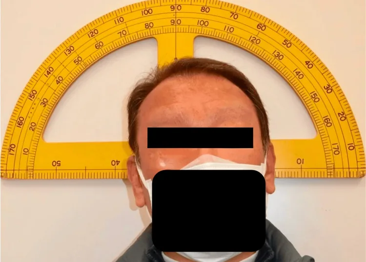
Kawamura J, et al. A Case of Bilateral Trochlear Nerve Palsy Induced by a Dorsal Midbrain Intracranial Lipoma. Cureus. 2025. Figure 1. PMCID: PMC12662652. License: CC BY.
Head tilt to the right, chin elevation, and face turn to the left are observed. This corresponds to the compensatory head posture discussed in the section “2. Main Symptoms and Clinical Findings.”
The main complaint is binocular vertical diplopia or torsional diplopia. The characteristics of symptoms differ depending on the type of palsy.
Congenital: Head tilt toward the unaffected side is the main sign. Since eye alignment is maintained with head tilt, patients often do not notice diplopia.
Decompensated: Head tilt has been present since childhood, and patients often notice vertical diplopia in their 20s to 30s. Before age 6, sensory adaptation makes torsional diplopia less noticeable.
Acquired: Patients often notice both vertical and torsional diplopia. In bilateral traumatic palsy, excyclotorsion of 10 degrees or more is observed.
Since the superior oblique muscle acts to depress the eye in adduction, diplopia worsens in downgaze. Symptoms are often noticed during reading or going up and down stairs. In mild or chronic cases, instead of diplopia, patients may experience blurred vision, difficulty focusing, or dizziness.
Clinical Findings (Findings Confirmed by Physician Examination)
Head tilt toward the healthy side: Abnormal head posture to compensate for eye position. Accompanied by facial asymmetry.
Large vertical fusion amplitude: Fusion amplitude of 10–15 prism diopters, no diplopia even with hypertropia of 10 PD or more.
Good binocular vision: Stereopsis is maintained as long as eye position is preserved with head tilt.
Decompensated
Severe hypertropia: Large deviation manifesting as exacerbation of congenital condition.
Facial asymmetry: Finding related to long-term ocular torticollis. The line connecting the eye and mouth corner on the healthy side intersects.
Superior oblique muscle atrophy: Atrophy of the superior oblique muscle on the affected side on MRI T1 coronal view is useful for diagnosis.
Acquired
Hypertropia + excyclotorsion on the affected side: Due to reduced intorsion and depression function of the superior oblique muscle.
Inferior oblique overaction: Secondary finding where the affected eye elevates on adduction.
Compensatory head tilt: Head tilt toward the healthy side to reduce diplopia.
Ocular movements often show no obvious abnormalities, and diagnosis is made by eye position testing. In bilateral trochlear nerve palsy, alternating hypertropia (left hypertropia on right gaze, right hypertropia on left gaze) occurs, and if excyclotorsion is 10 degrees or more, bilaterality should be considered.
QIn what situations is diplopia from trochlear nerve palsy likely to be noticed?
A
The superior oblique muscle acts strongly as a depressor during adduction, so diplopia worsens on downward gaze. Symptoms are easily noticed during daily activities such as reading, checking items at hand during meals, or going down stairs. It also worsens on lateral gaze to the side opposite the affected side.
Congenital: Caused by congenital hypoplasia of the trochlear nerve or morphological abnormalities of the superior oblique muscle itself. A considerable number of cases with CN4 deficiency have been reported on high-resolution 3T MRI. It is a common cause of congenital cranial nerve palsy.
Traumatic: The most common cause. Because the trochlear nerve has the longest intracranial course, it is susceptible to injury even from relatively minor trauma. Trauma to the midline to vertex of the head can cause contusion at the decussation in the anterior medullary velum, often resulting in bilateral involvement.
Ischemic (microvascular): Common in elderly patients with systemic diseases that cause arteriosclerosis, such as hypertension, diabetes, and hyperlipidemia. Many cases resolve spontaneously within 2 to 6 months.
Idiopathic: Reported in about 4% of cases in population-based studies. May improve or resolve within weeks.
In peripheral trochlear nerve palsy, as with other ocular motor nerve palsies, vascular disorders are often the cause, but aneurysms are rarely the cause. However, in acute-onset cases, the possibility of giant cell arteritis (GCA) should also be considered.
Occurs as part of a broader syndrome related to trauma, tumor, infection, inflammation, etc. Increased intracranial pressure can also be a cause. It is classified based on the affected site (midbrain, subarachnoid space, cavernous sinus, orbit).
QCan trochlear nerve palsy be caused by an aneurysm?
A
In peripheral trochlear nerve palsy, aneurysms are rarely the cause. This is a major difference from oculomotor nerve palsy. However, rare cases have been reported where a subarachnoid aneurysm presents as isolated fourth nerve palsy.
This is the standard method used to diagnose unilateral fourth nerve palsy.
Step 1 (Determine the hypertropic eye): Identify the eye with hypertropia, narrowing down the possible muscles to four. The ipsilateral depressors (superior oblique, inferior rectus) or contralateral elevators (superior rectus, inferior oblique) are candidates.
Step 2 (Determine the direction of gaze that worsens the hypertropia): Check whether the hypertropia worsens on right or left gaze, narrowing the candidates to two.
Step 3 (Bielschowsky head tilt test): If tilting the head toward the affected side worsens the hypertropia, this identifies palsy of the ipsilateral superior oblique muscle. This occurs because the superior rectus attempts to compensate for the weakened intorsion of the superior oblique, elevating the eye.
Skew deviation results from damage to fibers from the vestibular nerve to the interstitial nucleus of Cajal in the midbrain and may show a pattern similar to trochlear nerve palsy on the Parks three-step test. Differentiation is important.
Double Maddox rod test: Quantitatively evaluates excyclotorsion. Bilateral cases show large excyclotorsion of 10 degrees or more.
Fundus photography: Confirms excyclotorsion of the affected eye.
Hess red-green test / Large synoptophore: Determines the paretic muscle by examining eye positions in 9 directions. In mild cases, outward rotation in the tertiary position on the large synoptophore may be the key to diagnosis.
MRI/CT: In congenital cases, abnormal insertion or hypoplasia of the superior oblique muscle can be observed. To differentiate between decompensated and acquired cases, compare the presence or absence of superior oblique muscle atrophy on MRI T1 coronal sections.
Forced duction test: In congenital cases, the superior oblique muscle is often loose; in acquired cases, it is rarely loose. This serves as a guide for performing superior oblique muscle strengthening surgery.
Myasthenia gravis: Presents with pseudo-trochlear nerve palsy. Differentiate by checking for diurnal variation and easy fatigability.
Thyroid eye disease: Presents with vertical strabismus. If there is no history of trauma, in addition to anti-AChR antibodies, always perform blood tests for thyroid-related autoantibodies (TSAb, TRAb, etc.).
Brown syndrome: Limitation of elevation in adduction due to abnormality of the superior oblique tendon sheath.
Ischemic: Many cases resolve spontaneously within 2 to 6 months, so observation is the main approach. Oral vitamin B complex and circulation-improving drugs may be used.
Traumatic: Spontaneous recovery is often not achieved, and if torsional diplopia persists, surgery is indicated.
Prism glasses: Effective for correcting vertical deviation, with a practical correction range of up to about 10 prism diopters.
Occlusion (patch): An alternative for patients who do not wish to use prisms or undergo surgery.
Torsional deviation cannot be corrected with prisms, so surgery is necessary when torsional diplopia is the main symptom.
Inferior oblique weakening (inferior oblique myectomy, recession, or anterior transposition) is performed. If vertical deviation is large, ipsilateral superior rectus recession or contralateral inferior rectus recession is combined.
To eliminate cyclodiplopia, contralateral inferior rectus recession with nasal transposition is the first choice. It can correct both vertical and cyclotorsional deviation simultaneously. Nasal transposition of the inferior rectus by one muscle belly width provides approximately 6–7 degrees of excyclotorsion correction.
Surgical outcomes are good. However, correction of vertical deviation is subtle, and examination on the first postoperative day may require adjustment. The probability of adjustment is about 30%.
QWhat if prism glasses cannot correct the condition?
A
Cyclotorsional deviation (excyclotorsion) cannot be corrected with prism glasses. When cyclodiplopia is the main symptom, surgery is indicated. In acquired cases, contralateral inferior rectus recession with nasal transposition is the first choice, correcting both vertical and cyclotorsional deviation simultaneously. See the “Standard treatment” section for details.
The trochlear nucleus is located in the ventral part of the inferior colliculus in the dorsal midbrain, continuous with the caudal end of the oculomotor nucleus. It lies dorsal to the medial longitudinal fasciculus (MLF).
The nerve fibers emerging from the nucleus follow the following pathway:
They run dorsally and decussate to the contralateral side at the level of the anterior medullary velum (the roof of the cerebral aqueduct).
They curve around the cerebellar peduncle and run in the subarachnoid space between the superior cerebellar artery and the posterior cerebral artery.
They pass through the lateral wall of the cavernous sinus, below the oculomotor nerve.
They enter the orbit through the superior orbital fissure, passing lateral to the annulus of Zinn, and distribute to the superior oblique muscle.
When a strong external force (traffic accident, head trauma) is applied, the dorsal midbrain is pressed against the tentorial edge, and the decussation of the anterior medullary velum is damaged. Because the structure crosses at the midline, bilateral trochlear nerve palsy is likely to occur.
In congenital superior oblique palsy, high-resolution 3T MRI has reported that CN4 is absent in a considerable number of patients. Congenital superior oblique palsy may be classified as congenital cranial dysinnervation disorder (CCDD), but the causative gene has not been identified. Abnormalities of the muscle insertion and hypoplasia of the superior oblique muscle are often more severe than in acquired cases.
In non-isolated fourth nerve palsy, the site of the lesion can be inferred from accompanying symptoms.
Midbrain (nuclear/fascicular): May be accompanied by hemisensory loss, ataxia, internuclear ophthalmoplegia (INO), hemiparesis, and central Horner syndrome. Due to infarction or hemorrhage.
Subarachnoid space: Meningitis (fever, headache, nuchal rigidity). May also be caused by aneurysm.
Cavernous sinus: Involvement of cranial nerves III, V, VI and Horner syndrome.
Orbit: Involvement of cranial nerves II, III, V, VI, proptosis, conjunctival edema.
7. Latest research and future perspectives (research-stage reports)
Kawamura et al. (2025) reported a 67-year-old man with bilateral trochlear nerve palsy due to a lipoma in the dorsal midbrain. Since his 50s, he had symptoms of the road center line appearing tilted, and MRI revealed a lipoma in the dorsal midbrain. Excyclotorsion was 25 degrees, and stereopsis was absent. Bilateral inferior rectus nasal transposition (with 2 mm additional recession in the right eye) was performed, and postoperatively excyclotorsion improved to 5 degrees, with stereopsis of 60 seconds and disappearance of diplopia1).
Selective Plication of the Anterior Fibers of the Superior Oblique Muscle
Rao et al. (2025) performed selective plication of the anterior one-third fibers of the superior oblique muscle in a 33-year-old man with 20 degrees of excyclotorsion due to bilateral trochlear nerve palsy after a motorcycle accident. Graduated plication of 5 mm in the right eye and 10 mm in the left eye corrected the excyclotorsion, achieving orthophoria and resolution of diplopia in all directions at 3 months postoperatively. This method is non-transecting, reversible, and less invasive than the Harada-Ito procedure or the Fells modification 5).
Kiarudi et al. (2024) reported the first case of fourth cranial nerve palsy in a healthy 10-year-old boy with confirmed asymptomatic COVID-19 infection. He presented with an abnormal head posture and left eye hypertropia; brain MRI was normal. After one year, 10 prism diopters of left hypertropia and inferior oblique overaction persisted, suggesting that post-viral fourth nerve palsy may have a poorer prognosis than sixth nerve palsy 4).
Combined Cranial Nerve Palsy Associated with Herpes Zoster Ophthalmicus
Low et al. (2022) reported a 78-year-old woman with simultaneous oculomotor and trochlear nerve palsy complicating herpes zoster ophthalmicus (HZO). Complete ptosis and ocular movement limitation appeared 12 days after rash onset. After six weeks of oral acyclovir (800 mg five times daily) and steroids, ptosis resolved and ocular movements markedly improved at one year 2).
Subramaniam et al. (2023) reported a 56-year-old woman with isolated fourth nerve palsy due to a posterior fossa arachnoid cyst. She presented with one week of vertical diplopia, and MRI revealed an arachnoid cyst in the left posterior cerebellar region. The cyst was thought to compress the posterior cerebellum and affect the fourth nerve course in the dorsal brainstem. Diplopia showed improvement during follow-up 3).
Iida et al. (2024) reported a 19-year-old man with superior oblique muscle palsy (canine tooth syndrome) due to a dog bite. MRI on day 5 after injury showed inflammation extending from the superior oblique tendon to the trochlea and muscle belly, and early steroid therapy was administered. Although the development of pseudo-Brown syndrome was prevented, diplopia persisted; inferior oblique myectomy was performed at 7 months, improving cyclotorsional and vertical diplopia6).
Kawamura J, Shimizu Y, Tabuchi H. A Case of Bilateral Trochlear Nerve Palsy Induced by a Dorsal Midbrain Intracranial Lipoma. Cureus. 2025;17(10):e95704. doi:10.7759/cureus.95704. PMID:41322811; PMCID:PMC12662652.
Low KL, Nurul-Ain M, Che Hamzah J, Wan Hitam WH. Simultaneous Oculomotor and Trochlear Nerve Palsy in Herpes Zoster Ophthalmicus. Cureus. 2022;14(10):e30755. doi:10.7759/cureus.30755. PMID:36447705; PMCID:PMC9700447.
Subramaniam R, Wan Hitam WH, Sonny Teo KS, et al. Right Trochlear Nerve Palsy as an Uncommon Manifestation of Arachnoid Cyst. Cureus. 2023;15(1):e33579.
Kiarudi MY, Sharifi M, Gharouni A, et al. Presumed Fourth Nerve Palsy in a Healthy and Asymptomatic Child with COVID-19 Infection. Iran J Child Neurol. 2024;18(3):137-141.
Rao S, Raghunandan N. Selective plication and grading of plication for correction of torsion in superior oblique palsy. Saudi journal of ophthalmology : official journal of the Saudi Ophthalmological Society. 2025;39(1):107-109. doi:10.4103/sjopt.sjopt_201_23. PMID:40182973; PMCID:PMC11964351.
Iida K, Goseki T, Kunimi K, Hashimoto R. A Case of Superior Oblique Palsy without Brown Syndrome Induced by a Dog Bite. The American journal of case reports. 2024;25:e943299. doi:10.12659/AJCR.943299. PMID:38508873; PMCID:PMC10926234.
Copy the article text and paste it into your preferred AI assistant.
Article copied to clipboard
Open an AI assistant below and paste the copied text into the chat box.