Eye Trauma reference image

Eye Trauma

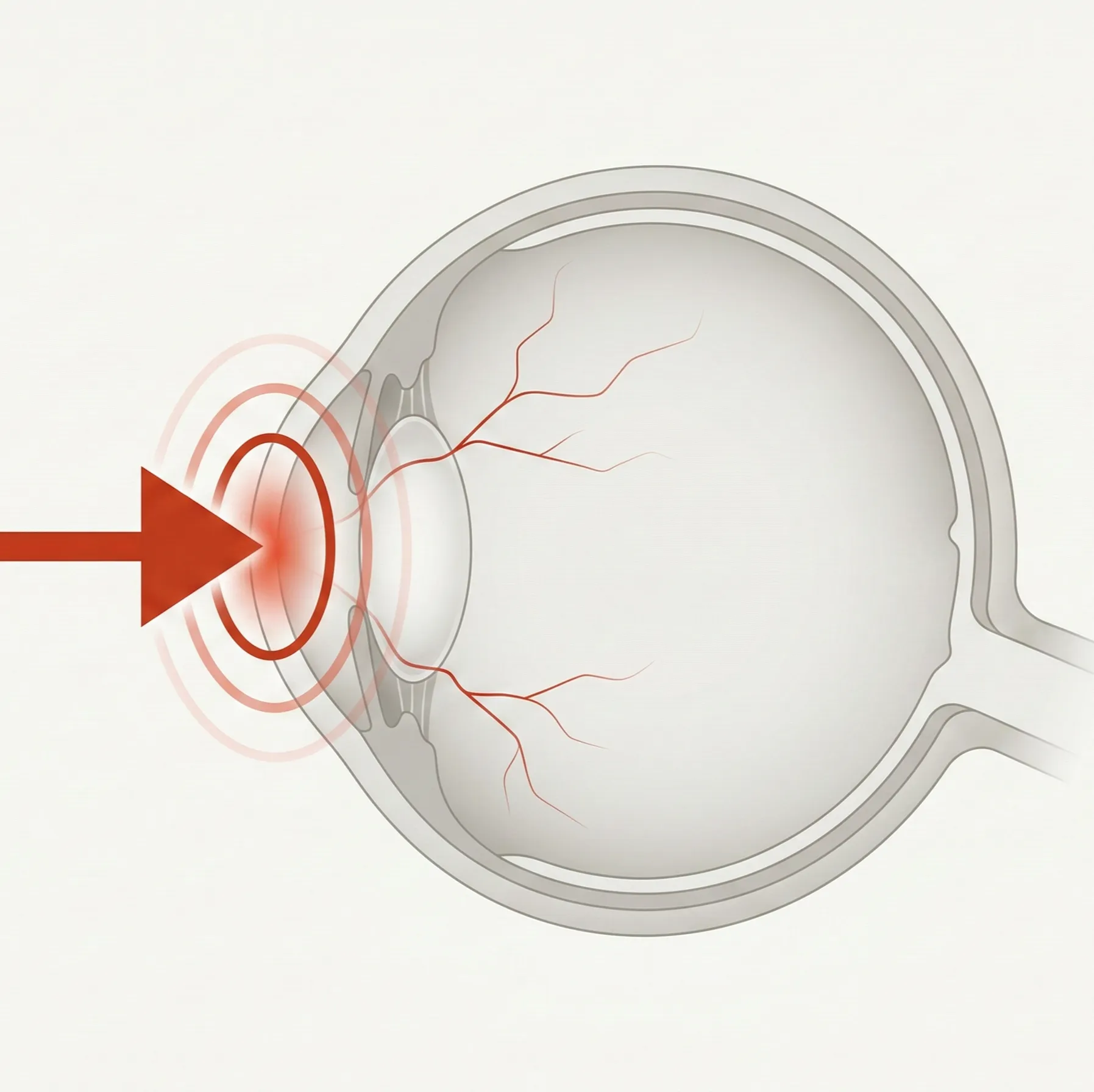
The eye is exposed to the outside environment and can be injured by blunt trauma, penetrating injury, foreign bodies, chemicals, heat, or radiation. This category covers classification and management principles for eye trauma.

61 English articles

Frequently referenced diseases

A

1 articles

B

1 articles

C

9 articles
Canalicular Laceration (Trauma)

Explains the causes, symptoms, diagnosis, and surgical treatment of traumatic canalicular laceration. Details the characteristics by injury mechanism such as dog bites and assault, repair techniques including lacrimal tube intubation, and postoperative management.

Chemical Injury (Acid Burn)

This article explains the definition, clinical features, severity assessment, and treatment from eye irrigation to surgery for ocular injuries caused by acidic chemicals. It clarifies the differences in pathophysiology from alkali injuries.

Chemical Injury (Alkali Burn)

This article explains the definition of ocular injury caused by alkaline chemicals, severity assessment using the Kinoshida classification, and treatment from emergency irrigation to ocular surface reconstruction.

Choroidal Rupture

A disease in which blunt trauma to the eye causes rupture of Bruch's membrane, the choriocapillaris, and the retinal pigment epithelium. Immediately after injury, it is hidden by hemorrhage, but after absorption, it is observed as a crescent-shaped white line. Attention must be paid to the development of choroidal neovascularization.

Commotio Retinae

A disease in which the outer segments of photoreceptors are damaged after blunt ocular trauma, resulting in characteristic white opacification of the retina. It often resolves spontaneously within about two weeks, but visual prognosis may be poor if the macula is affected.

Conjunctival foreign body

A condition in which a foreign body is attached to or embedded in the conjunctiva (tarsal conjunctiva, bulbar conjunctiva, or fornix conjunctiva). It accounts for 4.4% of new ophthalmology outpatients. Common foreign bodies can be removed with moist cotton, but toxic caterpillar hairs carry a risk of vitreous penetration and require complete removal and a retinal-vitreous examination.

Conjunctival laceration

A traumatic tear of the conjunctival tissue. Small lacerations may heal on their own with antibiotic eye drops, but large lacerations require suturing. The most important clinical point is to rule out a scleral laceration or globe rupture hidden beneath the conjunctival laceration.

Corneal Epithelial Abrasion

A condition in which the entire corneal epithelium is lost, and one of the most frequent ocular traumas in ophthalmic emergencies. Simple cases heal within a few days with antibiotic eye drops and hyaluronic acid eye drops, but persistent epithelial defects (PED) lasting more than one week require investigation of the cause.

Corneal Foreign Body (e.g., Iron Filings)

This article explains the types, diagnosis, and removal techniques (foreign body needle, drill, forceps) of corneal foreign bodies, as well as postoperative management. It covers essential knowledge for ophthalmic emergencies, from rust ring formation due to iron foreign bodies to infection risk from vegetative foreign bodies.

D

1 articles

E

4 articles

G

1 articles

I

3 articles

L

3 articles

M

2 articles

O

14 articles
Ocular Burns (Thermal Burns)

This article explains the classification of ocular burns caused by high-temperature substances (first to third degree), the Kinoshita classification for evaluating corneoconjunctival damage, and treatment from acute-phase cooling to ocular surface reconstruction.

Ocular evisceration

This article explains the indications, surgical technique, postoperative care, and comparison with enucleation for evisceration. It is a procedure that preserves the scleral shell and extraocular muscles while removing only the contents of the eye, and it offers advantages for prosthetic eye movement and appearance.

Ocular Trauma from Firearms

This article explains the epidemiology, diagnosis, treatment, and prevention of ocular and orbital trauma caused by firearms (handguns, rifles, airsoft guns, etc.).

Ocular Trauma from Fireworks and Fragments

This article explains the epidemiology, symptoms, diagnosis, treatment, and prognosis of ocular trauma caused by flying debris (fragments) from fireworks or explosions. Blunt trauma, burns, and chemical injuries occur in combination, and severe cases can lead to globe rupture or permanent vision loss.

Ocular Trauma Management Before Ophthalmologist Visit

This article explains the initial assessment and management of ocular trauma, including triage, emergency treatment, and criteria for ophthalmology referral that non-ophthalmologists should perform.

Ophthalmic artery pseudoaneurysm

A rare vascular lesion in which a false lumen forms in the ophthalmic artery due to trauma or postoperative complications. It is characterized by a pulsatile orbital mass, and endovascular coil embolization is the first-line treatment.

Optic Nerve Avulsion

A rare severe injury in which the optic nerve is separated from the eye at the level of the lamina cribrosa due to trauma. Complete avulsion results in no light perception, and there is no established effective treatment. Early diagnosis and avoidance of unnecessary treatment are important.

Optic nerve head avulsion

Optic nerve head avulsion (optic nerve head avulsion) is a rare condition in which trauma separates the optic nerve from the eyeball at the level of the lamina cribrosa. It causes severe visual impairment, and there is no established treatment.

Orbital Compartment Syndrome

An ophthalmic emergency in which a rapid increase in intraorbital pressure compromises blood flow to the optic nerve and retina, leading to irreversible blindness if urgent decompression is not performed. Immediate decompression via lateral canthotomy and inferior cantholysis is the mainstay of treatment.

Orbital Emphysema

A condition caused by abnormal accumulation of air within the orbit. The most common cause is nose blowing after an orbital wall fracture. In most cases, it resolves spontaneously within 7–10 days, but severe cases can lead to orbital compartment syndrome and potential blindness.

Orbital Floor Fracture

An orbital floor fracture (blowout fracture) is a fracture caused by blunt trauma to the eye area, with double vision, sunken eye, and impaired eye movement as the main symptoms. Treatment ranges from urgent surgery to observation depending on whether tissue is trapped and on the fracture type.

Orbital Floor Fracture (Blowout Fracture)

A fracture of the orbital floor caused by blunt trauma to the eye, with main symptoms of diplopia, enophthalmos, and impaired eye movement. Closed fractures with entrapment of extraocular muscles are common in children and require emergency surgery. Surgery reconstructs the orbital floor using titanium mesh or absorbable plates.

Orbital Foreign Body

This article explains the definition, classification, diagnosis, treatment, and prognosis of orbital foreign bodies, including imaging diagnosis centered on CT and management strategies based on material type.

Orbital Roof Fracture

Orbital roof fracture is a relatively rare orbital fracture caused by trauma to the forehead. In adults, it is often associated with high-energy trauma and severe head injury. Multidisciplinary collaboration with neurosurgery and otolaryngology is necessary.

P

4 articles

R

3 articles

S

2 articles

T

11 articles
Terson syndrome

Explains the definition, mechanism of onset, diagnosis, and treatment of Terson syndrome, a vitreous hemorrhage and preretinal hemorrhage that occurs secondary to subarachnoid hemorrhage. It develops in 3–20% of SAH cases, and deciding whether vitreous surgery is indicated affects the visual prognosis.

Traumatic Cataract

Classification of traumatic cataracts following ocular trauma (blunt, sharp, foreign body, non-mechanical), clinical findings such as Vossius ring, diagnosis, preoperative planning, surgical techniques, amblyopia management in children, and prognosis. Detailed explanation of prognosis prediction using the OTS score and selection criteria for primary versus secondary extraction.

Traumatic Hyphema

A condition in which blood accumulates in the anterior chamber due to blunt trauma. Classified into Grades 0 to IV. Basic treatment includes rest, cycloplegics, and steroid eye drops. Attention should be paid to rebleeding, elevated intraocular pressure, corneal blood staining, and angle recession glaucoma.

Traumatic Lens Dislocation

Classification, symptoms, diagnosis, and surgical indications (CTR, intrascleral fixation, vitrectomy) of traumatic lens dislocation/subluxation caused by rupture of the zonules of Zinn due to blunt ocular trauma, along with differential diagnosis from systemic diseases.

Traumatic Macular Hole

This article explains the definition, epidemiology, OCT diagnosis, possibility of spontaneous closure, vitrectomy (with or without ILM peeling), and prognosis of traumatic macular hole caused by blunt ocular trauma or laser injury. It also details the differentiation from idiopathic macular hole and management of choroidal neovascularization.

Traumatic Mydriasis (Sphincter Pupillae Rupture)

A dilated pupil caused by rupture of the sphincter pupillae muscle due to blunt trauma. Main symptoms are decreased near vision and photophobia. The diagnosis is suggested by the absence of miosis after instillation of 1% pilocarpine. Differentiation from oculomotor nerve palsy is important.

Traumatic Optic Neuropathy

A disease in which indirect force from blunt trauma to the eyebrow area acts on the optic canal, damaging the optic nerve. Early treatment within 24 to 48 hours after injury greatly affects prognosis. Steroid pulse therapy and optic canal decompression are treatment options.

Traumatic Phthisis Bulbi (End-Stage Ocular Atrophy)

This article explains the definition, causes, diagnosis, treatment, and artificial eye management of phthisis bulbi, the end-stage condition of an irreversibly atrophied eye following ocular trauma, endophthalmitis, chronic uveitis, etc.

Traumatic Retinal Detachment

Rhegmatogenous retinal detachment caused by open or closed ocular trauma. For open injuries, vitrectomy is the basic approach; for closed injuries, scleral buckling surgery is the standard. It is common in young people and children, and attention must be paid to progression to proliferative vitreoretinopathy.

Traumatic subconjunctival hemorrhage

Subconjunctival bleeding caused by blunt trauma to the eye and rupture of conjunctival blood vessels. The bleeding itself usually clears on its own within 1 to 4 weeks, but the most important part of care is ruling out a conjunctival laceration, scleral laceration, or globe rupture hidden beneath the subconjunctiva.

Traumatic Vitreous Hemorrhage

Explains the causes, symptoms, diagnosis using B-mode ultrasound, indications for vitrectomy, management of ghost cell glaucoma, and prognosis of traumatic vitreous hemorrhage following blunt or open eye injury.

U

1 articles

Z

1 articles