Cataract & Anterior Segment reference image

Cataract & Anterior Segment

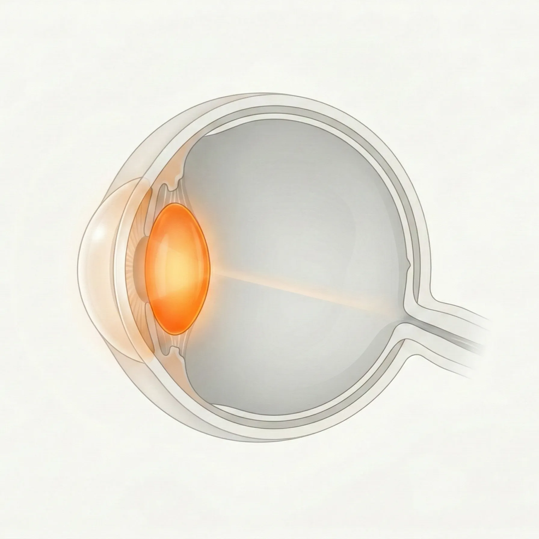
The lens focuses light inside the eye and can become cloudy with age or other causes. The anterior segment includes the region from the cornea to the lens. This category covers cataract, lens position abnormalities, iris disease, and anterior chamber conditions.

123 English articles

Frequently referenced diseases

A

12 articles
ADAMTSL4-Related Eye Disease

Congenital eye abnormalities caused by mutations in the ADAMTSL4 gene. This rare hereditary eye disease is characterized by isolated ectopia lentis, ectopia lentis et pupillae, and congenital cataract, with autosomal recessive inheritance.

Add-on Intraocular Lens (Add-on IOL)

This article explains the types, indications, procedures, and complications of add-on IOLs (piggyback IOLs) that are inserted in front of an existing intraocular lens after cataract surgery.

After-Cataract and Optic Capture

This article explains the pathology, prevention, and treatment of posterior capsule opacification (after-cataract) after cataract surgery, as well as the optic capture technique using posterior optic buttonhole fixation (POBH).

Aniridia

Congenital iris defect caused by PAX6 gene mutation. It is frequently associated with glaucoma, cataract, corneal limbal stem cell deficiency, and macular hypoplasia. This article explains the definition, diagnostic criteria, severity classification, and treatment strategies.

Aniridia Fibrosis Syndrome

A rare complication occurring in patients with congenital aniridia after intraocular surgery. A fibrous membrane forms from the rudimentary iris root, causing intraocular lens displacement, corneal endothelial failure, and hypotony.

Anterior Chamber Intraocular Lens

An anterior chamber-fixated intraocular lens (ACIOL) used when capsular or zonular support is lost. Modern flexible open-loop designs achieve better outcomes than older designs.

Anterior Chamber Washout

A procedure in which blood, pus, inflammatory substances, chemicals, viscoelastic materials, etc. in the anterior chamber are washed out with irrigation fluid. It is indicated for a wide range of anterior segment diseases such as endophthalmitis, hyphema, corneal blood staining, chemical burns, and post-cataract surgery complications.

Anterior Segment Developmental Anomalies (ASDA)

A collective term for congenital disorders caused by abnormal development of the anterior segment of the eye, including the cornea, iris, and lens. They are characterized by high genetic heterogeneity and a common tendency to complicate secondary glaucoma.

Artificial Iris Device (Prosthetic Iris)

Detailed explanation of types, indications, surgical techniques, and complications of artificial iris devices for aniridia, trauma, albinism, etc., and the risks of cosmetic use.

Astigmatism Correction During Cataract Surgery

A treatment strategy to simultaneously correct preexisting corneal astigmatism during cataract surgery. This article explains multiple methods such as toric intraocular lenses, femtosecond laser arcuate incisions, and limbal relaxing incisions.

Atopic Cataract

Juvenile cataract associated with atopic dermatitis. Characterized by star-shaped opacities under the anterior capsule. Eye rubbing behavior and influx of intraocular eosinophil granule proteins are involved in its development. Surgical planning must consider the risk of zonular weakness and retinal detachment.

Axenfeld nerve loop

A normal anatomical variant where the long posterior ciliary nerve forms a loop within the sclera. It is incidentally found during slit-lamp examination and is important to differentiate from scleral foreign bodies or tumors, but no treatment is required.

B

3 articles

C

18 articles
Capsule Support Devices (Capsular Tension Ring and Segment)

A group of devices used to maintain capsular stability during cataract surgery with zonular weakness or dehiscence. Includes capsular tension ring (CTR), modified CTR, capsular tension segment (CTS), and capsule support hooks.

Cataract

A disease in which the lens becomes cloudy, causing decreased vision. Aging is the most common cause, and the prevalence reaches 100% in people aged 80 and older. Phacoemulsification and intraocular lens implantation are the only curative treatments.

Cataract Surgery after Vitrectomy and Combined Cataract and Vitrectomy Surgery

Overview of surgery for cataract that progresses after vitrectomy, and combined cataract and vitrectomy surgery (phacovitrectomy). Preoperative, intraoperative, and postoperative points to note are explained.

Cataract Surgery in Patients with Keratoconus

This article explains preoperative planning, IOL power calculation, surgical techniques, and postoperative management for cataract surgery in patients with keratoconus (a progressive ectatic corneal disease). The main challenges are difficulty in IOL power calculation and postoperative refractive error.

Cataract Surgery Positioning for Patients Unable to Lie Flat

Positioning management for cataract surgery in patients who cannot assume the supine position (lying flat on the back). Explains three options for patients with ankylosing spondylitis, orthopnea, kyphosis, etc.

Cataract Surgery Training

Explanation of educational methods for acquiring cataract surgery (phacoemulsification). Shows an overview of modern surgical education including VR simulators, wet labs, and competency assessment.

Cataract Surgery Using the Bag-in-the-Lens (BIL) Fixation Technique

An innovative cataract surgery technique that prevents posterior capsule opacification by fitting both the anterior and posterior capsule edges into the groove of the intraocular lens. It is applicable to both adults and children.

Cerebrotendinous Xanthomatosis (CTX)

A rare autosomal recessive lipid storage disease caused by CYP27A1 gene mutation leading to sterol 27-hydroxylase deficiency, impairing bile acid synthesis and resulting in accumulation of cholestanol in the brain, tendons, and lens.

Clear Lens Extraction

A refractive surgery that removes the clear lens without cataract and inserts an intraocular lens. Performed for high refractive errors and angle-closure glaucoma.

Combined Cataract and Glaucoma Surgery (Filtration Surgery / MIGS)

This article organizes the concepts of combined surgery for cataract and glaucoma. It clearly explains the selection axes: cataract surgery alone, combined with MIGS, combined with trabeculectomy, and combined with drainage device.

Comparison of Intraocular Lens (IOL) Materials

A comparative explanation of the characteristics of intraocular lens (IOL) materials used in cataract surgery. Covers optical properties, biocompatibility, and complications of major materials such as hydrophobic acrylic, hydrophilic acrylic, silicone, PMMA, and collamer.

Complicated Cataract (Uveitis Cataract)

Cataract that occurs secondary to uveitis or systemic diseases (e.g., diabetes, atopic dermatitis, myotonic dystrophy). Preoperative inflammation control and perioperative management determine surgical outcomes.

Complications of Cataract Surgery

A general term for intraoperative and postoperative complications associated with cataract surgery. Includes posterior capsule rupture, posterior capsule opacification, cystoid macular edema, endophthalmitis, intraocular lens dislocation, etc. Appropriate prevention and management directly affect visual prognosis.

Congenital Cataract

Lens opacity that develops at birth or during infancy. It occurs in 1 in 1,000 to 10,000 births. Early surgery to prevent form deprivation amblyopia, along with long-term amblyopia treatment and refractive correction, determines visual prognosis.

Continuous Curvilinear Capsulorhexis (CCC)

Standard technique for anterior capsulotomy in cataract surgery. Creates a circular opening in the anterior lens capsule to achieve stable fixation of the intraocular lens.

Corneal Wound Hydration

A technique in which irrigation fluid is injected into the corneal stroma to swell the cornea, assisting in the closure of clear corneal incisions (CCI) during cataract surgery. It is widely used to prevent postoperative endophthalmitis.

Creation of Incisions in Cataract Surgery

Explains the methods for creating incisions in cataract surgery (phacoemulsification). Summarizes the techniques for clear corneal incision, sclerocorneal incision, and transconjunctival single-plane sclerocorneal incision, as well as criteria for selecting location, size, and structure, and key points for preventing complications.

Cystoid Macular Edema after Cataract Surgery

A postoperative complication in which cystoid fluid accumulation occurs in the outer plexiform and inner nuclear layers of the macula after cataract surgery, causing vision loss. Also called Irvine-Gass syndrome, it often resolves spontaneously, but if it becomes chronic, it can lead to permanent visual impairment.

D

4 articles

E

2 articles

F

1 articles

H

5 articles

I

20 articles
Immediate Sequential Bilateral Cataract Surgery (ISBCS)

This article explains the indications, contraindications, surgical techniques, complications, and safety protocols for simultaneous bilateral cataract surgery (Immediately Sequential Bilateral Cataract Surgery: ISBCS). It also details the comparison with sequential surgery performed one eye at a time, and the importance of complete sterile separation to prevent endophthalmitis.

Infection Prevention in Cataract Surgery

Perioperative strategies for preventing infectious endophthalmitis after cataract surgery. This article explains evidence-based prevention methods including preoperative disinfection, preoperative and postoperative antibiotic eye drops, intracameral antibiotic administration, and incision wound management.

Intracapsular Hematoma

A rare complication after cataract surgery in which blood accumulates between the posterior surface of the intraocular lens and the posterior lens capsule.

Intraocular Lens (IOL)

A comprehensive hub summarizing types, indications, power calculation, and postoperative visual function of intraocular lenses (IOLs). It organizes monofocal, trifocal, EDOF, toric, and phakic IOLs with comparison tables and power calculation formula selection charts.

Intraocular Lens Dislocation

A complication after cataract surgery where the intraocular lens shifts from its normal position. Exfoliation syndrome and zonular weakness are major risk factors, causing decreased vision and increased intraocular pressure.

Intraocular Lens Opacification

A complication in which the optic part of an intraocular lens (IOL) becomes cloudy after cataract surgery. There are several mechanisms, including calcium deposition and glistening. If visual function is impaired, IOL removal and exchange is required.

Intraoperative Aberrometry

A wavefront aberrometry technique used during cataract surgery to verify and optimize intraocular lens (IOL) power. It is particularly useful in eyes with prior refractive surgery and for toric IOL alignment.

Intraoperative Floppy Iris Syndrome (IFIS)

A syndrome characterized by iris flaccidity, prolapse, and progressive miosis that occurs during cataract surgery in patients with a history of taking α1-adrenergic receptor blockers (such as tamsulosin). Preoperative medication history review and appropriate intraoperative management are important.

Intratunnel Nucleus Fragmentation

A new nucleus processing technique for manual small-incision cataract surgery (MSICS). This procedure reduces induced astigmatism by dividing and removing the lens nucleus within a sclerocorneal tunnel smaller than 6 mm.

Intumescent Cataract

A progressive cataract in which the lens cortex liquefies and swells, increasing intra-lenticular pressure. Anterior capsulotomy during surgery is difficult, requiring special preoperative evaluation and intraoperative measures.

IOL Power Calculation in Eyes with Corneal Ectasia

This article explains the challenges and solutions for IOL power calculation during cataract surgery in patients with corneal ectasia, such as keratoconus and post-refractive surgery ectasia. The use of corneal tomography and keratoconus-specific formulas is key.

Iris Coloboma

Iris coloboma is a congenital condition caused by incomplete closure of the embryonic fissure, resulting in a defect in the lower part of the iris. It is characterized by a keyhole-shaped pupil and may be associated with choroidal or optic nerve involvement. This article explains the classification, diagnosis, treatment, and genetic background.

Iris cyst

An iris cyst is a fluid-filled sac that forms in the iris. It is classified as primary (pigment epithelial cyst or stromal cyst) or secondary (traumatic, postoperative, or drug-induced). Most are asymptomatic, but large cysts can cause glaucoma or corneal endothelial damage.

Iris mammillations

Congenital benign cystic lesions at the pupillary margin. Derived from iris pigment epithelium, usually follow a benign course, but can be an ocular marker for familial aortic disease associated with ACTA2 gene mutations.

Iris microhemangioma

A minute, tangled vascular lesion at the pupillary margin (also called Cobb tuft or iris vascular tuft). Usually asymptomatic, but can cause spontaneous hyphema or secondary glaucoma; a rare benign vascular lesion.

Iris Repair (Iris Suture and Pupilloplasty)

Detailed explanation of indications, techniques (Siepser method, McCannel method, SFT method), and postoperative outcomes of iris suture (pupilloplasty) for congenital iris coloboma, trauma, and iris damage after cataract surgery. Also explains the criteria for choosing between iris repair and artificial iris devices.

Iris retraction syndrome

A rare condition characterized by posterior bowing of the peripheral iris and adhesion of the iris to the anterior lens surface. It occurs in association with retinal detachment, hypotony, and intraocular inflammation, and presents with a deep anterior chamber, opposite to iris bombe.

Iris varix

Iris varix is a benign lesion characterized by abnormal dilation and tortuosity of iris blood vessels. It is often asymptomatic, unilateral, and follows a stable course over the long term.

Irrigation Misdirection Syndrome

A serious intraoperative complication during cataract surgery where irrigation fluid passes through the zonules and misdirects posteriorly, causing anterior chamber shallowing and elevated intraocular pressure via a ciliary block-like mechanism. Early recognition and appropriate intraoperative management determine visual prognosis.

Ivacaftor-induced cataract (in patients with cystic fibrosis)

Non-congenital cataract occurring in children taking ivacaftor, a treatment for cystic fibrosis (CF). Visual impact is often minor, but regular ophthalmic screening is recommended.

L

8 articles
Lens Capsule Complications after Cataract Surgery

A group of complications of the lens capsule that occur after cataract surgery. This article explains three conditions: anterior capsule contraction syndrome (excessive contraction and fibrosis of the capsule), capsular distension syndrome (fluid accumulation within the capsule), and dead bag syndrome (late IOL dislocation due to loss of lens epithelial cells).

Lens Capsule Rupture (Capsular Break) and Management

A rupture of the posterior lens capsule during cataract surgery. Early recognition and appropriate management are crucial for visual prognosis.

Lens Coloboma

Lens coloboma is a congenital condition in which a partial defect of the zonular fibers causes a notch-like indentation at the equator of the lens. It is associated with incomplete closure of the embryonic fissure and often coexists with iris and chorioretinal coloboma.

Lens Luxation and Subluxation

A condition in which the lens is displaced from its normal position. It is caused by weakening or rupture of the zonules of Zinn. Congenital cases are associated with systemic diseases such as Marfan syndrome and homocystinuria, while trauma is the most common cause of acquired cases. Mild cases are managed with refractive correction and observation, while advanced cases require lens extraction.

Lens Nucleus Division and Fragmentation Techniques

General term for techniques to divide and fragment the lens nucleus during cataract surgery. Multiple procedures exist, such as divide-and-conquer, phaco-chop, and stop-and-chop, selected based on nuclear hardness, surgeon experience, and complication risk.

Light-Adjustable Intraocular Lens (LAL)

An intraocular lens (LAL) whose power can be adjusted postoperatively by ultraviolet irradiation after cataract surgery. FDA approved in 2017. An advanced IOL technology that improves refractive accuracy and patient satisfaction.

Long Anterior Zonules

An anatomical variant in which the zonules extend anteriorly beyond their normal attachment to the lens equator. Estimated prevalence is approximately 2%. Associated with risks of pigment dispersion syndrome, angle-closure glaucoma, and complications during cataract surgery.

Lowe syndrome (oculocerebrorenal syndrome)

An inborn error of metabolism caused by X-linked recessive mutations in the OCRL gene. It is a rare disease in males characterized by the triad of congenital cataracts, glaucoma, intellectual disability, and renal dysfunction.

M

7 articles
Management of Dropped Nucleus

This article explains the diagnosis and treatment of a dropped nucleus, a condition where the lens nucleus falls into the vitreous cavity during cataract surgery. It details the indications and techniques of vitrectomy (PFCL method, kebab method), secondary IOL fixation, and visual prognosis for this intraoperative complication with an incidence of 0.1–0.28%.

Manual Small Incision Cataract Surgery (MSICS)

A type of extracapsular cataract extraction in which the nucleus is removed through a self-sealing corneoscleral tunnel incision. It requires no sutures, is low-cost and short-duration, and is widely used especially in developing countries.

Marfan Syndrome

An autosomal dominant connective tissue disorder caused by mutations in the FBN1 gene. Ocular manifestations frequently include ectopia lentis, with increased risk of glaucoma, cataract, and retinal detachment.

Microspherophakia / Lenticonus

Microspherophakia is a congenital malformation characterized by a decreased equatorial diameter and increased anteroposterior diameter of the lens. Lenticonus is a congenital anomaly in which a conical protrusion occurs on the anterior or posterior surface of the lens. Both are caused by abnormalities of the zonules or lens capsule development and are often complicated by secondary glaucoma, high myopia, and cataracts.

Monofocal Intraocular Lens

Explains the types, materials, optical designs, power calculation, surgical outcomes, and postoperative complications of monofocal intraocular lenses (IOLs), which are most commonly used in cataract surgery. Hydrophobic acrylic is the standard material, and there are various options such as aspheric design and toric design.

Morgagnian Cataract

A condition in which a hypermature cataract progresses and the cortex liquefies, causing the hard nucleus to sink to the bottom of the lens capsule. It is common in developing countries and carries a risk of complications such as phacolytic glaucoma and phacoanaphylactic uveitis.

Multifocal Intraocular Lens (Presbyopia Correction)

A comprehensive explanation of multifocal intraocular lenses (trifocal, EDOF, accommodating IOL) in cataract surgery, including classification, optical principles, selection criteria, visual outcomes, complication management, and mix-and-match strategies.

O

4 articles

P

16 articles
Persistent Pupillary Membrane

Persistent pupillary membrane (PPM) is a congenital anomaly resulting from the persistence of the anterior tunica vasculosa lentis. This article details its epidemiology, classification, clinical findings, diagnosis, and treatment (mydriatics, laser, surgery).

Phacoanaphylactic Uveitis

Granulomatous anterior uveitis caused by type III allergy to lens proteins. It develops after trauma, surgery, or hypermature cataract with lens capsule rupture, and early removal of lens material is the definitive treatment.

Phacodynamics

Fluid dynamics and physical principles of ultrasonic energy in phacoemulsification (PEA). Explains the parameters and mechanisms that surgeons should control for safe and efficient cataract surgery.

Phacolytic Uveitis

Endophthalmitis caused by spontaneous capsular rupture of hypermature cataract or leakage of lens proteins after cataract surgery or trauma. Macrophages phagocytose lens proteins and obstruct the trabecular meshwork, often leading to elevated intraocular pressure. Surgical removal of lens material is the definitive treatment.

Phakic Posterior Chamber Lens (ICL)

Phakic posterior chamber lens (ICL) is a refractive surgery that corrects myopia and astigmatism by inserting a Collamer lens into the eye. It does not involve corneal ablation, is reversible, and can treat a wide range from moderate to high myopia.

Pinhole Intraocular Lens

An intraocular lens that extends the depth of focus using the small aperture (pinhole) principle. It is used during cataract surgery to correct presbyopia and reduce irregular astigmatism.

Post-cataract surgery eye drops

This article explains the types, usage, and evidence of topical eye drops (antibiotics, steroids, NSAIDs, dry eye treatments) used after cataract surgery, as well as the dropless strategy that omits eye drops by intraoperative administration.

Posterior Capsule Opacification

The most common postoperative complication after cataract surgery, caused by proliferation and migration of residual lens epithelial cells leading to opacification of the posterior capsule. It can be effectively treated with Nd:YAG laser posterior capsulotomy.

Posterior Polar Cataract

A subtype of congenital cataract characterized by discoid opacity at the posterior pole of the lens. It shows autosomal dominant inheritance and cataract surgery is challenging due to posterior capsule fragility.

Postoperative Endophthalmitis

A review of postoperative endophthalmitis occurring after cataract surgery. It covers both acute-onset (within 1 week post-surgery) and delayed-onset (after 1 month, typically caused by Cutibacterium acnes) forms. Topics include epidemiology, causative organisms, clinical features, diagnosis, stepwise treatment, EVS recommendations, and prevention strategies.

Preoperative Corneal Topography

This article explains the purpose, types, and clinical significance of corneal topography performed before cataract surgery and refractive surgery. It covers improvement of IOL power calculation accuracy, the impact of dry eye, and practical aspects of preoperative evaluation.

Preparation for Cataract Surgery in RGP Contact Lens Wearers

Long-term wear of rigid gas permeable (RGP) contact lenses can alter corneal shape (corneal warpage), affecting the accuracy of intraocular lens power calculation. It is necessary to discontinue lens wear for a sufficient period before cataract surgery to stabilize the corneal shape.

Pseudoexfoliation Syndrome

A systemic disease in which fibrous abnormal material deposits on the lens capsule, iris, ciliary body, etc. It increases the risk of complications during cataract surgery and is an important cause of glaucoma. It is found in about 4% of people aged 70 and older, and 20-40% of those affected develop glaucoma.

Pupil Abnormalities (Summary of Miosis and Mydriasis)

A comprehensive review article covering the causes, differential diagnosis, and diagnosis of miosis (small pupil) and mydriasis, including management of small pupils during cataract surgery. It organizes the differential diagnosis and treatment of Horner syndrome, Adie pupil, oculomotor nerve palsy, IFIS, and others.

Pupil Expansion Devices and Mechanical Pupil Dilation

Explanation of techniques and devices for securing the pupil using iris retractors, pupil expansion rings, and mechanical stretching in cataract surgery for small pupils and intraoperative floppy iris syndrome (IFIS).

Pupillary Capture of Intraocular Lens (IOL Capture)

A complication after cataract surgery where the optic part of the intraocular lens prolapses in front of the iris. It is more likely to occur after sutured IOL or intrascleral fixation IOL, with an incidence of about 3.6% for scleral-sutured IOL. Characterized by glare and photophobia, and attention should be paid to recurrence due to reverse pupillary block.

R

3 articles

S

9 articles
Scleral Fixation of IOL (Suturing and Intrascleral Fixation)

Surgical procedures for dislocated or dropped intraocular lenses. Suturing fixes the IOL to the sclera with sutures, while intrascleral fixation (Yamane technique) embeds the haptic into a scleral tunnel. This article explains indications, surgical selection, techniques, and complications.

Secondary Intraocular Lens Implantation

Secondary IOL implantation performed in aphakic eyes where IOL could not be inserted during initial cataract surgery, or in eyes with IOL dislocation, opacification, refractive error, etc. The surgical technique is selected based on the status of capsular support.

Shallow Anterior Chamber

A basic article on anterior segment practice covering classification of causes of shallow anterior chamber (pupillary block, plateau iris, postoperative, inflammatory, etc.), diagnosis by van Herick method, gonioscopy, anterior segment OCT, and UBM, and from emergency management of acute attack to lens reconstruction surgery.

Single-piece Intraocular Lens (Single-piece IOL)

This article explains the structure, characteristics, indications, and complications of a one-piece intraocular lens (1-piece IOL), in which the optic and haptics are made of the same material and molded as a single unit. It also provides a detailed comparison with three-piece IOLs and the rationale for contraindication of ciliary sulcus fixation.

Slit-lamp examination (slit-lamp test)

This article explains examination methods for the anterior and posterior segments using a slit lamp. It covers illumination techniques, systematic observation procedures, clinical significance, and applications in treatment. It also describes the latest applications such as slit-lamp treatment for silicone oil-induced pupillary block.

Stop-and-Chop Technique

A nuclear processing technique in cataract surgery (phacoemulsification). After dividing the nucleus into two parts using the divide-and-conquer method, the remaining nucleus is processed by transitioning to the phaco-chop technique. It is highly versatile and widely used by surgeons from beginners to experts.

Sub-Tenon's Anesthesia

An ophthalmic surgical anesthesia method in which a local anesthetic is injected into the potential space between Tenon's capsule and the sclera. Because a blunt needle is used, serious complications such as globe perforation are rare, and it is widely used from cataract surgery to vitrectomy.

Sunrise Syndrome

A condition where the intraocular lens (IOL) is displaced upward due to asymmetric placement of the haptic in the capsular bag and ciliary sulcus after cataract surgery, causing visual dysfunction.

Surgical Management of Intumescent Cataract

Key points of surgical management for intumescent cataract (a complex cataract with lens opacification and swelling). Explains difficulties in capsulorhexis due to increased intralenticular pressure and loss of red reflex, and management techniques using trypan blue staining, decompression, viscoelastic substances, and AS-OCT.

T

3 articles

U

3 articles

V

2 articles

W

1 articles

Z

1 articles

Other

1 articles