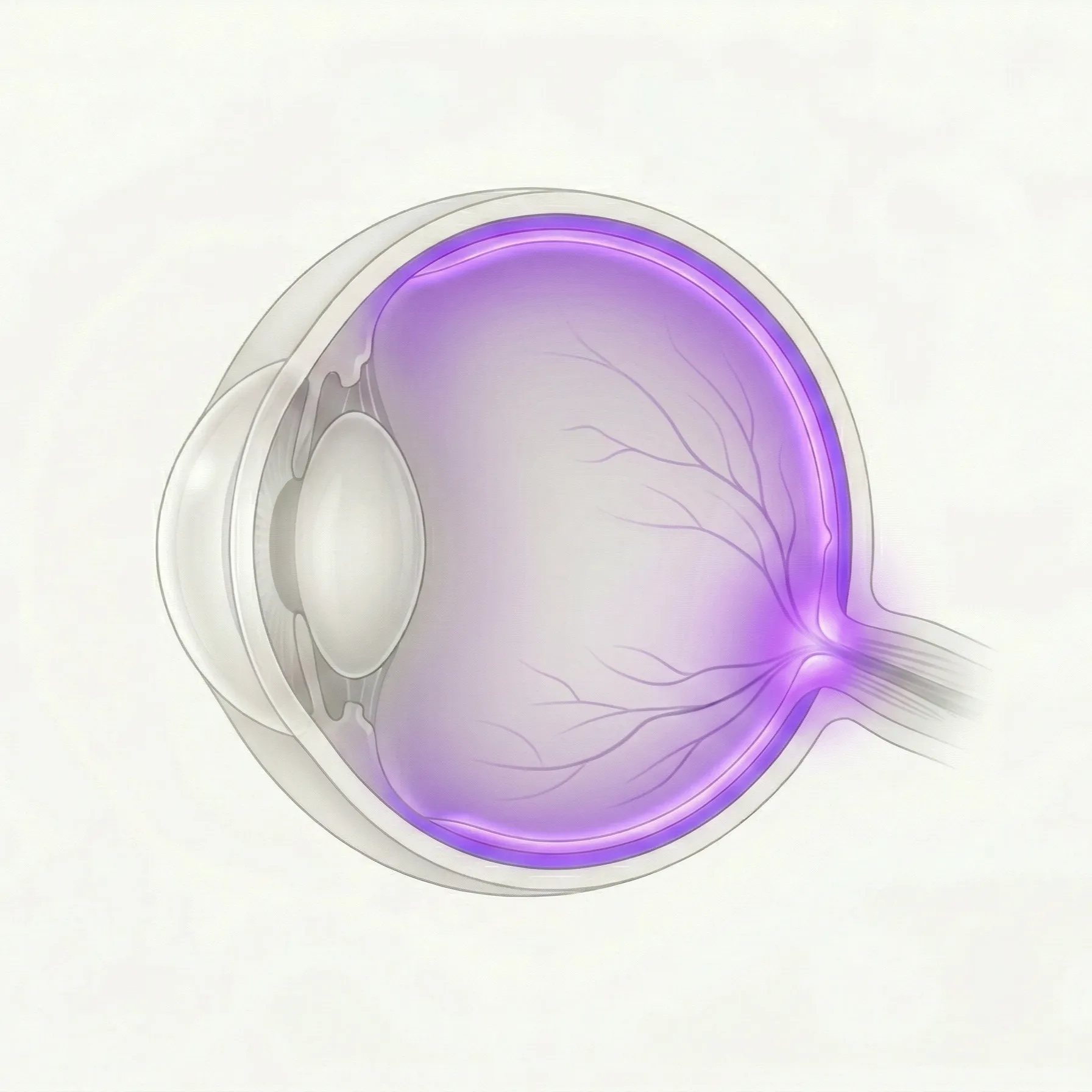
Retina & Vitreous reference image

Retina & Vitreous

The retina is the light-sensitive nerve tissue at the back of the eye, and the vitreous is the clear gel that fills the eye. This category covers vascular disease, degeneration, detachment, bleeding, and other retinal or vitreous conditions.

73 English articles

Frequently referenced diseases

A

7 articles
Abicipar Pegol

Abicipar pegol is a DARPin-based anti-VEGF agent. Clinical trials were conducted for neovascular age-related macular degeneration, but the FDA rejected approval due to a high incidence of intraocular inflammation.

Achromatopsia (Total Color Blindness)

Achromatopsia is an autosomal recessive retinal disease in which all three types of cone photoreceptor cells are dysfunctional. Its main features are reduced visual acuity, photophobia, nystagmus, and lack of color vision. Mutations in the CNGA3 and CNGB3 genes account for the majority of cases.

Acquired Retinal Macroaneurysm (RAM)

Explains the causes, symptoms, diagnosis, and treatment of acquired retinal macroaneurysm (RAM). Covers association with hypertension, FA/IA/OCT findings, and the latest insights on laser photocoagulation and anti-VEGF therapy.

Age-Related Macular Degeneration (AMD)

Definition, classification, diagnostic criteria, and standard treatment of age-related macular degeneration (AMD) based on Japanese clinical guidelines. Covers anti-VEGF therapy for neovascular AMD (typical AMD, PCV, RAP), pathology and latest treatments for atrophic AMD (geographic atrophy), and the pachychoroid concept.

Asteroid Hyalosis

An age-related degenerative disease in which calcium-phospholipid complexes deposit in the vitreous body. It is common in elderly individuals and usually asymptomatic, but acute vision loss may occur following posterior vitreous detachment.

Asteroid Hyalosis

A degenerative eye disease in which cholesterol crystals accumulate in the vitreous body. Secondary to trauma or vitreous hemorrhage, golden crystals settle due to gravity.

Avacincaptad Pegol (Izervay)

A complement C5 inhibitor for geographic atrophy (GA) secondary to age-related macular degeneration. Administered intravitreally as a pegylated RNA aptamer to slow the expansion of GA lesions.

B

5 articles

C

9 articles
Central Retinal Artery Occlusion

An ophthalmic emergency in which the central retinal artery suddenly occludes, causing rapid and severe vision loss. Irreversible changes begin approximately 100 minutes after occlusion, and it shares the same risk factors as stroke.

Central Retinal Vein Occlusion

A vascular disease in which the central retinal vein becomes occluded within the optic nerve, causing hemorrhage and edema throughout the retina. It is the second most common retinal vascular disease after diabetic retinopathy, and macular edema and neovascular glaucoma determine visual prognosis.

Central Serous Chorioretinopathy

A disease characterized by serous retinal detachment in the macula, presenting with metamorphopsia, micropsia, and central scotoma. It predominantly affects men in their 30s to 40s, with stress and steroids being major risk factors.

Choroidal Neovascularization: OCT Angiography Findings

Explains OCT angiography (OCTA) findings of choroidal neovascularization (CNV/MNV). Covers OCTA features of type 1, type 2, and type 3 CNV and PCV, comparison with conventional fluorescein angiography, and notes on artifacts.

Choroideremia

An X-linked recessive chorioretinal dystrophy caused by mutations in the CHM gene. It leads to progressive degeneration of the retinal pigment epithelium, photoreceptors, and choriocapillaris, progressing from night blindness to peripheral visual field loss and eventually severe visual impairment. Gene therapy clinical trials are ongoing.

Chromovitrectomy

A technique in vitreous surgery that visualizes semitransparent tissues such as the internal limiting membrane, vitreous, and epiretinal membrane using vital dyes. Brilliant Blue G, triamcinolone acetonide, and indocyanine green are used.

Coats disease

Idiopathic retinal vascular disease characterized by abnormal dilation and exudation of retinal capillaries. It predominantly affects young boys and progresses unilaterally.

Cone Dystrophy

Cone dystrophy is a hereditary disease in which the cone photoreceptors of the retina are progressively impaired. After the age of 20–30, patients develop decreased visual acuity, photophobia, and color vision abnormalities. ERG shows marked reduction of cone responses. Treatment mainly involves light-filtering lenses and low vision care.

Cystoid Macular Edema

A condition in which fluid accumulates mainly in the outer plexiform layer of the macula due to breakdown of the blood-retinal barrier, resulting in cystoid changes. Causes include diabetic retinopathy, retinal vein occlusion, post-cataract surgery, and drug-induced factors.

D

3 articles

E

2 articles

F

2 articles

G

1 articles

H

1 articles

I

2 articles

L

1 articles

M

2 articles

N

1 articles

O

1 articles

P

11 articles
Pachychoroid Neovasculopathy (PNV)

A disease in which new blood vessels form beneath the retinal pigment epithelium against a background of choroidal thickening. It accounts for about half of exudative age-related macular degeneration in Japan, and anti-VEGF therapy and PDT are the mainstays of treatment.

Pachychoroid Spectrum

A group of diseases characterized by abnormally dilated choroidal vessels (pachyvessels). This concept encompasses central serous chorioretinopathy, polypoidal choroidal vasculopathy, and retinal pigment epithelial detachment.

Pattern Dystrophy

A group of inherited macular degenerations characterized by lipofuscin accumulation in the retinal pigment epithelium (RPE). Mainly caused by PRPH2 gene mutations, leading to mild visual impairment in middle age and later.

Pediatric Vitreoretinal Surgery

A collective term for surgical treatments of vitreoretinal diseases occurring in childhood, such as retinopathy of prematurity (ROP), familial exudative vitreoretinopathy (FEVR), persistent fetal vasculature (PFV), and Coats disease. It requires a specialized approach considering anatomical and physiological characteristics different from adults.

Pegcetacoplan (Syfovre)

A complement C3/C3b inhibitor approved by the FDA in 2023 for geographic atrophy (GA). It is a first-in-class treatment that slows the expansion of GA lesions with monthly or every-other-month intravitreal injections.

Photodynamic Therapy (PDT)

A treatment for retinal and choroidal diseases combining the photosensitizer verteporfin with a 689 nm laser. Used for age-related macular degeneration, central serous chorioretinopathy, polypoidal choroidal vasculopathy, and others.

Polypoidal Choroidal Vasculopathy (PCV)

A disease characterized by polyp-like vascular dilations arising from an abnormal branching vascular network in the choroid. It is common in exudative age-related macular degeneration among Asians and Japanese, and definitive diagnosis by ICGA along with anti-VEGF therapy and PDT are important.

Port Delivery System (PDS / Susvimo)

A refillable ranibizumab sustained-release device permanently implanted in the sclera. It significantly reduces the injection burden of anti-VEGF therapy for wet age-related macular degeneration, maintaining continuous drug concentration with refills every 24 weeks.

Posterior Vitreous Detachment (PVD)

A physiological, age-related change in which the posterior vitreous cortex separates from the internal limiting membrane of the retina. It is the most common cause of floaters and photopsia, and differentiation from retinal tear and retinal detachment is important.

Proliferative Vitreoretinopathy (PVR)

An abnormal proliferative disease that occurs as a complication of retinal detachment. Fibrous membranes form on the retina, causing traction and making surgery difficult.

Pseudophakic Cystoid Macular Edema (Irvine-Gass Syndrome)

Cystoid macular edema (CME) occurring after cataract surgery. The main mechanism is disruption of the blood-retinal barrier mediated by prostaglandins and VEGF, and it is one of the common causes of postoperative visual acuity loss.

R

11 articles
Retinal Angiomatous Proliferation (RAP)

A special type of age-related macular degeneration characterized by neovascularization originating from the intraretinal capillary plexus (type 3 MNV). It predominantly affects elderly women, has a high rate of bilateral involvement, and tends to be treatment-resistant. Intravitreal anti-VEGF injection is the first-line treatment, and combination with PDT is also an option.

Retinal Displacement After Rhegmatogenous Retinal Detachment Repair

A phenomenon in which the retina shifts relative to the RPE after surgery for rhegmatogenous retinal detachment (RRD). It causes metamorphopsia and aniseikonia, and is diagnosed by a hyperautofluorescent line on fundus autofluorescence imaging.

Retinal Photocoagulation (Laser Treatment) Explanation

Comprehensive explanation of the principles, types, and indications of laser treatment for retinal diseases. Covers various techniques such as panretinal photocoagulation (PRP), focal photocoagulation, and subthreshold micropulse laser, along with the latest evidence.

Retinal Pigment Epithelial Detachment (PED)

A condition in which the retinal pigment epithelium (RPE) separates from Bruch's membrane. It is an important associated finding in age-related macular degeneration and central serous chorioretinopathy, and significantly affects visual prognosis.

Retinal Prosthesis (Artificial Retina)

An implantable device that activates remaining retinal neurons via electrical or chemical stimulation in patients with photoreceptor loss due to retinitis pigmentosa or age-related macular degeneration. It aims to partially restore vision.

Retinal Tear, Hole, and Lattice Degeneration

Explains retinal tears caused by traction at sites of vitreoretinal adhesion, retinal holes due to atrophy, and lattice degeneration as a peripheral degeneration. Comprehensively describes risk factors, diagnosis, laser treatment, and indications for prophylactic retinal photocoagulation for rhegmatogenous retinal detachment.

Retinal Vasculitis

A condition characterized by inflammation of retinal blood vessels. It is associated with various underlying diseases such as SLE, Behçet's disease, sarcoidosis, and GPA. Stepwise treatment with steroids, immunosuppressants, and biologics is the standard approach.

Retinal Vein Occlusion

Explanation of the definition, classification (BRVO, CRVO), symptoms, diagnosis, and treatment (anti-VEGF therapy, laser photocoagulation) of retinal vein occlusion (RVO).

Retinitis Pigmentosa

Retinitis pigmentosa (RP): symptoms, causative genes, tests, treatments, and latest research. Onset with night blindness and visual field constriction; an inherited retinal dystrophy involving over 100 genes.

Retinopathy Associated with Pathologic Myopia (Myopic Degeneration)

A disease involving fundus degeneration in high myopia (≥ -6D or axial length ≥ 26.5 mm). Comprehensive explanation of myopic maculopathy (choroidal atrophy, MNV, MTM). It is the second leading cause of adult blindness in Japan. Anti-VEGF drugs and vitrectomy are the main treatments.

Robot-Assisted Vitreoretinal Surgery

Advanced technology that utilizes robotic techniques to enhance the precision of vitreoretinal surgery. It supports delicate procedures such as ILM peeling, subretinal injection, and retinal vein cannulation through tremor filtering and motion scaling.

S

7 articles
S-cone enhancement syndrome

An autosomal recessive retinal degeneration caused by mutations in NR2E3/NRL genes. It presents with night blindness, macular schisis, and characteristic ERG findings, and belongs to the same spectrum as Goldmann-Favre syndrome.

Sickle Cell Retinopathy

Retinal vascular disorder associated with sickle cell disease. Explains non-proliferative and proliferative stages according to Goldberg classification, laser treatment, and gene therapy (Casgevy/Lyfgenia).

Solar Retinopathy

Explains the symptoms, causes, OCT diagnosis, and treatment of solar retinopathy. Introduces the pathophysiology and prevention of foveal retinal damage due to sunlight and phototoxicity.

Sorsby Fundus Dystrophy

An autosomal dominant macular dystrophy caused by mutations in the TIMP3 gene. It typically presents in the 3rd to 4th decade of life and is characterized by choroidal neovascularization and macular atrophy.

Stargardt Disease (Fundus Flavimaculatus)

Explains symptoms, causes (ABCA4 gene, etc.), diagnostic methods (dark choroid, FAF, OCT), current treatments, and latest research on Stargardt disease. Also details genotype-phenotype correlation, differential diagnosis, and genetic counseling.

Stickler Syndrome

An inherited connective tissue disease caused by collagen gene mutations. Characterized by ocular complications such as high myopia, retinal detachment, cataract, and glaucoma, as well as systemic features including cleft palate and hearing loss.

Suprachoroidal Hemorrhage

Explanation of the causes, symptoms, diagnosis, and treatment of suprachoroidal hemorrhage (SCH). Introduction of risk factors and management as a complication of cataract and glaucoma surgery.

T

2 articles

U

1 articles

V

2 articles

W

1 articles

#

1 articles