Tumor & Pathology
Benign and malignant tumors can arise in the eyeball, eyelids, conjunctiva, orbit, and related tissues. This category covers tumor classification, differential diagnosis, and pathologic features.
69 English articles
Benign and malignant tumors can arise in the eyeball, eyelids, conjunctiva, orbit, and related tissues. This category covers tumor classification, differential diagnosis, and pathologic features.
69 English articles
Actinic keratosis (Actinic Keratosis) is a precancerous squamous lesion caused by ultraviolet exposure. It often appears on sun-exposed areas as a red, scaly patch or papule, and it carries a risk of progressing to squamous cell carcinoma.
A rare malignant epithelial tumor arising in the lacrimal gland, frequently associated with perineural invasion and distant metastasis. Standard treatment involves surgery combined with radiotherapy, but the long-term prognosis is poor, with a 10-year survival rate of 20–30%.
A benign cystic tumor that arises from apocrine sweat glands. In the eyelid, it develops from the glands of Moll and appears as a blue-gray, dome-shaped nodule. Prognosis is good after complete excision.
Basal cell carcinoma (BCC) is a malignant tumor originating from the basal cell layer of the epidermis and is the most common eyelid malignancy. It is locally invasive but extremely rarely metastasizes, and surgical excision offers a good prognosis.
Benign lobular inner nuclear layer proliferations (BLIPs) are a new benign intra-retinal tumor arising from the inner nuclear layer of the retina and may be accompanied by congenital hypertrophy of the retinal pigment epithelium (CHRPE). It is an extremely rare disease first reported in 2022, and no intervention is considered necessary.
A rare systemic vascular disorder characterized by multiple venous malformations, mainly in the skin and gastrointestinal tract. It often causes iron-deficiency anemia due to chronic gastrointestinal bleeding and may involve multiple organs, including the orbit and the central nervous system.
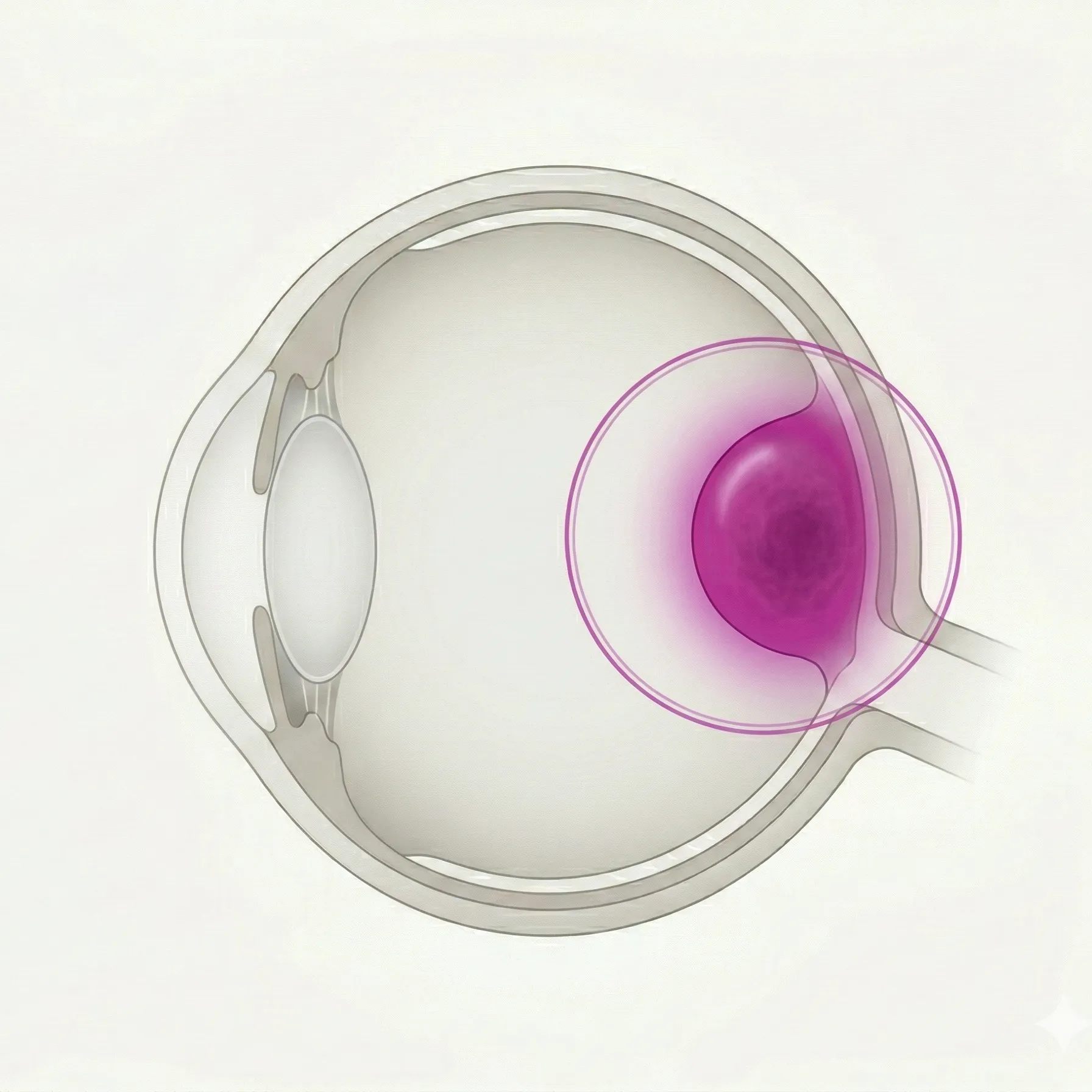
This article explains the diagnosis, treatment, and prognosis of posterior uveal melanoma arising from the choroid and ciliary body, which is the most common primary intraocular malignant tumor in adults.
This article explains the evaluation of malignant transformation risk factors (TFSOM-UHHD) and follow-up strategies for benign pigmented lesions derived from choroidal melanocytes.
A rare benign tumor with ectopic bone formation in the choroid. It commonly occurs in the posterior pole and is slightly more frequent in young women. High CT attenuation equivalent to bone is key for definitive diagnosis. This article also discusses treatment including management of choroidal neovascularization.
This article explains the diagnosis, differential diagnosis including transillumination, and treatment strategy for primary ciliary body malignant melanoma, which accounts for approximately 7% of uveal melanomas.
A localized (solitary) benign vascular tumor of the choroid. It appears as an orange-red elevated lesion in middle-aged and older adults, and when it causes vision loss due to serous retinal detachment, PDT or laser treatment is indicated.
Combined hamartoma of the retina and retinal pigment epithelium (CHRRPE) is a rare benign tumor made up of glial, vascular, and retinal pigment epithelial tissue in the retina and RPE, mainly occurring near the optic disc and macula in children, and causing decreased vision and strabismus.
Congenital hypertrophy of the retinal pigment epithelium (CHRPE) is a congenital hamartoma of the retinal pigment epithelium that is usually benign and asymptomatic. Atypical variants are associated with familial adenomatous polyposis (FAP) and play an important role as an early screening marker for colorectal cancer.
A comprehensive overview of benign tumors (e.g., papilloma), precancerous lesions (conjunctival intraepithelial neoplasia: CIN), and malignant tumors (invasive squamous cell carcinoma: SCC) arising from the conjunctival epithelium. Details include epidemiology, clinical findings, diagnostic methods, TNM classification, treatment options including surgical excision and topical chemotherapy, and pathophysiology.
Conjunctival intraepithelial neoplasia (CIN) is a spectrum from conjunctival epithelial dysplasia to carcinoma in situ, while invasive squamous cell carcinoma (SCC) is a malignant tumor that invades beyond the basement membrane. Ultraviolet exposure is the greatest risk factor, and surgical excision with no-touch technique and cryotherapy is the first-line treatment.
Conjunctival malignant lymphoma is a malignant tumor caused by monoclonal proliferation of B cells, with extranodal marginal zone lymphoma (EMZL / MALT lymphoma) being the most common. It is characterized by a salmon-pink conjunctival mass, and radiation therapy is the first-line treatment for localized cases.
Conjunctival malignant melanoma is a malignant tumor derived from conjunctival melanocytes, with approximately 60-75% arising from PAM. Surgical excision using the no-touch technique and cryotherapy are the mainstays of treatment. BRAF, NF1, and NRAS mutations are major drivers, and the application of immune checkpoint inhibitors is under investigation.
A comprehensive overview of melanocytic tumors arising in the conjunctiva. It details the classification, diagnosis, treatment, and prognostic factors from benign nevi to premalignant primary acquired melanosis (PAM) and malignant conjunctival melanoma.
Conjunctival nevus is the most common benign pigmented tumor of the conjunctiva, characterized by proliferation of nevus cells in the conjunctival basal cells or subepithelium. Tapioca-like cysts are a key diagnostic feature, and the risk of malignant transformation is low, approximately 1%. Rapid growth or color change is a warning sign of malignancy.
A benign cauliflower-shaped tumor of the conjunctiva caused by HPV infection. It is typically pedunculated, but sessile types require differentiation from squamous cell carcinoma. Adding cryotherapy after excision reduces recurrence.
Corneoconjunctival dermoid is a congenital choristoma, a benign tumor commonly occurring at the limbus. Attention should be paid to association with Goldenhar syndrome. Standard treatment involves early visual management to prevent amblyopia and surgery combined with superficial keratoplasty.
This article explains diffuse choroidal hemangioma, which almost always occurs with Sturge-Weber syndrome, covering the characteristic fundus finding known as "tomato ketchup fundus", management of associated glaucoma, and treatment with PDT and low-dose radiation.
A condition characterized by multiple atypical melanocytic nevi and an increased risk of skin and ocular melanoma. It includes FAMMM syndrome and is known to be associated with CDKN2A gene mutations.
An eyelid nevus is a benign tumor caused by proliferation of nevus cells, and is the most common benign eyelid tumor. It is classified into junctional nevus, compound nevus, and intradermal nevus. Junctional and compound nevi rarely may transform into malignant melanoma, so caution is required.
Eyelid papilloma is a benign epithelial tumor associated with HPV, forming a pink cauliflower-like mass. It is often pedunculated, but sessile types require differentiation from squamous cell carcinoma. Excision plus cryocoagulation is the standard treatment.
A malignant tumor that arises from the squamous layer of the eyelid and is the second most common malignant eyelid tumor. UV exposure and immunosuppression are risk factors, and complete surgical excision is the standard treatment.
A systemic disease in which fibroinflammatory lesions rich in IgG4-positive plasma cells occur in the orbit. Painless swelling of the lacrimal gland is most common, and immunosuppressive therapy with steroids or rituximab is the mainstay of treatment.
A minimally invasive diagnostic test in which cellulose acetate filter paper is applied to the ocular surface to collect and analyze superficial epithelial cells. It is widely used for diagnosing dry eye, limbal stem cell deficiency, and ocular surface squamous neoplasia.
This article explains the indications, procedures, drugs, and clinical outcomes of selective ophthalmic artery infusion chemotherapy (IAC) for retinoblastoma, as well as its position in the treatment system in Japan.
This article explains the diagnosis and treatment of primary intraocular lymphoma (PIOL) / vitreoretinal lymphoma (VRL), including diagnosis using the IL-10/IL-6 ratio, treatment outcomes of intravitreal MTX injection, and risk of CNS progression.
This article explains the definition, diagnosis, and treatment of cystic lesions occurring in the iris, focusing on two types: iris stromal cyst and iris pigment epithelial cyst.
This article explains the diagnosis, genetic mutations, treatment, and prognosis of primary malignant melanoma of the iris, which accounts for about 2% of uveal melanomas. It tends to have a lower malignancy compared to those arising from the choroid and ciliary body.
This article explains the definition, differential diagnosis, follow-up, and treatment strategy for benign pigmented tumors derived from iris melanocytes.
Explanation of the types, symptoms, diagnosis, and treatment of tumors that occur in the lacrimal gland. This overview covers the characteristics and management strategies for each classification, from epithelial tumors such as pleomorphic adenoma (about 70% of lacrimal gland epithelial tumors) and adenoid cystic carcinoma to malignant lymphoma.
A general term for benign and malignant tumors arising in the lacrimal sac. Epithelial tumors are the most common, with about 55% being malignant. It is often misdiagnosed as chronic dacryocystitis, and delayed diagnosis leads to poor prognosis.
A malignant tumor arising from melanocytes in the eyelid skin. It is rare, accounting for less than 1% of all cutaneous melanomas, but pigmented lesions with a diameter of 7 mm or more require referral to a specialist. Prognosis depends largely on tumor thickness and stage.
This article explains a rare intraocular tumor arising from the nonpigmented ciliary epithelium, including its clinical features in children, differentiation from retinoblastoma, and treatment strategies.
A rare and highly malignant neuroendocrine tumor derived from Merkel cells. It commonly occurs in the head and neck region, with 5–10% arising on the eyelid. It grows rapidly and tends to metastasize via the lymphatic system.
A rare condition in which systemic malignant tumors metastasize hematogenously to the extraocular muscles. Primary tumors include breast cancer, lung cancer, and cutaneous melanoma, causing restricted eye movement and diplopia. Prognosis is poor, and treatment is mainly palliative.
A condition in which systemic malignant tumors, such as lung cancer and breast cancer, metastasize hematogenously to the choroid. It is characterized by yellowish-white flat lesions and marked serous retinal detachment, with radiation therapy and systemic chemotherapy being the main treatment options.
Muir-Torre syndrome (MTS) is a subtype of Lynch syndrome, an autosomal dominant inherited disorder that combines sebaceous skin tumors and cancers of internal organs. It is caused by mutations in DNA mismatch repair genes, and early diagnosis and surveillance of multiple organs are important.
Necrobiotic xanthogranuloma (NXG) is a type of non-Langerhans cell histiocytosis. It is a rare granulomatous disease characterized by yellow to orange nodules that commonly appear around the orbit, and it has a strong association with paraproteinemia and lymphoproliferative disorders.
Necrotizing fasciitis is a severe infection that affects the superficial fascia and leads to rapid skin necrosis. In periorbital cases, reported mortality is 8–15% and vision loss is 13.8%. Early surgical debridement and antibiotic therapy are the mainstays of treatment.
Xeroderma pigmentosum (XP) is an autosomal recessive genetic disorder caused by a defect in DNA repair, and ophthalmic abnormalities are seen in 93% of patients. It causes a wide range of ocular manifestations, from photophobia and corneal opacity to conjunctival squamous cell carcinoma, and UV protection and early tumor detection are central to management.
Approaches to orbital tumor excision (anterior, lateral, transcranial, and transsinus) and surgery plans by disease. For benign tumors, complete excision without breaking the capsule is the rule. For malignant lymphoma, treatment after biopsy; for adenocarcinoma and adenoid cystic carcinoma, orbital exenteration is chosen. Radiation therapy, carbon ion therapy, and chemotherapy are combined as supportive treatment.
This article explains the symptoms, diagnosis, pathological features, and treatment of benign oncocytoma (oncocytoma) arising in the ocular adnexa. It most often occurs in the caruncle, and the prognosis after complete excision is extremely good.
This article explains the definition, imaging diagnosis, chemotherapy (carboplatin + vincristine), association with NF1, and prognosis of optic pathway glioma.
This article explains the definition, imaging diagnosis (tram-track sign), and management including stereotactic radiotherapy of optic nerve sheath meningioma (ONSM).
A rare benign soft tissue tumor in the orbit that arises from Schwann cells. It often develops in the extraocular muscles, especially the inferior rectus, and can cause proptosis and diplopia. Complete removal is the first-choice treatment.
Orbital melanoma is a malignant tumor derived from melanocytes that occurs within the orbit. It is classified into primary and secondary types. Primary orbital melanoma is extremely rare, accounting for less than 1% of all orbital tumors. Standard treatment involves surgery and adjuvant radiation therapy.
Orbital rhabdomyosarcoma is the most common orbital malignant tumor in children. It is characterized by rapidly progressive proptosis. Standard treatment is a combination of surgery, chemotherapy (VAC regimen), and radiation therapy (proton beam therapy has been covered by insurance since 2016). The 5-year survival rate for primary orbital cases is over 90%.
Orbital schwannoma is a rare benign tumor derived from Schwann cells, accounting for 1-2% of all orbital tumors. The main symptom is slowly progressive proptosis, and complete surgical excision is the standard treatment.
This article explains the symptoms, clinical findings, and management of acute and chronic complications of the eyelids, lacrimal drainage system, orbit, and cornea-conjunctiva associated with radiation to the periorbital area.
Primary acquired melanosis (PAM) is an acquired flat pigmented conjunctival lesion caused by abnormal proliferation of melanocytes. PAM with atypia is a major precursor of conjunctival malignant melanoma, and biopsy for atypia assessment and regular follow-up are essential.
Pyogenic granuloma is a reactive capillary proliferative lesion that often develops after a chalazion or trauma (lobular capillary hemangioma). It appears as a red pedunculated mass and is treated by excision or local steroid injection.
Describes the types of radiation therapy used for eye tumors and the radiation doses used for each disease. This overview explains the features and side effects of external beam radiation (30–70 Gy), stereotactic radiotherapy, proton beam therapy (covered by insurance for rhabdomyosarcoma in 2016), heavy ion therapy (adenoid cystic carcinoma and uveal melanoma), and plaque therapy (¹⁰⁶Ru/¹²⁵I).
This article explains the definition, clinical findings, diagnosis, treatment, and prognosis of retinal astrocytic hamartoma associated with tuberous sclerosis.
This article explains the diagnosis, treatment, and surveillance of retinal capillary hemangioma (retinal hemangioblastoma) associated with VHL disease, including the latest knowledge such as the VHL Disease Clinical Practice Guidelines (2024 edition).
Retinal cavernous hemangioma is a low-flow venous malformation, usually non-progressive. This article discusses clinical findings, differential diagnosis, and management.
A malignant tumor that develops in the retina of infants and young children. Caused by mutations in the RB1 gene, with 70-80 new cases per year in Japan. The most common initial symptom is leukocoria (white pupil). The 5-year survival rate in developed countries is over 95%. Hereditary cases carry a risk of secondary cancers.
A rare, highly malignant tumor arising from the sebaceous glands of the eyelid (mainly the meibomian glands). It resembles chalazion and blepharitis and is often diagnosed late, so it is called the 'great masquerader'.
A highly malignant tumor arising from the sebaceous glands of the eyelid (mainly the meibomian glands). It resembles chalazion or blepharitis and is called "the great masquerader." It is the second most common eyelid malignancy after basal cell carcinoma.
Seborrheic keratosis is the most common benign eyelid tumor in middle-aged and elderly people, also called senile wart. It does not become malignant, but differentiation from basal cell carcinoma and malignant melanoma is important, and histopathological examination is essential for definitive diagnosis.
This article describes the indications, techniques, and outcomes of sentinel lymph node biopsy for detecting micrometastases in periorbital malignant tumors (melanoma, sebaceous carcinoma, squamous cell carcinoma, Merkel cell carcinoma, etc.).
A slowly progressive meningioma arising from the sphenoid wing, causing proptosis and visual impairment due to extension into the orbit and cavernous sinus. This article explains the WHO classification-based grading, imaging diagnosis, and treatment centered on surgery and radiotherapy.
A malignant tumor arising from the spinous layer of the eyelid. There are two types: conjunctival and cutaneous. In Japan, it accounts for about half of all malignant eyelid tumors. Standard treatment is complete excision with postoperative cryocoagulation.