Merkel cell carcinoma is a rare neuroendocrine tumor derived from Merkel cells in the basal layer of the epidermis. Merkel cells are sensory cells discovered by Friedrich Merkel in 1875, involved in light touch and discrimination of shape and texture.
The annual incidence is 0.23 per 100,000 population. Approximately 43–50% of all cases occur in the head and neck, of which 5–10% arise on the eyelid 1). From 2000 to 2013, the number of Merkel cell carcinoma cases increased by 95%, far exceeding the increase in all solid tumors (15%) and melanoma (56%) 1). This is attributed to an aging population, an increase in immunosuppressed patients, and improved diagnostic techniques.
On the eyelid, it characteristically occurs along the lash line, appearing red with tense, shiny skin. Because it grows rapidly and tends to metastasize via lymphatics, prompt diagnosis and treatment are necessary.
QHow rare is Merkel cell carcinoma?
A
The annual incidence is 0.23 per 100,000 population, making it rare among skin malignancies. However, it has been increasing rapidly, with a 95% increase in cases from 2000 to 2013 1).
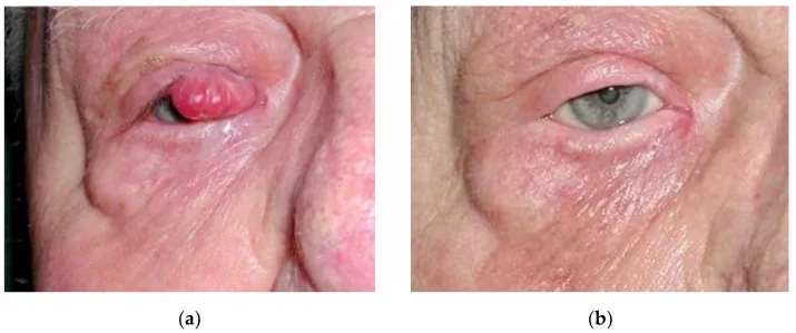
Boileau M, et al. An Effective Primary Treatment Using Radiotherapy in Patients with Eyelid Merkel Cell Carcinoma. Curr Oncol. 2023. Figure 2. PMCID: PMC10377768. License: CC BY.
Four years after curative radiotherapy for a 20 mm Merkel cell carcinoma of the right eyelid, showing (a) before treatment and (b) after treatment. This corresponds to the eyelid tumor discussed in the section “2. Main symptoms and clinical findings”.
Merkel cell carcinoma often appears as a violaceous, painless, solitary nodule. The AEIOU criteria have been proposed to aid diagnosis.
Criterion
Description
Asymptomatic
Asymptomatic (no tenderness)
Expanding rapidly
Rapid expansion
Immunosuppression
Immunosuppressed state
Older than 50
Age over 50
UV-exposed site
UV-exposed site
89% of Merkel cell carcinoma patients meet 3 or more criteria, and 52% meet 4 or more.
Periocularly, it most commonly occurs near the eyelash line of the upper eyelid. It forms a red, dome-shaped nodule with dilated tumor vessels. Partial or complete madarosis and superficial telangiectasias are characteristic.
At diagnosis, up to 37% have lymph node metastasis and 6-12% have distant metastasis. Palpation of preauricular, submandibular, and cervical lymph nodes is essential.
Merkel cell polyomavirus: Approximately 80% of Merkel cell carcinomas are positive (USA). In Japan, about 90%1). The viral genome integrates into the host chromosome, and the large T antigen is mutated and truncated to become an oncoprotein.
Immunosuppression: Organ transplant recipients, chronic lymphocytic leukemia, HIV, etc.
Advanced age: Over 50 years old. Incidence increases particularly over 701).
Fair skin: Predominantly in Caucasians.
Virus-positive Merkel cell carcinoma
Merkel cell polyomavirus-associated: About 80% of all cases. Viral oncoproteins inactivate RB1 and p53. Highly immunogenic, forming the basis for T-cell targeted therapy1).
Cell origin: Various theories include dermal fibroblasts, pro/pre-B lymphocytes, and epidermal progenitor cells1).
Virus-negative Merkel cell carcinoma
UV mutation-related: High mutational burden. Expresses UV-induced DNA neoantigens and is highly immunogenic1).
Cell of origin: Arises from keratinocytes/epidermal progenitor cells with numerous UV mutations. Collision tumors with squamous cell carcinoma have been reported1).
QDoes infection with Merkel cell polyomavirus cause Merkel cell carcinoma?
A
Merkel cell polyomavirus is a nearly ubiquitous virus present in normal skin, but progression to Merkel cell carcinoma is extremely rare, occurring in about 1 in 3000 people over a lifetime1). Two events—integration of the viral genome into the host and mutation of the large T antigen—must occur in the same cell.
Tumor cells have scant cytoplasm, large round nuclei with finely dispersed chromatin, and numerous mitotic figures. They form large nests beneath the epidermis. They resemble lymphoma at first glance but are differentiated by immunostaining.
Dot-like (perinuclear dot pattern) positivity for cytokeratin 20 is the most characteristic finding in Merkel cell carcinoma, and it shows co-expression with pan-cytokeratin2). Cytokeratin 20 is reported to be 100% positive in Merkel cell carcinoma. Electron microscopy reveals neuroendocrine granules with electron-dense cores in the cytoplasm.
Even in patients without clinical lymph node metastasis, sentinel lymph node biopsy identifies micrometastases in one-third of cases1). Patients with positive sentinel lymph node biopsy have approximately three times the risk of recurrence compared to negative patients (3-year recurrence rate: 60% vs 20%), which is important for determining surveillance plans1).
Baseline imaging with FDG PET/CT upstages approximately one in six patients (16.8%)1). The frequency of occult metastases is significantly higher compared to melanoma (<1%).
Metastatic small cell lung carcinoma: Most difficult to differentiate morphologically
QHow to differentiate Merkel cell carcinoma from small cell lung carcinoma?
A
Immunohistochemical staining is key. Perinuclear dot pattern positivity for cytokeratin 20 is specific to Merkel cell carcinoma, while small cell lung carcinoma is cytokeratin 20-negative and TTF-1-positive2).
The mainstay of management is surgical excision and pathologic lymph node evaluation. While 1-2 cm margins are recommended for other body sites, a conservative margin of about 5 mm is acceptable in the periorbital area2). Mohs micrographic surgery or frozen section is used to confirm negative margins.
In cases with adhesion and infiltration to the eyelid skin and tarsus, full-thickness eyelid resection and reconstruction are required, similar to sebaceous carcinoma. After confirming negative margins, 2-3 cycles of cryotherapy (freeze and thaw) may be added to the resection bed. For patients who cannot tolerate radical resection, radiotherapy is an option.
Indications: NCCN guidelines recommend adjuvant radiotherapy within 4-6 weeks after surgical resection for all stages, significantly reducing the 5-year local recurrence risk.
Dose: Typically 50-66 Gy2). Periorbital areas require caution regarding effects on ocular structures.
Definitive Radiotherapy
Indications: Primary treatment for patients not eligible for surgery. Merkel cell carcinoma is highly radiosensitive and may be treated with radiotherapy alone.
Single fraction radiotherapy: A single dose of 8 Gy achieves a response rate of over 94%. It is an alternative for elderly or frail patients 1).
Conventional chemotherapy with platinum agents plus etoposide has a high initial response rate, but the duration of response is short (most progress within 90 days) and no improvement in survival has been demonstrated 1).
Immune checkpoint inhibitors have become the standard systemic therapy for advanced Merkel cell carcinoma:
Avelumab (anti-PD-L1 antibody): FDA approved in 2017. In the JAVELIN Merkel 200 trial, response rate 33%, complete response 11%, 2-year progression-free survival 26%, overall survival 36% 2). Approved in many regions including Japan.
Pembrolizumab (anti-PD-1 antibody): FDA approved in 2018. Response rate as first-line therapy 55–62% 1).
Similar response rates are observed regardless of Merkel cell polyomavirus infection status or PD-L1 expression 1).
In second-line or later after chemotherapy, the response rate drops to approximately 30% 1).
QHow effective are immune checkpoint inhibitors for Merkel cell carcinoma?
A
As first-line therapy, the response rate is 55–62%, significantly higher than chemotherapy 1). Unlike chemotherapy, responses are often durable (lasting several years). Efficacy can be expected regardless of viral status.
There are two major pathways in the development of Merkel cell carcinoma.
In virus-positive Merkel cell carcinoma, the oncoproteins of Merkel cell polyomavirus (truncated large T antigen and small T antigen) inactivate RB1 and p53 and activate Myc signaling 1). In virus-negative Merkel cell carcinoma, ultraviolet-induced mutations directly dysregulate these pathways. In both types, the “PARCB” factors (abnormalities of p53, Akt1, RB1, c-Myc, and Bcl2) drive neuroendocrine differentiation 1).
Merkel cell carcinoma tends to spread discontinuously, and local recurrence can occur even with pathologically negative margins 1). Six factors that increase recurrence risk have been reported: 1) chronic T-cell immunosuppression, 2) tumor diameter >1 cm, 3) lymphovascular invasion, 4) positive sentinel lymph node biopsy, 5) positive margins, and 6) primary site in the head and neck 1).
Measurement of circulating antibodies against Merkel cell polyomavirus oncoproteins is useful as a prognostic indicator. Seronegative patients have approximately 42% higher risk of recurrence than seropositive patients 1). In seropositive patients, changes in antibody titers can be used for early detection of recurrence, and this method is included in the NCCN guidelines as a surveillance tool 1).
7. Latest Research and Future Perspectives (Investigational Reports)
Preoperative (neoadjuvant) immune checkpoint inhibitors have been reported to achieve rapid tumor shrinkage in approximately 50% of locally advanced Merkel cell carcinomas 1). Case-by-case evaluation is necessary.
Challenges in Immunotherapy-Resistant Merkel Cell Carcinoma
Approximately half of patients with advanced Merkel cell carcinoma do not achieve a durable response to anti-PD-1/PD-L1 therapy 1). Addressing primary or acquired resistance is the greatest unmet need, and many clinical trials are ongoing.
Analysis of circulating tumor DNA (ctDNA) is being developed as a promising new tool for early detection of recurrence in patients with virus-negative Merkel cell carcinoma 1). A stage-specific recurrence risk calculator (merkelcell.org/recur) also contributes to personalized surveillance.