Eyelid hemangioma is a benign tumor caused by proliferation of vascular endothelial cells. When it occurs in infancy, it is called infantile hemangioma. It was previously referred to as “strawberry hemangioma” or “capillary hemangioma,” but is now classified as infantile hemangioma based on the ISSVA classification (International Society for the Study of Vascular Anomalies, 2018 revision)2). In contrast, the former “cavernous hemangioma” is a different condition and is classified as a “venous malformation,” a type of vascular malformation.
Infantile hemangioma is congenital and begins to proliferate around 2 weeks after birth. It reaches a peak of proliferation at 1–2 months and continues to enlarge until about 1.5 years of age. Thereafter, it gradually shrinks over several years. 70% regress spontaneously by around school age. Rarely, it may enlarge rapidly and infiltrate the entire eyelid, causing difficulty opening the eye.
The ISSVA classification (2018) 2) broadly divides vascular lesions into “vascular tumors” and “vascular malformations.” Infantile hemangioma is a tumor due to true proliferation of vascular endothelial cells and is classified as a “vascular tumor.” In contrast, venous malformation is a vascular abnormality and is classified as a “vascular malformation.” Since the two differ in pathogenesis, natural course, and treatment strategy, accurate classification is important for treatment selection.
QDoes eyelid hemangioma resolve on its own?
A
70% of infantile hemangiomas regress spontaneously by around preschool age. Growth begins around 2 weeks after birth, continues until about 1.5 years of age, and then gradually shrinks over several years. However, large lesions that cause difficulty in opening the eyelid can lead to form deprivation amblyopia, so early treatment may be necessary without waiting for spontaneous regression. Venous malformations (formerly called cavernous hemangiomas) do not regress spontaneously, so management strategies differ.
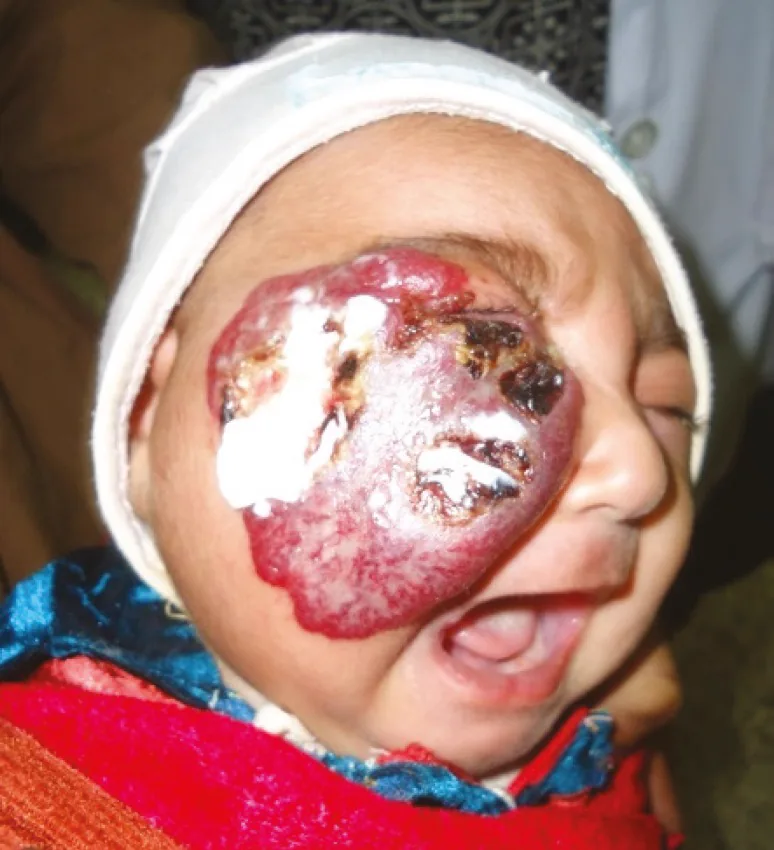
Hossain MA, Shamsuddin AHM, Haque ME, et al. Successful Propranolol Treatment of a Large Size Infantile Hemangioma of the Face Causing Recurrent Bleeding and Visual Field Disruption. World J Plast Surg. 2015;4(1):79-83. Figure 1. PMCID: PMC4298869. License: CC BY.
Initial visit photograph of a large infantile hemangioma occupying the right cheek and most of the right eyelid (with inability to open the eye due to ptosis). Corresponds to infantile hemangioma (superficial type with strawberry-like appearance and risk of form deprivation amblyopia due to mechanical ptosis) discussed in the section “2. Main Symptoms and Clinical Findings.”
Infantile hemangiomas are classified by location into superficial, deep, and mixed types.
Superficial type (strawberry hemangioma)
Well-defined, dark red mass: A hemispherical elevation on the skin surface with a bright red color, a typical appearance.
Reticular vascular pattern on skin and conjunctiva: A network of blood vessels is seen on the skin surface and conjunctiva. This is an important finding for differentiating from chalazion.
Atypical cases: Some types have minimal color change and slight elevation.
Deep type (subcutaneous type)
Deep subcutaneous origin: Redness on the skin surface is not prominent, but a mass is palpable under the skin.
Bluish-purple skin discoloration: The skin appears bluish-purple due to visible venous blood. The borders are indistinct.
Mixed type: Some cases combine superficial and deep types (mixed type).
The typical clinical picture of infantile hemangioma is a red, well-defined, raised lesion that appears after birth. It is important for diagnosis to evert the eyelid and observe the conjunctival side for the presence of a reticular vascular pattern.
QHow to distinguish between chalazion and hemangioma?
A
Infantile hemangioma develops early after birth (within weeks), appears as a red raised lesion with a reticular vascular pattern on the skin and conjunctiva, which is a key point for differentiation. Chalazion does not show vascular patterns on the skin surface. Also, infantile hemangioma gradually enlarges, while chalazion often has a relatively acute onset. If diagnosis is uncertain, ultrasound or histopathological examination may be performed.
Infantile hemangiomas arise from the proliferation of vascular endothelial cells. Vascular endothelial growth factor (VEGF) has been shown to be involved in this proliferation, which also provides the basis for the mechanism of action of propranolol.
GLUT-1 (glucose transporter 1) is a characteristic immunohistochemical marker for infantile hemangiomas 3). It shares an immunophenotype with placental microvessels, and is negative in venous malformations. This property is used for differentiation in histopathological diagnosis.
PHACE syndrome: A syndrome characterized by large facial infantile hemangioma associated with posterior fossa malformations, arterial anomalies, cardiac defects, and eye abnormalities 4). Large facial hemangiomas require exclusion of this syndrome.
Kasabach-Merritt syndrome: A condition in which a giant hemangioma is complicated by consumptive coagulopathy 5). It occurs mainly in kaposiform hemangioendothelioma and tufted angioma, and is rare in infantile hemangioma.
Propranolol, a beta-blocker, is currently the standard first-line drug 7). Its mechanism of action is thought to involve suppression of VEGF and bFGF production via beta-2 receptor blockade, vasoconstriction, and induction of apoptosis in vascular endothelial cells.
Dosage: 2–3 mg/kg/day, divided into three doses 7)
Start timing: Optimal between 5 weeks and 5 months of age 7)
Side effects: Hypoglycemia, bradycardia, hypotension, bronchospasm (contraindicated in children with asthma)
Management: Initiation in hospital under the guidance of a pediatric specialist is recommended 7)
A randomized controlled trial on oral propranolol (NEJM, 2015) showed that the propranolol group (3 mg/kg/day for 6 months) had significantly better treatment outcomes than the control group7).
Timolol ophthalmic solution (0.5%) is applied to the surface of the hemangioma twice daily (off-label use). Efficacy has been reported for superficial and small lesions11). It is an option for cases where systemic propranolol is difficult. Discontinue once effect is confirmed.
Pulsed dye laser (585–595 nm) is effective for superficial infantile hemangiomas8). Early irradiation can promote early regression and suppress protrusive changes. It is effective for red surface lesions but not for deep components.
For patients intolerant to propranolol or for specific localized lesions, intralesional injection of triamcinolone (3–5 mg/kg) is used9). This was previously the first-line treatment, but its indications have become limited since the introduction of propranolol.
Attention is needed for both form deprivation amblyopia due to difficulty opening the eyelid and astigmatic amblyopia caused by eyeball compression and corneal deformation from the hemangioma. Continuously assess tumor size and risk of form deprivation amblyopia, and provide refractive correction and occlusion therapy.
QWhat are the side effects of propranolol?
A
Main side effects include hypoglycemia, bradycardia, hypotension, and bronchospasm. Hypoglycemia is more likely during poor feeding or insufficient food intake; administration at the same time as feeding or meals is recommended. Asthma and reactive airway disease are contraindications due to the risk of bronchospasm. For risk management, inpatient initiation under the guidance of a pediatric specialist is recommended.
QWhen should treatment be started?
A
Early intervention is important if there is a risk of amblyopia. The optimal timing to start propranolol is between 5 weeks and 5 months of age; the earlier it is started during the proliferative phase (up to 18 months of age), the more effective it is. For large lesions with risk of form deprivation amblyopia due to difficulty opening the eyelid or corneal astigmatism, prompt treatment should be considered. Small lesions without amblyopia risk can be observed.
Infantile hemangioma is a true proliferative tumor of vascular endothelial cells, fundamentally different from venous malformations (vascular malformations). VEGF (vascular endothelial growth factor) plays a central role in tumor growth. Propranolol’s beta-2 receptor blockade is thought to reduce tumor size by suppressing VEGF and bFGF production, inducing vasoconstriction, and promoting apoptosis7).
GLUT-1 (glucose transporter 1) positivity is a specific immunophenotype of infantile hemangioma, indicating commonality with placental microvessels 3). This characteristic supports the hypothesis that the tumor originates from placental-derived vascular endothelial progenitor cells. GLUT-1 is negative in other vascular lesions such as venous malformations, lymphangiomas, and pyogenic granulomas, making it a practical marker for differential diagnosis.
Venous malformations (formerly called cavernous hemangiomas) are vascular malformations, not true tumors. They consist of dilated venous channels lined by flat endothelial cells. They are GLUT-1 negative and show little proliferative response to growth factors. Since they do not regress spontaneously, treatment indications differ from infantile hemangioma.
The ISSVA classification (2018 revision) 2) provides a framework dividing vascular anomalies into “vascular tumors” (proliferation of endothelial cells) and “vascular malformations” (abnormal vessel formation). Infantile hemangioma is classified as a vascular tumor, while venous malformations and lymphatic malformations are vascular malformations. This classification directly impacts treatment decisions. Drugs that suppress endothelial proliferation, such as propranolol, are effective for vascular tumors but have limited efficacy for vascular malformations.
Large eyelid hemangiomas can compress the eyeball, causing asymmetric corneal deformation and leading to irregular astigmatism and anisometropia. If this astigmatism is not properly corrected, visual development is impaired, resulting in astigmatic amblyopia or anisometropic amblyopia. Form deprivation amblyopia occurs when light stimulation is blocked due to difficulty opening the eyelid. If intervention is delayed during the sensitive period of vision (from birth to 7–8 years of age), amblyopia may become irreversible.
Atenolol, a selective β1-blocker, is being investigated as an alternative to propranolol (a non-selective β-blocker). A retrospective non-inferiority study 10) showed that atenolol (1 mg/kg/day) was non-inferior to propranolol (2 mg/kg/day) in treatment outcomes. It may be safely used in infants with asthma or reactive airway disease for whom propranolol is contraindicated, and the results of future prospective trials are awaited.
A randomized controlled trial 11) of topical timolol maleate gel (0.5%) evaluated its efficacy and safety for superficial infantile hemangiomas in infants aged 5 to 24 weeks. The risk of systemic side effects from local absorption was low, but accumulation of long-term safety data is needed.
Application of sirolimus (mTOR inhibitor) to vascular malformations
Sirolimus (rapamycin) inhibits the mTOR pathway and suppresses vascular endothelial cell proliferation. A prospective study 12) examining the efficacy and safety of sirolimus for refractory complex vascular malformations (combined venous, lymphatic, and arteriovenous malformations) reported symptom improvement in 84% of cases. Although its indication for infantile hemangiomas is limited, it is expected as a new treatment option for refractory vascular malformations.
Wassef M, Blei F, Adams D, Alomari A, Baselga E, Berenstein A, Burrows P, Frieden IJ, Garzon MC, Lopez-Gutierrez JC, Lord DJ, Mitchel S, Powell J, Prendiville J, Vikkula M, ISSVA Board and Scientific Committee. Vascular Anomalies Classification: Recommendations From the International Society for the Study of Vascular Anomalies. Pediatrics. 2015;136(1):e203-14. doi:10.1542/peds.2014-3673. PMID:26055853.
North PE, Waner M, Mizeracki A, Mihm MC.. GLUT1: a newly discovered immunohistochemical marker for juvenile hemangiomas. Hum Pathol. 2000;31(1):11-22. doi:10.1016/s0046-8177(00)80192-6. PMID:10665907.
Metry D, Heyer G, Hess C, Garzon M, Haggstrom A, Frommelt P, Adams D, Siegel D, Hall K, Powell J, Frieden I, Drolet B, PHACE Syndrome Research Conference. Consensus Statement on Diagnostic Criteria for PHACE Syndrome. Pediatrics. 2009;124(5):1447-1456. doi:10.1542/peds.2009-0082. PMID:19858157.
Kasabach HH, Merritt KK. Capillary hemangioma with extensive purpura. Am J Dis Child. 1940;59:1063-1070. doi:10.1001/archpedi.1940.01990160135009.
Dubois J, Patriquin HB, Garel L, et al. Soft-tissue hemangiomas in infants and children: diagnosis using Doppler sonography. AJR Am J Roentgenol. 1998;171(1):247-252. doi:10.2214/ajr.171.1.9648798. PMID:9648798.
Léauté-Labrèze C, Hoeger P, Mazereeuw-Hautier J, Guibaud L, Baselga E, Posiunas G, Phillips RJ, Caceres H, Lopez Gutierrez JC, Ballona R, Friedlander SF, Powell J, Perek D, Metz B, Barbarot S, Maruani A, Szalai ZZ, Krol A, Boccara O, Foelster-Holst R, Febrer Bosch MI, Su J, Buckova H, Torrelo A, Cambazard F, Grantzow R, Wargon O, Wyrzykowski D, Roessler J, Bernabeu-Wittel J, Valencia AM, Przewratil P, Glick S, Pope E, Birchall N, Benjamin L, Mancini AJ, Vabres P, Souteyrand P, Frieden IJ, Berul CI, Mehta CR, Prey S, Boralevi F, Morgan CC, Heritier S, Delarue A, Voisard JJ.. A randomized, controlled trial of oral propranolol in infantile hemangioma. N Engl J Med. 2015;372(8):735-746. doi:10.1056/nejmoa1404710. PMID:25693013.
Kapila Batta, Helen M Goodyear, Celia Moss, Hywel C Williams, Louise Hiller, Ruth Waters. Randomised controlled study of early pulsed dye laser treatment of uncomplicated childhood haemangiomas: results of a 1-year analysis. The Lancet. 2002;360(9332):521-527. doi:10.1016/s0140-6736(02)09741-6.
Avery H. Weiss, John P. Kelly. Reappraisal of Astigmatism Induced by Periocular Capillary Hemangioma and Treatment with Intralesional Corticosteroid Injection. Ophthalmology. 2008;115(2):390-397.e1. doi:10.1016/j.ophtha.2007.03.077.
Bayart CB, Tamburro JE, Vidimos AT, Wang L, Golden AB.. Atenolol Versus Propranolol for Treatment of Infantile Hemangiomas During the Proliferative Phase: A Retrospective Noninferiority Study. Pediatr Dermatol. 2017;34(4):413-421. doi:10.1111/pde.13177. PMID:28556385.
Chan H, McKay C, Adams S, Wargon O.. RCT of timolol maleate gel for superficial infantile hemangiomas in 5- to 24-week-olds. Pediatrics. 2013;131(6):e1739-47. doi:10.1542/peds.2012-3828. PMID:23650294.
Adams DM, Trenor CC 3rd, Hammill AM, Vinks AA, Patel MN, Chaudry G, et al. Efficacy and Safety of Sirolimus in the Treatment of Complicated Vascular Anomalies. Pediatrics. 2016;137(2):e20153257. doi:10.1542/peds.2015-3257. PMID:26783326; PMCID:PMC4732362.
Copy the article text and paste it into your preferred AI assistant.
Article copied to clipboard
Open an AI assistant below and paste the copied text into the chat box.