Amblyopia is a condition in which best-corrected visual acuity in one or both eyes does not develop sufficiently due to visual deprivation or abnormal binocular interaction during the sensitive period of visual development. It is characterized by visual impairment that cannot be explained by organic disease and can be improved with appropriate treatment 1).
Social low vision refers to visual impairment that cannot be corrected with glasses and is a social term, distinct from medical amblyopia. There have been several definitions of amblyopia over time. Bangerter (1955) defined it as “a visual impairment without organic changes or with changes insufficient to explain the impairment.” von Noorden (1977) described it as “a decrease in visual acuity in one or both eyes caused by form deprivation or abnormal binocular interaction, with no detectable ocular abnormality, and reversible in many cases with treatment.” In Japan, the definition by Uemura (1993) is widely accepted: “a decrease in visual acuity in one or both eyes caused by visual deprivation or abnormal binocular interaction during the visual development period, with no organic disease found on ocular examination, and preventable or treatable in appropriate cases.” Amblyopia is considered a “developmental disorder of vision” and is diagnosed by exclusion after ruling out organic disease.
The prevalence of amblyopia varies widely, from 0.14% to 4.8% in reports including overseas data. In Japan, it is estimated at 0.58% based on a meta-analysis of 3-year-old health checkups, and population-based studies (children aged 30–71 months) show a range of 0.7% to 2.6% 1). By type, anisometropic amblyopia is the most common, followed by refractive amblyopia, strabismic amblyopia, and form deprivation amblyopia. Unilateral amblyopia is associated with strabismus in 19–50% of cases and with refractive error in 46–79% of cases 1).
Factors that increase the risk of amblyopia include prematurity, low birth weight, developmental delay (including Down syndrome), and a family history of amblyopia or strabismus in first-degree relatives. Some reports also suggest an association with smoking and alcohol consumption during pregnancy.
QCan amblyopia develop in adulthood?
A
Amblyopia is a developmental disorder that occurs during the sensitive period of visual development (generally from birth to around 8 years of age). It does not newly develop in adults, but untreated childhood amblyopia may persist. There are reports of visual improvement with treatment even after age 12, so treatment should not be abandoned based on age alone.
Unilateral amblyopia is often asymptomatic. Since the healthy eye compensates for daily vision, affected children rarely notice visual impairment.
Decreased visual acuity: Often noticed only when the healthy eye is covered. Most cases are discovered incidentally during vision screening.
Impaired stereopsis (depth perception): Loss of stereopsis has been reported even with anisometropia of 1D or more. Difficulty in judging distances may occur.
Crowding phenomenon: In the amblyopic eye, identifying letters in a row is more difficult than identifying single letters.
The diagnostic criteria for amblyopia are shown below1).
Age
Unilateral Amblyopia
Bilateral Amblyopia
3–4 years
Interocular difference ≥2 lines
Both eyes <20/50
4–5 years
Interocular difference ≥2 lines
Both eyes <20/40
≥5 years
Interocular difference ≥2 lines
Both eyes <20/30
Interocular difference in corrected visual acuity: A difference of 2 or more lines on the logMAR scale is a diagnostic criterion.
Fixation abnormality: Strabismic amblyopia may be accompanied by eccentric fixation (parafoveal, paramacular, or peripheral fixation).
Reduced contrast sensitivity: Anisometropic amblyopia is characterized by reduced contrast sensitivity in the medium to high spatial frequency range, affecting both central and peripheral visual fields. In strabismic amblyopia, only the central visual field is reduced.
Loss or reduction of stereopsis: Assessed using the Worth 4-Dot Test, Titmus stereo test, Lang stereo test, etc. 1)
QWhat is crowding phenomenon?
A
It is a phenomenon in which the amblyopic eye has difficulty identifying letters when they are crowded together. Since single letters are seen better than a line of letters, testing with single optotypes may underestimate the severity of amblyopia. It is recommended to use optotypes with crowding bars for visual acuity testing.
Amblyopia is broadly classified into four types based on cause.
Refractive Amblyopia
Ametropic amblyopia: A condition in which both eyes have a similar high degree of refractive error, preventing clear image formation on the fovea and hindering visual development. Risk increases with hyperopia ≥3D and astigmatism ≥1.5D.
Meridional amblyopia: A special type occurring in eyes with high astigmatism. Sensitivity to stripe patterns in specific orientations is reduced.
Anisometropic Amblyopia
Anisometropic amblyopia: A condition in which there is a large difference in refractive error between the two eyes, and the eye with the stronger refractive error fails to develop normal vision. Generally, a difference of ≥2D poses a risk, but in hyperopia, even a 1D difference can lead to amblyopia.
This is the most common type of amblyopia. About one-third of children with 2D anisometropia have amblyopia, and even a 1–2D difference increases the odds of amblyopia by 4.5 times 1).
Strabismic Amblyopia
Strabismic amblyopia: A unilateral amblyopia caused by strabismus, where the deviated eye does not receive a clear retinal image on the fovea, leading to suppression of the non-dominant eye. More common in esotropia. If alternating fixation is present, amblyopia is less likely. It rarely occurs in intermittent exotropia.
Form Deprivation Amblyopia
Form deprivation amblyopia: Caused by obstruction of visual stimuli due to congenital cataract, corneal opacity, severe ptosis, etc. It is the most treatment-resistant type and has the poorest prognosis. Unilateral cases tend to be more severe than bilateral. During the sensitive period of visual development, even one week of deprivation can cause amblyopia.
Microstrabismic amblyopia is sometimes considered a fifth type. It occurs in primary microstrabismus due to strong eccentric fixation, resulting in amblyopia that is not deep, typically achieving vision around 0.7.
By type of anisometropia, hyperopic anisometropia (1–1.5 D or more) is the most common. In myopic anisometropia, the more myopic eye obtains a clearer image at near, making amblyopia less likely; generally, the risk increases with a difference of 3 D or more. In astigmatic anisometropia, the risk of developing amblyopia exists with a difference of 2 D or more.
Hyperopic Anisometropia
Amblyopia risk: Can develop with a difference of 1–1.5 D or more between the two eyes.
Characteristics: The most common type of anisometropic amblyopia. The eye with higher hyperopia does not receive a clear image on the fovea, making it prone to amblyopia.
Myopic Anisometropia
Amblyopia risk: Can develop with a difference of 3 D or more between the two eyes.
Characteristics: At near, the more myopic eye obtains a clearer image, making amblyopia less likely.
Astigmatic Anisometropia
Amblyopia risk: Can develop with a difference of 2 D or more between the two eyes.
Characteristics: The axis direction affects visual development. The greater the difference in astigmatism, the higher the risk of developing amblyopia.
Main risk factors:
Family history: Increased risk with a first-degree family history of amblyopia or strabismus
Prematurity or low birth weight
Developmental delay: Developmental disorders including Down syndrome
Environmental factors: Reports suggest an association with smoking and alcohol consumption during pregnancy
In Japan, the 3-year-old vision screening consists of three stages. First, a primary screening conducted by parents at home (vision test using picture charts, etc.), followed by a secondary screening performed by physicians, public health nurses, or orthoptists at health centers, and finally a detailed examination at an ophthalmology clinic. The success rate of 5-meter visual acuity testing using a single Landolt C chart is reported to be 73.3% at 3 years 0 months and nearly 95% at 3 years 6 months. In recent years, screening devices such as binocular open-field infrared video refractometers have become available for use from 6 months of age.
Choose a method appropriate for the child’s age. In addition to the Landolt C chart, picture charts, Lea chart, and HOTV are available. The use of crowded optotypes is recommended.
Can be used on the first visit. Can be combined with oxybuprocaine hydrochloride 0.4%. Side effects: facial flushing, tachycardia, and very rarely hallucinations and ataxia
Atropine sulfate
After 5 to 7 days of instillation
Approximately 3 weeks
Strongest cycloplegia. Recommended for initial spectacle prescription. 1% ophthalmic solution, 1% ophthalmic ointment. Dilute to 0.25-0.5% for young children. Instill 2-3 times daily for 5-7 days before examination. Side effects: facial flushing, tachycardia
For children under 5 years old, the refractive value obtained under atropine sulfate is used as the spectacle prescription (full correction). For children 5 years and older with esotropia, prescribe full correction.
Differentiation from organic visual impairment is important, including optic neuritis (22.4%), refractive error (21.2%), trauma (10.6%), dominant optic atrophy (7.0%), and accommodative spasm (5.8%). Evaluation of the pupillary light reflex (RAPD) is important. Retinal dystrophies (Stargardt disease may present with normal fundus initially) and prechiasmal lesions are also considered in the differential.
QHow is amblyopia detected during the 3-year-old health checkup?
A
In Japan, the visual screening for 3-year-olds consists of three stages: ① Primary screening at home (visual acuity test using picture charts, etc.), ② Secondary screening at health centers (evaluation by physicians, public health nurses, or orthoptists), and ③ Detailed examination at an ophthalmology clinic. Children with low visual acuity in one eye or high refractive error are referred for detailed examination. Recently, binocular open-field photoscreeners have enabled screening from 6 months of age. The rate of successful testing with a single Landolt C optotype reaches nearly 95% by 3 years and 6 months.
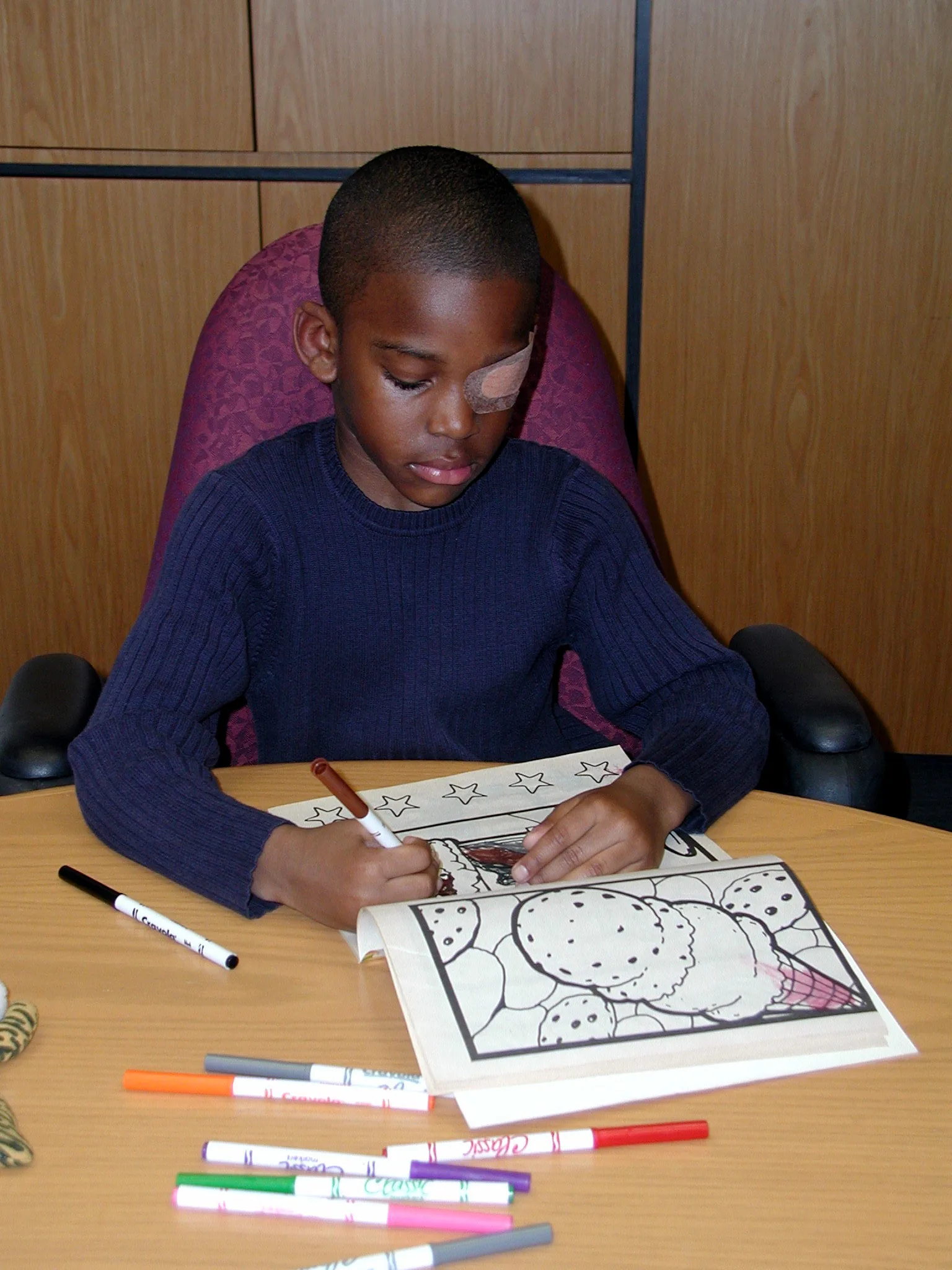
Wikimedia Commons. File:Child_eyepatch.jpg. License: CC BY-SA.
A child undergoing occlusion therapy with an eye patch on the healthy eye, actively using the amblyopic eye. Near vision tasks such as coloring are used to stimulate the amblyopic eye.
Since the publication of large-scale RCT results by the Pediatric Eye Disease Investigator Group (PEDIG) in 2002, evidence from multicenter studies has accumulated for amblyopia treatment 9). The basics of treatment are removal of the underlying cause and promotion of visual stimulation to the amblyopic eye.
Removal of obstacles along the visual axis is the highest priority. For congenital cataracts, if surgery is not performed by 6–8 weeks of age for unilateral cases and by 10–12 weeks (up to 3 months for bilateral cases) to allow light stimulation of the retina, significant impairment of visual development will remain. In severe ptosis, early surgery or taping is used to secure the visual axis. During the sensitive period of visual development, even one week of deprivation can cause amblyopia.
The first step in treatment is spectacle prescription based on cycloplegic refraction. Approximately 75% of patients achieve an improvement of two or more lines in visual acuity with spectacle wear alone.
In a prospective PEDIG study, 27% of children aged 3–6 years with anisometropic amblyopia were cured with spectacle correction alone, with a mean improvement of 0.29 logMAR, and 77% showed improvement of 0.2 logMAR or more 10). In strabismic and mixed amblyopia, improvement was 0.26 logMAR, with 75% improving by 0.2 or more and 32% cured 10). Another report indicates that after 18 weeks of constant spectacle wear, more than two-thirds of untreated anisometropic amblyopia cases showed improvement 1). Currently, it is standard practice to observe with spectacles alone until visual acuity stabilizes 10).
In Japan, a medical expense benefit system for therapeutic spectacles for children under 9 years of age has been in place since April 2006. Renewal conditions are at least one year after the previous benefit for children under 5 years, and at least two years for those 5 years and older.
When anisometropia is large (3D or more), spectacles alone have limitations, so occlusion of the healthy eye is added. After spectacle prescription, re-examination should occur within one month until visual acuity stabilizes.
When visual acuity does not improve sufficiently with spectacles alone, the non-amblyopic (healthy) eye is occluded. The basic method is complete occlusion using an adhesive patch.
Key results from the PEDIG ATS study group 1):
Study
Content
Result
ATS1
Occlusion vs. Atropine (6 months)
3.16 lines vs. 2.84 lines improvement, equivalent
ATS2A
Full-time vs 6 hours (severe amblyopia)
No significant difference
ATS2B
6 hours vs 2 hours (moderate amblyopia)
No significant difference
ATS3
Treatment for ages 7-17
Effective for ages 7-12; also effective for ages 13-17 if untreated
ATS15
Stagnation at 2 hours → increase to 6 hours
Improvement seen
Guidelines for occlusion time:
Severe amblyopia (0.05–0.2): No significant difference in improvement between 6-hour and full-time occlusion
Moderate amblyopia (0.25–0.5): No significant difference between 2-hour and 6-hour occlusion
Microstrabismic amblyopia: Full occlusion about 4.5 hours per day, typically achieving around 0.7
Electronic occlusion time monitoring studies have reported that the occlusion time required for a one-line improvement changes from 58 hours/line in the first month to an average of 169 hours/line at 4 months 11). Starting occlusion of the sound eye after the child has become accustomed to wearing glasses can reduce stress.
In addition to wearing fully corrective glasses, atropine sulfate is instilled into the sound eye, and distigmine bromide is instilled into the amblyopic eye. The sound eye experiences near blur due to cycloplegia from atropine, while the amblyopic eye becomes near-dominant due to miosis and increased accommodative response from distigmine. Patients are instructed to perform near work within 30 cm for about 30 minutes per day.
This method involves attaching a Bangerter (translucent) filter to the spectacle lens of the sound eye. In moderate amblyopia, the difference in visual acuity improvement compared to patching is within 0.5 lines, and it has been reported to cause less burden and stress for parents 1). Amblyz liquid crystal shutter glasses (intermittent occlusion for 30 seconds per minute) have shown equivalent efficacy to patching 3).
Refractive surgery (PRK) has been reported to be effective in cases of anisometropic amblyopia that are noncompliant with spectacle correction, but sufficient evidence for its use in children has not been established 1).
QHow many hours of occlusion therapy is appropriate?
A
The PEDIG ATS studies showed that for severe amblyopia, full-time occlusion and 6-hour occlusion, and for moderate amblyopia, 6-hour and 2-hour occlusion, showed no significant difference in improvement 1). Depending on the severity of amblyopia, 2 to 6 hours of occlusion is recommended; excessive occlusion increases the risk of occlusion amblyopia. If improvement stalls, consider increasing to 6 hours.
Up to age 12, there is potential for improvement of 2 or more lines of visual acuity. Even in untreated cases aged 13 and older, improvement is possible, but response diminishes with age. PEDIG ATS3 showed improvement of 0.2 logMAR or more in 53% of children aged 7–12 and 25% of those aged 13–17; in untreated 13–17-year-olds, glasses plus occlusion improved 47% by 0.2 or more (glasses alone 20%) 3).
At the end of treatment, do not stop abruptly; gradually reduce occlusion time. It has been reported that 24% of cases show a decrease of 0.2 logMAR within one year after achieving a 0.3 logMAR improvement 1), and follow-up for at least 2 years after treatment termination is necessary. About 30% experience visual decline within one year, and recurrence occurs within 2 years. Response to retreatment after recurrence is good 12).
Human visual acuity at birth is about 0.02–0.05, reaching 0.1 at 1 year, 0.5 at 2 years, and 1.0 at 3 years. Some studies indicate that actual testing reaches 1.0 in the latter half of 4 years. Visual sensitivity is highest from 1 to 18 months of age, then gradually declines but persists until around 8 years of age.
Visual development involves multiple critical periods 2).
Critical period for normal development: The period during which visual input is necessary for normal development.
Critical period for deprivation: The period during which abnormal visual input causes permanent adverse effects (including after normal development is complete).
Critical period for recovery: The period during which recovery from the adverse effects of deprivation is possible.
Different visual functions have different sensitive periods, with distinct periods for visual acuity, peripheral vision, global motion, and OKN asymmetry 2).
Abnormal visual stimulation during the sensitive period of visual development leads to structural and functional changes in the lateral geniculate nucleus (LGN) and the striate cortex (primary visual cortex, V1).
Lateral geniculate nucleus (LGN): Shrinkage of cell bodies of neurons corresponding to the amblyopic eye. fMRI studies have confirmed reduced LGN response.
Functions primarily involving the striate cortex, such as visual acuity and contrast sensitivity, are impaired by competitive interactions between the deprived and non-deprived eyes 2). Functions involving extrastriate cortex, such as global motion, involve binocular cooperative interactions 2). In the amblyopic eye, reduced contrast sensitivity and accommodative function are observed 1). Subtle functional deficits may also exist in the fellow eye 1).
Anisometropic amblyopia: Two mechanisms are involved: blurring of the retinal image in one eye (direct effect) and interocular competition/suppression (indirect effect). Contrast sensitivity loss occurs at medium to high spatial frequencies and affects both central and peripheral visual fields, distinguishing it from strabismic amblyopia.
Strabismic amblyopia: Competitive and suppressive interactions between neurons processing non-fusible binocular inputs lead to dominance of the fixating eye and chronic reduction of cortical response in the deviating eye. Contrast sensitivity loss is limited to the central visual field.
Form deprivation amblyopia: Deterioration of the retinal image due to complete or partial occlusion of the visual axis. In congenital cataracts, there is a time limit for treatment: approximately 6–8 weeks for unilateral and 10–12 weeks for bilateral cases.
While conventional occlusion therapy passively stimulates the amblyopic eye, dichoptic treatment presents different images with adjusted contrast to both eyes, actively balancing binocular vision.
The digital therapeutic “Luminopia One” using a VR headset was evaluated in an RCT involving 105 children aged 4–7 years with anisometropic or strabismic amblyopia. The amblyopic eye improved by 1.8 lines in the treatment group compared to 0.8 lines in the spectacle-only group. It received FDA approval in October 2021 4). A recent analysis showed that dichoptic movie viewing (with 15% contrast in the fellow eye) resulted in a 0.15 logMAR improvement after 72 hours of viewing 3).
“CureSight,” which uses eye-tracking technology, demonstrated non-inferiority to patching (2 hours daily, 7 days per week) in a multicenter trial of 103 children aged 4 to under 9 years 4).
Arnold et al. (2024) administered CureSight to a 6-year-old girl with severe anisometropic amblyopia who had poor compliance with conventional patching and atropine treatment 4). After one month of treatment, logMAR improved from 0.9 to 0.7, and stereoacuity improved from 400 to 140 arcseconds. After 5 months, visual acuity reached 0.6.
In a PEDIG RCT (dichoptic therapy using a falling blocks game in 385 children aged 5–12 years), patching showed superior results 3), but device improvements have led to better outcomes.
Molina-Martin et al. (2023) conducted 18 sessions (30 minutes each) of immersive VR Gabor patch stimulation in 4 children with anisometropic amblyopia5). Younger children (≤8 years) showed a 3–4 line improvement in distance visual acuity, and all cases had at least a one-step improvement in stereoacuity, with 3 cases reaching 60 arcseconds. Contrast sensitivity at 3 cpd also improved by approximately 0.5 CS units 5).
Traditionally, amblyopia in adults beyond the sensitive period has been considered difficult to treat. However, a report showed that spectacles plus 1 hour of occlusion daily (ages 21–61) improved logMAR by 0.24, with 31% improving by 3 or more lines 3). Perceptual learning has also been shown to improve visual acuity and contrast sensitivity in adult amblyopic eyes 3).
Halicka et al. (2021) reported that a 22-year-old adult with anisometropic amblyopia underwent 44 hours of dichoptic training in a VR environment, and the corrected visual acuity of the amblyopic eye improved from 0.05 to 0.5 6). One year after training, visual acuity remained at 0.4. fMRI showed changes in visual cortex activity patterns.
In a prospective study of 100 cases by Jost et al. (2023), the risk of amblyopia recurrence after binocular dichoptic treatment was 24% (95% CI: 16–35%) at 36 months by Kaplan-Meier analysis 7). The mean time to recurrence was 11.8 months, similar to reported values after cessation of patching or atropine treatment. There was no significant difference between the group that received additional treatment (19%) and the group that did not (32%).
Drews-Botsch et al. (2025) followed 105 children with unilateral congenital cataract (UCC) and showed that visual acuity at age 4 predicts prognosis at age 10.5 (Spearman r=0.83) 8). There was no correlation between the amount of patching after age 4 and change in visual acuity; children with acuity 20/200 or worse did not reach 20/100 or better even with additional patching.
Regarding attempts to enhance treatment efficacy by combining levodopa (dopamine precursor) with patching, PEDIG is conducting a multicenter RCT, but its effectiveness for residual amblyopia is currently considered limited 1). Some reports also exist on the application of citicoline for amblyopia 3).
At present, it does not completely replace standard treatment, but improved devices (Luminopia One, CureSight) have been reported to have equivalent or superior effects 1)3). They offer advantages in terms of compliance and are promising options for cases where patching is difficult. The FDA approved Luminopia One in 2021.
Cruz OA, Repka MX, Hercinovic A, Cotter SA, Lambert SR, Hutchinson AK, et al. Amblyopia Preferred Practice Pattern. Ophthalmology. 2023;130(3):P136-P178. doi:10.1016/j.ophtha.2022.11.003. PMID:36526450; PMCID:PMC10701408.
Terri L. Lewis, Daphne Maurer. Multiple sensitive periods in human visual development: Evidence from visually deprived children. Developmental Psychobiology. 2005;46(3):163-183. doi:10.1002/dev.20055.
Meier K, Tarczy-Hornoch K. Recent treatment advances in amblyopia. Annu Rev Vis Sci. 2022;8:1-25.
Arnold RW. Dichoptic Rescue for Spectacle-Flip Sabotage of Anisometropic Amblyopia Therapy. Clinical optometry. 2024;16:83-87. doi:10.2147/OPTO.S454342. PMID:38476961; PMCID:PMC10929207.
Molina-Martín A, Leal-Vega L, de Fez D, Martínez-Plaza E, Coco-Martín MB, Piñero DP.. Amblyopia Treatment through Immersive Virtual Reality: A Preliminary Experience in Anisometropic Children. Vision (Basel). 2023;7(2):42. doi:10.3390/vision7020042. PMID:37218960; PMCID:PMC10204412.
Juraj Halicka, Michal Bittsansky, Stefan Sivak, David P. Piñero, Peter Ziak. Virtual Reality Visual Training in an Adult Patient with Anisometropic Amblyopia: Visual and Functional Magnetic Resonance Outcomes. Vision. 2021;5(2):22. doi:10.3390/vision5020022.
Jost RM, Kelly KR, Birch EE. Risk of recurrence after cessation of dichoptic, binocular treatment of amblyopia. Journal of AAPOS : the official publication of the American Association for Pediatric Ophthalmology and Strabismus. 2023;27(5):298-300. doi:10.1016/j.jaapos.2023.06.009. PMID:37619861; PMCID:PMC10592044.
Drews-Botsch CD, Cotsonis G, Celano M, et al. Is patching after age 4 beneficial for children born with a unilateral congenital cataract? Ophthalmology. 2025;132:389-396. doi:10.1016/j.ophtha.2024.11.005. PMID:39522734; PMCID:PMC11930621.
Pediatric Eye Disease Investigator Group. A randomized trial of atropine vs. patching for treatment of moderate amblyopia in children. Archives of ophthalmology (Chicago, Ill. : 1960). 2002;120(3):268-78. doi:10.1001/archopht.120.3.268. PMID:11879129.
Cotter SA, Pediatric Eye Disease Investigator Group, Edwards AR, Wallace DK, Beck RW, Arnold RW, et al. Treatment of anisometropic amblyopia in children with refractive correction. Ophthalmology. 2006;113(6):895-903. doi:10.1016/j.ophtha.2006.01.068. PMID:16751032; PMCID:PMC1790727.
Fronius M, Cirina L, Ackermann H, Kohnen T, Diehl CM. Efficiency of electronically monitored amblyopia treatment between 5 and 16 years of age: new insight into declining susceptibility of the visual system. Vision research. 2014;103:11-9. doi:10.1016/j.visres.2014.07.018. PMID:25130409.
Holmes JM, Beck RW, Kraker RT, Astle WF, Birch EE, Cole SR, et al. Risk of amblyopia recurrence after cessation of treatment. Journal of AAPOS : the official publication of the American Association for Pediatric Ophthalmology and Strabismus. 2004;8(5):420-8. doi:10.1016/S1091853104001612. PMID:15492733.
Copy the article text and paste it into your preferred AI assistant.
Article copied to clipboard
Open an AI assistant below and paste the copied text into the chat box.