Conjunctival surface (palpebral conjunctiva) type is common in Japan. It forms a flat, reddish tumor on the palpebral conjunctiva with visible fireworks-like tumor vessels. When keratinization is strong, it appears whitish. As the tumor grows and elevates, it presents as a nodular lesion. The initial lesion must be differentiated from benign conjunctival papilloma. While papilloma grows from the conjunctival surface via a thin vascular stalk, squamous cell carcinoma develops with a broad base.
Skin and eyelash line type occurs on the skin near the eyelid or the eyelash line (rare). Some cases are thought to be malignant transformation of actinic keratosis (next section). Depending on the degree of keratinization, it appears white, yellowish-white, or red, with a rough surface. Eosinophilic polygonal tumor cells with a tendency for keratinization proliferate in a cobblestone pattern and may form cancer pearls.
Note that if a lesion appears histologically like squamous cell carcinoma but seems to originate from inside the eyelid, the possibility of poorly differentiated sebaceous carcinoma should be considered.
Relationship with ocular surface squamous neoplasia (OSSN)
Squamous cell carcinoma arising from the palpebral conjunctiva overlaps with the concept of ocular surface squamous neoplasia (OSSN). OSSN is a general term for intraepithelial to invasive lesions centered on the limbus, bulbar conjunctiva, and cornea. The eyelid skin type belongs to the AJCC eyelid classification, while the palpebral conjunctival type belongs to OSSN (AJCC conjunctival classification)4).
The incidence is reported to be 0.09 to 2.42 per 100,000 population2). In the United States and Canada, the age-adjusted incidence has increased by 50-200% over the past few decades. In Western countries, it accounts for only 5-10% of eyelid malignancies, but in a study of 536 cases in India, it comprised 18% (third after sebaceous carcinoma 53% and basal cell carcinoma 24%), and one Japanese study reported it accounted for about 48%, indicating a higher proportion in Asia1).
In a retrospective study of 536 cases in India (Kaliki 2019), the mean age at diagnosis was 55 years (range 8-90 years), the male-to-female ratio was 1:1.1 with a slight female predominance, and the most common sites were the upper eyelid (40%) and lower eyelid (41%)1). The metastasis rate ranges from 1 to 21% in reports, showing a more aggressive biological behavior than basal cell carcinoma.
QHow often does eyelid squamous cell carcinoma occur?
A
It is estimated at 0.09 to 2.42 per 100,000 population. In Western countries, it accounts for 5-10% of eyelid malignancies, but the proportion is higher in Asia, with some reports indicating it accounts for about half of eyelid malignancies in Japan.
Tzili N, El Yamouni O, El Khaoua M, et al. [Verrucous squamous carcinoma of the eyelid: report of a case]. Pan Afr Med J. 2014;18:93. Figure 1. PMID: 25400860; PMCID: PMC4231312; DOI: 10.11604/pamj.2014.18.93.4248. License: CC BY.
Typical clinical appearance of squamous cell carcinoma: a white, keratotic, exophytic tumor occupying the entire right upper eyelid, with crust formation and eyelid margin destruction. Corresponds to the nodular-ulcerative type of cutaneous SCC discussed in section “2. Main symptoms and clinical findings”.
Clinical appearance differs greatly between precursor lesions and invasive squamous cell carcinoma.
Precursor lesions
Actinic keratosis: Hyperkeratotic lesion common in fair-skinned individuals over 40. Round to oval with an erythematous base. Considered squamous cell carcinoma in situ.
Bowen’s disease (carcinoma in situ): Appears as a persistent brown to red patch. Often misdiagnosed as psoriasis or eczema. Strongly associated with HPV type 16. If left untreated, may progress to invasive squamous cell carcinoma.
Cutaneous horn: A papule to nodular base with a keratin cap. May be associated with carcinoma in situ or invasive squamous cell carcinoma at the base, and always requires excision.
Keratoacanthoma: A cup-shaped nodule with a central keratin crater. It is sometimes classified as a subtype of squamous cell carcinoma.
Invasive squamous cell carcinoma
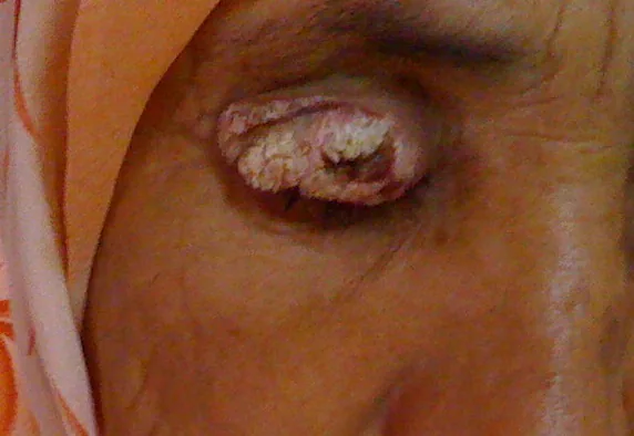
Conjunctival type: A flat reddish tumor with fireworks-like tumor vessels. If keratinization is strong, it appears whitish. When it proliferates and elevates, it becomes a nodular lesion.
Cutaneous type: White, yellowish-white, or red with a rough surface. It presents as nodular (52%) or ulcerative (40%) forms.
Note the direction of tumor invasion: If the tumor extends beyond the eyelid margin and invades anterior to the tarsal plate, full-thickness eyelid resection and reconstruction are required, similar to sebaceous carcinoma.
Eyelid findings: Eyelash loss, telangiectasia, distortion of eyelid structure, and eyelid malposition.
The development of squamous cell carcinoma is promoted by the overlap of multiple risk factors.
Ultraviolet exposure: The most important reversible risk factor. Cumulative exposure to ultraviolet A and B waves directly (base transversion) or indirectly (reactive oxygen species) damages DNA, promoting carcinogenesis via p53 mutations 2)
Precursor lesions: Gradual progression from actinic keratosis to squamous cell carcinoma in situ to invasive squamous cell carcinoma to metastatic squamous cell carcinoma.
Human papillomavirus (HPV) infection: In Bowen’s disease, there is a strong association with HPV type 16. Involvement of HPV types 6/11, 16, and 18 has been reported 4)
Immunosuppression: Organ transplantation, immunosuppressive drug use, and HIV infection/AIDS increase the risk 2)
Xeroderma pigmentosum: Autosomal recessive mutations in DNA repair genes (XPA to XPF) result in a 10,000-fold increased risk of non-melanoma skin cancer compared to the general population 2)
Others: Exposure to petroleum derivatives and arsenic, smoking, albinism, old burn scars (Marjolin’s ulcer), chronic ulcers
QDoes the risk of eyelid squamous cell carcinoma increase after organ transplantation?
A
Yes, significantly. Cutaneous squamous cell carcinoma is one of the most common malignancies after solid organ transplantation, with a 5-year incidence reaching 30% in lung transplant recipients and up to 26% in heart transplant recipients. Regular skin and ophthalmic examinations are important after transplantation.
The concordance rate between clinical diagnosis and pathological diagnosis is low for squamous cell carcinoma at 46% (compared to 86% for basal cell carcinoma and 91% for sebaceous carcinoma), making histopathological examination (excisional biopsy) essential for definitive diagnosis1).
Record of general appearance, size, ulceration, madarosis (eyelash loss), and telangiectasia of the lesion
Complete ophthalmic examination including eye movement and proptosis
Evaluation of the entire face and sun-exposed areas, and assessment of facial sensation
Palpation of regional lymph nodes (preauricular, sublingual, submandibular, cervical)
Observation of the entire eyelid during opening and closing, and the conjunctival surfaces of the upper and lower eyelids
For large tumors, orbital CT/MRI to assess internal structure and orbital extension
Preoperatively, head and neck CT or MRI is performed to check for metastasis.
The following non-invasive diagnostic adjuncts are useful for conjunctival lesions4).
High-resolution OCT (HR-OCT): Visualizes epithelial thickening, hyperreflective changes, and abrupt transition from normal epithelium. Functions as an optical biopsy
Impression cytology: Collects cells from the corneal and conjunctival surface for cytologic examination
In vivo confocal microscopy: Non-invasively evaluates epithelial dysplasia
When clinical distinction from basal cell carcinoma or poorly differentiated sebaceous carcinoma is difficult, immunohistochemical staining is useful as an adjunctive diagnostic tool.
Complete surgical excision with histologically confirmed tumor-free margins is the standard treatment with the strongest evidence.
For early lesions (confined to the palpebral conjunctiva), total excision including part of the tarsal plate is performed. After confirming negative resection margins, 2–3 cycles of cryocoagulation (freeze and thaw) are added to the resection surface.
If the tumor extends beyond the eyelid margin and infiltrates anterior to the tarsal plate, full-thickness eyelid resection and reconstruction are required, similar to sebaceous carcinoma.
As with sebaceous carcinoma, check for metastasis (head and neck CT/MRI) before surgery.
The main surgical procedures are as follows:
Mohs micrographic surgery: A technique that excises while evaluating margins in real time pathologically. Allows complete removal while minimizing tumor tissue.
Excision with intraoperative frozen section pathology: Intraoperative confirmation of resection margins.
For extensive full-thickness eyelid resection: Reconstruction using a switch flap or Cutler-Beard procedure.
Sentinel lymph node biopsy: Considered for extensive lesions, perineural invasion, or recurrent lesions.
Orbital exenteration: Performed when there is orbital invasion, poor visual prognosis, and the cavernous sinus is not involved. It was performed in 19% of squamous cell carcinoma cases1).
In Kaliki 2019, wide excisional biopsy was performed in 76% of squamous cell carcinoma cases1).
Tissue destruction using liquid nitrogen. Indicated only for actinic keratosis and squamous cell carcinoma in situ (not for invasive carcinoma). Also used as additional freeze-thaw on intraoperative resection margins for early lesions.
Used as monotherapy for patients at too high surgical risk, or as postoperative adjuvant therapy for cancers with nerve/lymph node extension or unclear margins. Irradiation is given 3-5 times a week for about 1-2 months. Squamous cell carcinoma is radiosensitive and is an option when curative resection is difficult.
Topical therapy for precursor lesions and intraepithelial localized lesions
Imiquimod cream: Immune modulator. Indicated for actinic keratosis and Bowen’s disease (precancerous lesions). Apply 3 times a week for 4-6 weeks.
Mitomycin C eye drops (0.04%): Indicated when conjunctival lesions such as pagetoid spread are confined to the epithelium. Instill 4 times daily, 1 week on, 1 week off, repeated for 2-3 cycles4).
5-Fluorouracil eye drops (1%): Similarly indicated for intraepithelial localized lesions. Instill 4 times daily, 4 days on and 30 days off, repeating up to 6 cycles4)
IFNα-2b eye drops: Fewer side effects and increasingly used in outpatient settings. Useful for conjunctival OSSN4)
Treatment outcomes in 99 patients with squamous cell carcinoma in India are shown below1).
Indicator
Percentage
Tumor recurrence
8%
Regional lymph node metastasis
8%
Distant metastasis
4%
Death from disease
4%
Eye preservation
79%
In the 5-year Kaplan-Meier estimate, local lymph node metastasis was 22%, distant metastasis 11%, and metastasis-related death 11%1).
QWhat is the likelihood of recurrence after surgery for eyelid squamous cell carcinoma?
A
In the Kaliki 2019 study, postoperative tumor recurrence was observed in 8%. The 5-year Kaplan-Meier estimate showed that local lymph node metastasis reached 22%, indicating that regular follow-up after surgery is important.
Ultraviolet radiation damages DNA directly (base transitions) or indirectly (oxidative damage via reactive oxygen species)2). Apoptosis induction by sunburn acts as a defense mechanism, but mutations accumulate when DNA repair cannot keep up.
p53 inactivation: The p53 tumor suppressor is directly damaged and inactivated by UV radiation. The functions of cell cycle arrest and apoptosis regulation are lost, allowing mutant cells to proliferate2)
Genomic instability: Genomic instability in keratinocytes is likely due to p53 inactivation
Eyelid skin-derived type (cutaneous type) involves stepwise malignant transformation of spinous layer cells and is managed by the AJCC eyelid TNM classification. The palpebral conjunctival type is a form of OSSN that progresses from conjunctival epithelial dysplasia (CIN: conjunctival intraepithelial neoplasia) to invasive SCC, and the AJCC conjunctival classification applies 4).
The stage distribution in Kaliki 2019 was T1: 26%, T2: 37%, T3: 7%, T4: 29%1). Histological grade ranges from G1 (well differentiated) to G4 (undifferentiated), with lower grades associated with better prognosis.
7. Latest Research and Future Perspectives (Investigational Reports)
Ye et al. (NEJM 2025) reported a case of a 34-year-old woman with a germline mutation in ZAP70 causing T-cell receptor signaling impairment, who developed treatment-resistant invasive cutaneous squamous cell carcinoma with genomic integration of β-HPV193). Typical squamous cell carcinoma driver mutations (TP53, NOTCH1/2, CDKN2A) were not detected, and the UV mutation signature was low at 26% (compared to an average of 77% in typical cutaneous squamous cell carcinoma). After allogeneic hematopoietic stem cell transplantation restored T-cell receptor signaling, HPV-specific T-cell responses recovered, and all HPV-related diseases, including squamous cell carcinoma, showed stable regression over 35 months of follow-up.
This report suggests that adaptive immune T-cell responses are involved in controlling the development and progression of squamous cell carcinoma3).
Cemiplimab (anti-PD-1 antibody) is FDA-approved for unresectable or metastatic cutaneous squamous cell carcinoma, and further expansion of indications is expected for cases involving orbital or lymph node extension. The involvement of immune surveillance mechanisms is becoming clearer, and the increased risk of squamous cell carcinoma in immunosuppressed states (e.g., after organ transplantation, hematologic malignancies) is thought to be related to dysregulation of T-cell responses2)3).
QIs immunotherapy effective for eyelid squamous cell carcinoma?
A
The anti-PD-1 antibody cemiplimab is FDA-approved for unresectable or metastatic cutaneous squamous cell carcinoma. There are also case reports of tumor regression in HPV-driven squamous cell carcinoma after immune reconstitution via hematopoietic stem cell transplantation. However, the latter is an investigational finding and not standard treatment.
Prognosis is good with early detection and complete resection. However, it exhibits more aggressive biological behavior than basal cell carcinoma, with risk of metastasis to the orbit, lymph nodes, and distant organs. Cases with malignant orbital invasion are managed through multidisciplinary collaboration with medical oncology, radiation oncology, etc.
Key points of follow-up are as follows.
Regular postoperative ophthalmic examinations and palpation of regional lymph nodes
Regular observation of the skin including sun-exposed areas of the entire face
Use of sunscreen and lifestyle guidance to reduce sun exposure
More frequent follow-up in immunocompromised patients
The 5-year Kaplan-Meier estimates show regional lymph node metastasis 22%, distant metastasis 11%, and metastasis-related death 11% 1), requiring long-term surveillance.
Kaliki S, Bothra N, Bejjanki KM, Nayak A, Ramappa G, Mohamed A, et al. Malignant Eyelid Tumors in India: A Study of 536 Asian Indian Patients. Ocular oncology and pathology. 2019;5(3):210-219. doi:10.1159/000491549. PMID:31049330; PMCID:PMC6489076.
Ye P, Bergerson JRE, Brownell I, Starrett GJ, Abraham RS, Anderson MV, et al. Resolution of Squamous-Cell Carcinoma by Restoring T-Cell Receptor Signaling. The New England journal of medicine. 2025;393(5):469-478. doi:10.1056/NEJMoa2502114. PMID:40742260; PMCID:PMC12370287.
Tsatsos M, Delimitrou C, Tsinopoulos I, Ziakas N. Update in the Diagnosis and Management of Ocular Surface Squamous Neoplasia (OSSN). Journal of clinical medicine. 2025;14(5). doi:10.3390/jcm14051699. PMID:40095695; PMCID:PMC11900158.
Copy the article text and paste it into your preferred AI assistant.
Article copied to clipboard
Open an AI assistant below and paste the copied text into the chat box.