Epithelial tumors of the conjunctiva are broadly classified into conjunctival intraepithelial neoplasia (CIN), where the basement membrane is preserved, and invasive squamous cell carcinoma (SCC), where the tumor extends beyond the basement membrane.
CIN is further classified by severity.
Mild CIN (dysplasia): Abnormal growth is confined to part of the epithelial layer.
Severe CIN (carcinoma in situ): Abnormal growth involves the full thickness of the epithelium; the basement membrane is preserved.
Invasive SCC: Invades the subconjunctival tissue beyond the basement membrane.
The concept of ocular surface squamous neoplasia (OSSN) is also widely used. It is a collective term for the spectrum of epithelial tumors from dysplasia to CIN and invasive SCC. All commonly arise at the limbus (corneal edge) and extend to the adjacent corneal surface and bulbar conjunctiva.
The incidence of conjunctival squamous cell carcinoma varies greatly by geographic region. It is reported to range from 0.02 to 3.5 per 100,000 people (depending on latitude and UV exposure)1). 75% of patients are male, 75% are aged 60 or older, and 75% arise from the corneal limbus1).
In Shields et al.’s series of 771 non-melanocytic conjunctival tumors, ocular surface squamous neoplasia accounted for 23% (179 cases), making it the most common non-pigmented tumor1). The worldwide age-standardized incidence rate of ocular surface squamous neoplasia is 0.26 per 100,000 per year, with the highest rate in Africa (3.4 per 100,000 per year)1).
QHow often do conjunctival epithelial tumors occur?
A
The incidence of squamous cell carcinoma varies greatly by region, ranging from 0.02 to 3.5 per 100,000 people1). The age-standardized incidence rate of ocular surface squamous neoplasia is 0.26 per 100,000 per year worldwide, but is notably higher in Africa at 3.4 per 100,000 per year1).
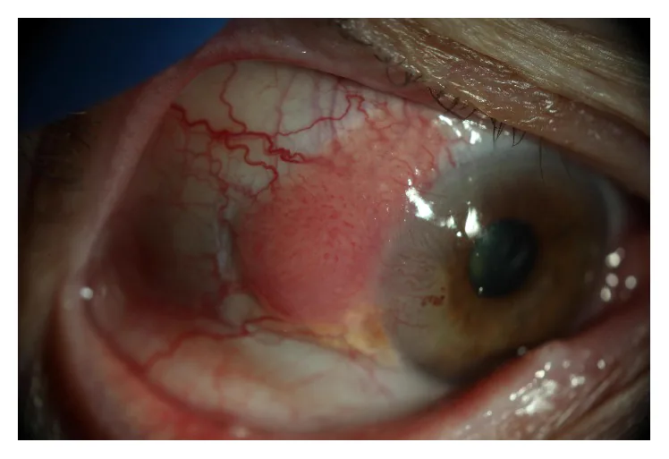
Ong SS, Vora GK, Gupta PK. Anterior Segment Imaging in Ocular Surface Squamous Neoplasia. J Ophthalmol. 2016;2016:5435092. Figure 1. PMCID: PMC5069377. DOI: 10.1155/2016/5435092. License: CC BY.
Slit-lamp photograph showing gelatinous and papillary lesions of corneal conjunctival intraepithelial neoplasia with characteristic feeding vessels. This corresponds to the characteristic gelatinous limbal lesion and feeding vessels of OSSN discussed in the section “2. Main Symptoms and Clinical Findings”.
Hyperemia and foreign body sensation: Most common complaint
Visual impairment: Occurs when the lesion involves the pupillary area
Asymptomatic: May be discovered incidentally
Redness and ocular discomfort: Symptoms of conjunctival squamous cell carcinoma are nonspecific; visual impairment occurs when the visual axis is involved 1)
Conjunctival intraepithelial neoplasia (CIN) is observed as a sessile, slightly opaque, flat elevated lesion. It appears white to pale red, with characteristic abnormal vascular patterns described as “firework-like”.
Invasive squamous cell carcinoma (SCC) takes various forms.
Cauliflower-like papillomatous lesion or white elevated lesion with an irregular surface
May be accompanied by leukoplakia due to hyperkeratosis on the surface
Pale red to reddish-pink, gelatinous, irregular appearance
Keratin may adhere to the surface
The morphological variations of the lesion and their clinical significance are shown below.
Gelatinous: Most common form
Leukoplakic: Reflects hyperkeratosis
Papillomatous/nodular: Associated with more aggressive pathological grade1)
Nodular ulcerative: Rare but strong indicator of invasive tumor1)
Abnormal tortuous dilated feeding vessels on the tumor: Important finding suggesting malignant growth1)
Elevated lesions tend to have higher malignancy than flat lesions1). Common sites are the interpalpebral fissure and limbus, while the palpebral conjunctiva is less frequently involved1).
QCan conjunctival intraepithelial neoplasia and invasive squamous cell carcinoma be distinguished clinically?
A
Clinical differentiation between conjunctival intraepithelial neoplasia and squamous cell carcinoma is difficult, and histopathological examination is essential for definitive diagnosis. High-resolution optical coherence tomography is useful for distinguishing invasive from non-invasive types1), but final diagnosis relies on histological examination.
Ultraviolet exposure: The greatest risk factor. Carcinogenesis mechanism via p53 gene mutation1)
Human papillomavirus: Types 16 and 18 have been implicated1). However, the association between HPV and ocular surface squamous neoplasia varies by region and is debated1)
Male sex and older age: Mean age at onset 56 years1)
Immunodeficiency: Occurs frequently in HIV/AIDS patients. Related to high prevalence in young women in Africa
Others: Smoking, chemical exposure (petroleum products, beryllium, arsenic, etc.), vitamin A deficiency, ocular surface trauma1)
Recurrence risk factors: Large tumor size, positive resection margins, HIV infection, high tumor grade, presence of feeder vessels, high proliferative index1)
QWhat are the risk factors other than UV?
A
HPV types 16/18, immunodeficiency (HIV/AIDS), xeroderma pigmentosum, smoking, chemical exposure (petroleum products, beryllium, arsenic, etc.), and vitamin A deficiency have been reported1). HIV infection and positive resection margins are strongly associated with tumor recurrence1).
Slit-lamp microscopy: Observe tumor size, borders, color, and surface irregularity. Photographic documentation is recommended.
Fluorescein staining: Utilizes increased permeability of abnormal epithelium to clearly delineate the border between lesion and healthy tissue. Useful for preventing oversight of flat or small lesions.
Scleral scattering: Clarifies the extent of flat lesions on the cornea.
Special stains: Rose bengal, lissamine green, methylene blue, etc., are also used to stain necrotic squamous epithelial cells1).
High-resolution optical coherence tomography (HR-OCT): Non-invasive tool. Characterized by a steep transition between hyperreflective thickened epithelium and normal epithelium. Epithelial thickness >140 μm is considered an indicator of potential tumor. Useful for differentiating invasive from non-invasive types1).
In vivo confocal microscopy: Useful for differentiating epithelial from subepithelial lesions1).
Impression cytology and exfoliative cytology: Minimally invasive but limited in assessing depth of invasion1).
Metastasis workup: Palpation of preauricular lymph nodes is basic. For extensive tumors, perform whole-body search with gallium scintigraphy and FDG-PET
Topical chemotherapy is used as first-line or adjuvant therapy. A cycle of “one week on, one week off” is typical.
Low-concentration mitomycin C or 5-fluorouracil eye drops have been reported to achieve tumor eradication. However, some reports indicate efficacy only for intraepithelial lesions, and long-term recurrence rates and complications are not fully understood.
Interferon alpha-2b is used as eye drops or subconjunctival injection. It has lower toxicity and better tolerability compared to mitomycin C and 5-fluorouracil, but is more costly.
Additional Treatment for Malignant Tumors (Invasive Squamous Cell Carcinoma)
Metastasis is rare and the life prognosis is good. The local recurrence rate of invasive squamous cell carcinoma is reported as 5%, and the regional lymph node metastasis rate as 2% 1). On the other hand, the mortality rate of untreated squamous cell carcinoma is 8–24%, and orbital invasion occurs in about 10% of cases 1).
QAre there treatments other than surgery?
A
Topical chemotherapy with mitomycin C, 5-fluorouracil, interferon alpha-2b, etc. is used as primary or adjuvant therapy. However, reports indicate it is useful only for intraepithelial lesions 1), and long-term outcomes and complications are not well established. Radiation therapy is used adjuvantly for unresectable cases or eyelid invasion.
Mild conjunctival intraepithelial neoplasia: Part of the surface epithelium is replaced by abnormal cells lacking normal maturation.
Severe conjunctival intraepithelial neoplasia: The full thickness of the epithelium is replaced by abnormal cells lacking maturation. Epithelial cells lose polarity and show atypia throughout the entire layer.
The basement membrane remains intact: This is a key difference from invasive squamous cell carcinoma.
Invasive squamous cell carcinoma
Basement membrane breach: Malignant squamous epithelial cells proliferate beyond the basement membrane into the stroma1).
Histological features: Atypical cells with mitotic figures infiltrate the lamina propria.
Mucoepidermoid carcinoma: An aggressive subtype of squamous cell carcinoma. It occurs more often in older individuals and contains yellow cystic components from mucus-secreting cells1).
7. Latest research and future perspectives (investigational reports)
The application of bevacizumab and ranibizumab to conjunctival lesions has been reported 1).
According to a review by Tsatsos et al., a study using ranibizumab (1.25–2.5 mg, subconjunctival injection 1–2 times per month) achieved complete regression in 34% and partial regression in 66% of cases, with no recurrence observed during 6 months of follow-up 1). Bevacizumab is promising for conjunctival lesions, but its efficacy for corneal lesions is unclear, and a risk of delayed corneal epithelial healing has been noted. Large-scale studies are needed for both.
External beam radiation therapy (EBRT): Irradiation with proton or electron beams. Useful for avoiding enucleation in cases of large tumors or intraocular invasion 1)
Postoperative proton therapy: Reported to reduce recurrence of squamous cell carcinoma1)
Brachytherapy: Sr-90, I-125, Ru-106. Good tumor control has been reported even in cases with positive resection margins 1)
According to a review by Tsatsos et al., a pilot study combining verteporfin and laser reported 100% tumor regression and no recurrence in conjunctival squamous cell carcinoma1). High cost, need for specialized training, and limited availability are challenges to widespread use.
A case report has shown a remarkable effect of the HPV vaccine on HPV type 16-positive conjunctival intraepithelial neoplasia. Verification through large-scale studies is needed.
Tsatsos M, Delimitrou C, Tsinopoulos I, Ziakas N. Update in the Diagnosis and Management of Ocular Surface Squamous Neoplasia (OSSN). J Clin Med. 2025;14(5):1699. doi:10.3390/jcm14051699.
Copy the article text and paste it into your preferred AI assistant.
Article copied to clipboard
Open an AI assistant below and paste the copied text into the chat box.