Corneoconjunctival dermoid (limbal dermoid) is a type of choristoma that occurs on the cornea, limbus, and conjunctiva. It is a congenital benign tumor derived from ectodermal dysplasia. A choristoma refers to ectopic tissue that is not normally present at that site. It is thought to be caused by abnormal development of the first and second branchial arches during the embryonic period, and it is a representative congenital tumor among periocular tumors.
This condition is not hereditary but sporadic, and it is usually unilateral and present at birth. The size of the tumor ranges from about 3 mm in diameter to as large as 10 mm.
Limbal dermoid is the most common type, with a characteristic predilection for the inferotemporal limbus. Corneal dermoid extends from the limbus to the central cornea and carries a high risk of visual impairment.
Limbal dermoid may be associated with ear abnormalities such as accessory auricles and preauricular fistulas, as well as spinal abnormalities. This combination of multiple malformations is known as Goldenhar syndrome (oculoauriculovertebral spectrum). Since it may also involve mandibular hypoplasia, a systemic examination is necessary when a limbal dermoid is observed.
Goldenhar syndrome is reported to occur in approximately 10–20% of patients with dermoid, and it is often first noticed based on ophthalmic findings alone. Collaboration with otorhinolaryngology, orthopedics, and oral surgery is important.
QCan a corneoconjunctival dermoid become malignant?
A
Corneoconjunctival dermoid is a benign choristoma, and malignant transformation is extremely rare. However, if the mass enlarges or shows surface changes, reevaluation including histological examination may be necessary. Ophthalmologically, amblyopia, corneal astigmatism, and cosmetic issues are the main management concerns, and the impact on visual development is prioritized over malignant change.
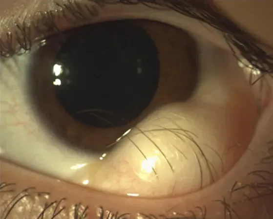
Triolo G, Ferrari G, Doglioni C, Rama P. In vivo confocal microscopy in Goldenhar syndrome: a case report. BMC Ophthalmol. 2013;13:55. Figure 1. PMID: 24131730; PMCID: PMC3853195. doi:10.1186/1471-2415-13-55. License: CC BY 2.0.
Clinical photograph showing a pale yellowish-white, dome-shaped, solid dermoid tumor at the limbus in a 15-year-old female with Goldenhar syndrome. This corresponds to the typical appearance of limbal dermoid (pale yellowish-white, dome-shaped, inferotemporal limbal predilection) discussed in the section “2. Main symptoms and clinical findings.”
Dermoid is a solid tumor. Limbal dermoid tends to occur particularly at the inferotemporal limbus and is observed as a hemispherical or flat, pale yellowish-white mass. The tumor diameter is 3–10 mm and is present from birth.
The surface of the dermoid is keratinized skin tissue, causing the tear film to break up at that site, preventing normal ocular surface coverage. Hair-like tissue may also be observed protruding from the tumor surface.
Dermoids alter the corneal shape, resulting in irregular and regular astigmatism. This corneal astigmatism can cause anisometropia and may lead to amblyopia. Since amblyopia progresses irreversibly during the sensitive period of visual development (from 3 months to about 8 years of age), regular visual acuity and refraction assessments from infancy are essential.
In large dermoids extending to the central cornea, the opaque tumor may obstruct the visual axis, causing form deprivation amblyopia.
Dermoids originate from dysplasia that occurs during embryonic development when ectoderm forms the cornea and conjunctiva. Specifically, abnormal development of the first branchial arch (mandibular arch) and second branchial arch (hyoid arch) is thought to be the starting point, leading to ectodermal tissue becoming entrapped and persisting on the ocular surface.
This condition is not hereditary but occurs sporadically. Familial clustering is rare, and cases requiring genetic counseling are limited.
Goldenhar Syndrome: Checklist for Systemic Complications
Goldenhar syndrome (oculoauriculovertebral spectrum) is a syndrome characterized by multiple congenital malformations of the eyes, ears, jawbone, and spine. Main symptoms include limbal dermoid, accessory auricle, preauricular fistula, micrognathia, and spinal anomalies, often occurring unilaterally. The frequency is approximately 1 in 5,600 to 1 in 26,550 births. Detection of limbal dermoid by an ophthalmologist often leads to diagnosis, and multidisciplinary systemic management is important.
Clinical diagnosis is possible based on the typical finding of a solid mass on the inferotemporal limbus present at birth. Slit-lamp examination can confirm surface keratinization, hair structures, and the solid nature of the mass.
The most important differential diagnosis is dermoid cyst. Dermoid cysts are cystic and contain secretions (sebum, hair) internally, whereas dermoids are solid, allowing differentiation by palpation, slit-lamp findings, and ultrasound.
Anterior segment optical coherence tomography (AS-OCT) is useful for noninvasively assessing the depth of dermoid infiltration into the corneal stroma. Preoperative assessment of infiltration depth is important for determining the depth (lamellar thickness) of superficial keratoplasty performed concurrently with excision.
Refraction, visual acuity testing, and amblyopia evaluation
Since visual acuity testing is difficult in infants, refraction tests such as corneal curvature measurement and autorefractometry (under cycloplegia) are performed to quantify the degree of astigmatism. Visual acuity is evaluated using the cover test (peek test), Teller acuity cards, and VEP (visual evoked potentials).
Spectacle Correction: Prescribe astigmatism-correcting glasses early according to corneal astigmatism
Contact Lenses: Consider hard contact lenses for irregular astigmatism
Occlusion (Eye Patch) Therapy: Visual training of the amblyopic eye by occluding the dominant eye
Pharmacological occlusion (atropine eye drops): Use atropine 1% eye drops in the dominant eye as an occlusion aid.
After the sensitive period for amblyopia (up to age 8), treatment effectiveness decreases significantly, so early detection and early intervention determine the prognosis.
Indications: Dermoids confined to the limbus, without growth tendency, and not involving the visual axis.
Timing: Since surgery is for cosmetic purposes, it is often performed after infancy (school age or later).
Management: Continue spectacle correction and amblyopia training while performing regular follow-up.
Early Surgery (Active Intervention)
Indications: Cases extending to the central cornea and obstructing the visual axis, or cases with rapid growth.
Timing: Early surgery is necessary to prevent form deprivation amblyopia. In young children, it is performed under general anesthesia.
Preoperative Preparation: Begin amblyopia management (astigmatism correction and occlusion) before surgery.
Cosmetic Surgery
Indications: Cases confined to the limbus with mild impact on vision and cornea, but where cosmetic improvement is desired.
Timing: Preferably after infancy (to avoid risks of general anesthesia in young children).
Note: Even for cosmetic purposes, superficial keratectomy is combined to prevent postoperative pseudopterygium and recurrence.
QWhen is the best time to perform surgery?
A
For large dermoids that obscure the visual axis or those with rapid growth, early surgery is necessary to prevent form deprivation amblyopia. On the other hand, if the dermoid is confined to the limbus and shows no growth, it is considered cosmetic surgery, and surgery is generally performed after infancy. In either case, it is important to start astigmatism correction and amblyopia training before surgery, as vision recovery may not be achieved with postoperative treatment alone. Surgery in young children requires general anesthesia.
Surgical treatment: Concomitant lamellar keratoplasty is essential
Simple excision alone of a dermoid frequently leads to recurrence and pseudopterygium. Moreover, because limbal dermoids extend into the corneal stroma, simple excision results in corneal thinning, posing a risk of corneal perforation. Therefore, the standard procedure is to combine excision with lamellar keratoplasty.
Outline of the standard surgical procedure:
Dissect and excise the dermoid from the limbus toward the cornea
Preoperatively assess the depth of the corneal bed at the excision site using AS-OCT
Suture a donor cornea (lamellar button) into the defect
Suture while preserving limbal stem cells
Preoperative planning using anterior segment OCT allows accurate determination of lamellar thickness according to the depth of invasion. The use of femtosecond laser for lamellar dissection has also been reported, and it is expected to improve the precision of the incision plane.
Surgery in young children (preschool age) requires general anesthesia, and continued amblyopia training is necessary after surgery.
QCan the dermoid recur after surgery?
A
Simple excision alone frequently leads to recurrence and pseudopterygium. Combining lamellar keratoplasty significantly reduces these risks. However, if limbal stem cell function remains impaired, it may affect long-term corneal epithelial stability, and regular anterior segment evaluation is necessary after surgery.
Dermoids are classified as choristomas. A choristoma is a mass of histologically normal tissue that is ectopically located in a site where it is not normally found. Unlike teratomas, which contain a disorganized mixture of tissues from multiple germ layers, dermoids consist of normal tissues (skin, hair follicles, sebaceous glands, fat, cartilage) that maintain their normal histological structure but are located in an abnormal site, such as the ocular surface.
During the embryonic period, the first pharyngeal arch (mandibular arch) and second pharyngeal arch (hyoid arch) are responsible for the development of the face, auricle, and mandible. Abnormalities in these structures can disrupt the migration of neural crest cells, leading to ectopic entrapment and retention of ectodermal tissue on the ocular surface. This results in dermoids containing a mixture of ectodermal tissues (skin, hair, tooth primordia) and mesodermal tissues (fat, cartilage).
In Goldenhar syndrome, these developmental abnormalities are more extensive, often involving multiple malformations of the auricle, jawbone, and vertebrae.
The presence of a dermoid alters the corneal shape, causing asymmetry in corneal curvature. When a limbal dermoid attaches to the inferotemporal quadrant, it flattens the cornea in that direction, often resulting in against-the-rule or oblique astigmatism. The degree of astigmatism depends on the size of the dermoid, the area of attachment to the limbus, and the depth of corneal stromal invasion.
High astigmatism (e.g., cylindrical difference of 3D or more) is a significant factor causing anisometropic amblyopia during the sensitive period of visual development, necessitating early refractive correction.
The high incidence of pterygium-like lesions after dermoid excision is attributed to damage to limbal stem cells. The limbus contains stem cells responsible for corneal epithelial regeneration, but the dermoid itself, located at the limbus, disrupts the stem cell niche. If residual stem cell function is compromised after excision, conjunctival epithelium may invade the cornea postoperatively, forming a pterygium-like lesion. Supplementation with donor tissue through lamellar keratoplasty reduces this risk.
Quantitative assessment of the depth of corneal stromal invasion by dermoids using high-resolution anterior segment OCT (AS-OCT) is advancing. Preoperative visualization of the depth of the dermoid’s base and the extent of limbal stem cell involvement improves the accuracy of incision depth setting in lamellar keratoplasty, contributing to a reduction in postoperative complications.
The application of lamellar dissection using a femtosecond laser (femtosecond lamellar keratoplasty) for dermoid excision has been reported. By precisely creating a lamellar button of uniform thickness, the regularity of the corneal shape after excision is improved, and better stability of postoperative refraction is expected.
Comparison of superficial lamellar keratoplasty vs. deep lamellar keratoplasty
In cases where the dermoid extends into the deep corneal stroma, the application of deep anterior lamellar keratoplasty (DALK) in addition to conventional superficial keratoplasty is being considered. DALK replaces the stroma up to just above Descemet’s membrane, allowing treatment of deep infiltrative cases, but it is technically challenging and the indication for pediatric cases must be carefully evaluated.
Accumulating reports have identified genetic variants in MYT1L, FOXI3, ZIC3, and other genes in cases of Goldenhar syndrome. Chromosomal copy number variations (CNVs) have also been detected in some cases. The role of comprehensive genetic testing, including genetic counseling and next-generation sequencing, may expand in the future.
Research is being conducted on the application of autologous or allogeneic limbal stem cell transplantation for limbal stem cell dysfunction following dermoid excision. When limbal stem cell deficiency (LSCD) occurs, cultured limbal epithelial transplantation (CLET) or cultured oral mucosal epithelial transplantation (COMET) may be options.
Scott JA, Tan DT.. Therapeutic lamellar keratoplasty for limbal dermoids. Ophthalmology. 2001;108(10):1858-1867. doi:10.1016/s0161-6420(01)00705-9. PMID:11581063.
Robb RM.. Refractive errors associated with hemangiomas of the eyelids and orbit in infancy. Am J Ophthalmol. 1977;83(1):52-58. doi:10.1016/0002-9394(77)90191-x. PMID:835667.
Mattos J, Contreras F, O’Donnell FE Jr. Ring dermoid syndrome. A new syndrome of autosomal dominantly inherited, bilateral, annular limbal dermoids with corneal and conjunctival extension. Arch Ophthalmol. 1980;98(6):1059-1061. doi:10.1001/archopht.1980.01020031049007.
Mader TH, Stulting RD. Keratorefractive surgery in the presence of corneal dermoids. Refractive Corneal Surg. 1992;8(6):498-499.
Liesegang TJ. Limbal dermoids revisited. Ophthalmology. 1999;106(12):2277-2279.
Ramos M, Krueger RR, Ang E. Laser-assisted in situ keratomileusis and photorefractive keratectomy for the treatment of anisometropia. J Refract Surg. 2001;17(2):S219-S222.
Watts P, Michaeli-Cohen A, Abdolell M, Rootman D.. Outcome of lamellar keratoplasty for limbal dermoids in children. J AAPOS. 2002;6(4):209-215. doi:10.1067/mpa.2002.124651. PMID:12185344.
Baum JL, Feingold M.. Ocular aspects of Goldenhar’s syndrome. Am J Ophthalmol. 1973;75(2):250-257. doi:10.1016/0002-9394(73)91020-9. PMID:4697181.
Elsas FJ, Maumenee IH, Kenyon KR, Yoder F.. Familial aniridia with preserved ocular function. Am J Ophthalmol. 1977;83(5):718-724. doi:10.1016/0002-9394(77)90139-8. PMID:868970.