Deep Anterior Lamellar Keratoplasty (DALK) is a surgical procedure in which almost all of the recipient’s corneal stroma is removed and only the donor stroma is transplanted. The recipient’s own Descemet membrane and corneal endothelium are preserved 6).
The concept of selective corneal stromal transplantation while preserving the endothelium dates back over 150 years. In 1959, Hallerman first performed deep stromal dissection near Descemet membrane. In 1974, Anwar reported that removing the donor’s Descemet membrane and endothelium resulted in a smoother interface and improved visual outcomes. In 1984, Archila introduced the air injection technique, laying the foundation for the current big bubble method.
No endothelial rejection: Since donor endothelium is not transplanted, endothelial rejection does not occur 6)
Improved graft survival: Long-term survival is superior to PK, especially in younger patients
Closed-system surgery: Lower risk of expulsive hemorrhage and traumatic wound dehiscence during surgery
Relaxed donor requirements: Donor corneas with low endothelial cell counts can also be used6)
Reduced steroid burden: The duration of postoperative steroid eye drops can be shortened
Disadvantages of DALK
Technical difficulty: The procedure is more difficult than PK and has a learning curve6)
Risk of Descemet’s membrane perforation: Intraoperative perforation may require conversion to PK
Interface opacity: Residual stroma can limit visual acuity
Stromal rejection: Although endothelial rejection does not occur, stromal rejection occurs in 2–12% of cases6)
The overall rejection rate is reported as 1.9% for DALK versus 7.8% for PK. Meta-analysis shows that the rejection rate for DALK is significantly lower than for PK (OR 0.28, 95% CI 0.15–0.50, P<0.001)6).
Period
DALK overall
PK overall
DALK high risk
PK high risk
1 year
95.8%
94.4%
84.6%
90.3%
5 years
93.9%
80.4%
82.1%
59%
10 years
93.9%
72.1%
82.1%
48.7%
In high-risk eyes (e.g., deep neovascularization, ocular surface disease, glaucoma), graft survival rate of DALK is significantly higher than PK.
The prerequisite for DALK is normal endothelial function6).
Corneal ectasia: Keratoconus is the most common indication. Candidates include patients intolerant to contact lenses or those who cannot achieve adequate visual function even with CL wear6). It is also applied to pellucid marginal degeneration and corneal ectasia after refractive surgery.
Corneal scar: Stromal scars that do not reach Descemet’s membrane are indicated. This includes scars after herpetic keratitis, trauma, and infection.
Tectonic purposes: For Descemetocele or corneal perforation, DALK is performed to preserve the structural integrity of the eye and restore vision 4). Tectonic DALK after chemical injury in children has also been reported 4). There are also reports of DALK combined with peripheral lamellar keratoplasty for severe peripheral ulcerative keratitis5).
Relative contraindications: History of Descemet’s membrane rupture due to acute hydrops, deep scars involving Descemet’s membrane, deep corneal neovascularization, severe corneal thinning 6)
QIs DALK suitable for macular corneal dystrophy?
A
DALK is not recommended for macular corneal dystrophy. Because glycosaminoglycan accumulation also affects the corneal endothelium, progressive endothelial cell loss may occur after DALK. Full-thickness penetrating keratoplasty (PK) is often chosen for macular dystrophy.
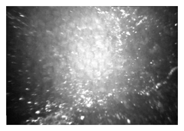
Domenico Schiano-Lomoriello, Rossella Annamaria Colabelli-Gisoldi, Mario Nubile, Francesco Oddone, et al. Descemetic and Predescemetic DALK in Keratoconus Patients: A Clinical and Confocal Perspective Study 2014 Aug 26 Biomed Res Int. 2014 Aug 26; 2014:123156 Figure 2. PMCID: PMC4160628. License: CC BY.
In vivo confocal microscopic image of a descemetic (D-DALK) interface with bright microdots
Trephination: Perform partial-thickness trephination to a depth of 60–80% of corneal thickness
Air injection: Insert a 27–30 gauge needle into the stroma and inject air forcefully
Bubble formation: A Type 1 bubble (between the posterior stroma and Descemet’s membrane) or Type 2 bubble (between Descemet’s membrane and the pre-Descemet layer) is formed.
Removal of anterior stroma: The anterior wall of the bubble is punctured, and the stroma is divided into four quadrants and excised.
Suturing of the donor: The donor button, with Descemet’s membrane stripped, is secured using a suturing method similar to penetrating keratoplasty (PK).
The success rate of Descemet’s membrane exposure using the big bubble technique is reported to be 47–82%, depending on the surgeon’s experience 6). The average rate of Descemet’s membrane perforation is 11.7%, and the conversion rate to PK is 2.4% 1).
Melles technique (air-guided deep stromal dissection)
This technique involves injecting an air bubble into the anterior chamber and using the mirror image of the dissecting spatula to assess the depth of stromal dissection 6). The stroma is dissected in layers.
Manual dissection is recommended for cases with deep corneal scars or Descemet’s membrane scars 6). The stroma is excised stepwise using a crescent knife. This may result in an irregular stromal bed, which can cause interface haze and astigmatism.
iOCT is useful for visualizing the depth of stromal dissection 1). Since metal instruments cause OCT shadow artifacts, a technique using an 8-0 nylon suture passed through the stromal tunnel as a depth marker has been reported 1). Success rates for big bubble formation are reported as 84% with Pentacam guidance, 81.8% with ultrasound pachymetry guidance, and 70% with AS-OCT guidance 1).
For DALK in post-PK corneas (e.g., hematocornea), the stromal peeling technique can be applied because the interlamellar adhesion of the PK graft is weak 3). This technique utilizes the natural cleavage plane along the pre-Descemet layer without requiring air injection 3).
QWhat is the difference between Type 1 and Type 2 in the big bubble technique?
A
Type 1 bubble forms between the posterior stroma and the pre-Descemet layer, resulting in a large bubble of 8 mm or more in diameter. Type 2 bubble forms between the Descemet membrane and the pre-Descemet layer, with a smaller diameter (about 6 mm) and a thinner wall. Type 2 bubble carries a higher risk of Descemet membrane perforation. Both types may coexist; if Type 2 is identified, the stroma within Type 1 should be removed first, and careful manipulation is required to avoid Descemet membrane perforation 2).
Descemet membrane perforation: The most common intraoperative complication in DALK. It occurs in approximately 10–30% of cases. Small perforations may allow continuation of surgery, but large perforations require conversion to PK 1). The perforation rate decreases with surgeon experience 6).
Double anterior chamber: The most common early postoperative complication 2). Aqueous humor accumulates between the Descemet membrane and the donor stroma. It is more likely to occur in cases with intraoperative Descemet membrane perforation, but can also arise from unrecognized Type 2 bubble even without perforation 2). It is usually treated with intracameral air injection (descemetopexy), but spontaneous resolution has also been reported 2)5). Since air tamponade for Descemet membrane detachment can cause endothelial cell loss of 20% or more 2), observation may be an option.
Stromal rejection: The incidence is reported to be 2–12% 6). It is usually reversible with topical steroids 6). Endothelial rejection does not occur.
Endothelial cell loss: Endothelial cell loss after DALK shows a biphasic pattern: acute loss due to intraoperative manipulation (especially during Descemet membrane perforation) and chronic gradual loss of approximately 3.9% per year. In DALK without Descemet membrane perforation, endothelial cell loss is significantly lower compared to PK.
Others: Interface opacity (due to residual stroma), suture-related complications, infection, Urrets-Zavalia syndrome (more common in keratoconus), pupillary block due to air/gas, and graft dehiscence.
Complication
Characteristics
DM perforation
Intraoperative 10–30%. Large perforation requires PK conversion
Double anterior chamber
Early postoperative. Air injection or spontaneous resolution
Stromal rejection
2–12%. Reversible with steroids
Interface opacity
Reduced visual acuity if residual stroma ≥80 μm
QIs treatment always necessary for double anterior chamber after DALK?
A
Treatment is not always necessary. Intracameral air injection (descemetopexy) is the standard treatment, but it can cause more than 20% endothelial cell loss 2). If there is a small amount of fluid accumulation that does not affect vision, spontaneous resolution has been reported after about 3 months of observation 2). In cases of non-riegmatogenous Descemet detachment due to an unrecognized Type 2 bubble without Descemet perforation, observation may be a reasonable option.
The cornea consists of six layers from anterior to posterior: epithelium, Bowman’s layer, stroma, pre-Descemet’s layer (Dua’s layer), Descemet’s membrane, and endothelium. In DALK, almost the entire stroma is removed, exposing the pre-Descemet’s layer or Descemet’s membrane.
The pre-Descemet’s layer is an acellular collagen layer approximately 10 μm thick located in the deepest stroma 3). In corneas after PK, a natural cleavage plane exists along this layer, providing the anatomical basis for the stromal peeling method 3).
Residual stromal thickness is the most important factor determining visual acuity after DALK 6). When Descemet’s membrane is fully exposed, visual acuity comparable to PK can be achieved. The thicker the residual stroma, the greater the scattering at the donor-host interface and the lower the contrast sensitivity.
The main target of corneal rejection is endothelial cells. Since DALK does not transplant donor endothelium, endothelial rejection does not occur 6). This reduces the postoperative steroid burden and also lowers the risk of steroid-induced glaucoma and cataract.
Use of intraoperative OCT: iOCT enables real-time visualization of the depth of stromal dissection, contributing to improved success rates of the big bubble technique 1). Methods using nylon sutures as depth markers have been developed to avoid artifacts from metal instruments 1). In tectonic DALK, iOCT is also useful for confirming the donor-host interface 4).
Stromal peeling technique: When selecting DALK for regrafting after PK, stromal peeling is attracting attention as a safe dissection method that does not require injection of gas or viscoelastic substances 3).
Expanding indications: The indications for DALK are expanding. Applications of DALK in areas where PK was previously chosen have been reported, such as tectonic DALK for severe chemical trauma in children 4) and combined DALK with peripheral lamellar keratoplasty for autoimmune peripheral ulcerative keratitis5).
In high-risk eyes (deep vascularization, ocular surface disease, glaucoma), DALK has been shown to have significantly better graft survival than PK, and appropriate patient selection and optimization of surgical technique will become even more important in the future.
Lin CC, Lee WS. Intraoperative optical coherence tomography-guided deep anterior lamellar keratoplasty. Taiwan journal of ophthalmology. 2023;13(1):106-109. doi:10.4103/tjo.TJO-D-22-00151. PMID:37252175; PMCID:PMC10220450.
Luangprasert P, Jongkhajornpong P, Lekhanont K, Nonpassopon M, Chuckpaiwong V. Delayed spontaneous resolution of a double anterior chamber following deep anterior lamellar keratoplasty (DALK). BMC ophthalmology. 2024;24(1):553. doi:10.1186/s12886-024-03819-6. PMID:39736594; PMCID:PMC11684058.
Scorcia V, Giannaccare G, Pellegrini M, et al. Stromal peeling for deep anterior lamellar keratoplasty in a post-penetrating keratoplasty eye with hematocornea. Am J Ophthalmol Case Reports. 2023;29:101808.
Lata S, Bari A, Agarwal T. Tectonic Deep Anterior Lamellar Keratoplasty in Severe Ocular Chuna Particle Injury in a Child. Cureus. 2023;15(7):e41712. doi:10.7759/cureus.41712. PMID:37575863; PMCID:PMC10415958.
Yokoyama K, Nakamura R, Otsuka T, Kimoto K, Kubota T. Deep Anterior Lamellar Keratoplasty and Peripheral Lamellar Keratoplasty for a Case of Severe Peripheral Ulcerative Keratitis. Case reports in ophthalmology. 2022;13(1):9-16. doi:10.1159/000521198. PMID:35221974; PMCID:PMC8832184.
American Academy of Ophthalmology Cornea/External Disease Preferred Practice Pattern Panel. Corneal Ectasia PPP — 2024. Ophthalmology. 2024.
Copy the article text and paste it into your preferred AI assistant.
Article copied to clipboard
Open an AI assistant below and paste the copied text into the chat box.