Branch Retinal Vein Occlusion (BRVO) is a vascular disease in which a vein becomes occluded at an arteriovenous crossing in the retina, causing retinal hemorrhage, macular edema, and capillary nonperfusion in the affected area. It is the most common type of retinal vein occlusion, occurring about five times more frequently than central retinal vein occlusion (CRVO) 1).
The global prevalence in 2015 was approximately 0.77%, affecting an estimated 28 million people aged 30–89 years 1). Among those aged 40 years and older, the prevalence is about 2.0%. In Japan, the Hisayama Study reported the cumulative incidence and risk factors over 9 years 2). The Beaver Dam Eye Study reported a 15-year cumulative incidence of 2.3% for all RVO1). Prevalence increases significantly with age and is higher after age 60 4). The incidence in East Asia is reported to be similar to that in the United States, with some suggestion of a slightly higher rate in Korea 12).
The most common occlusion site is the superotemporal quadrant (58–66%), followed by the inferotemporal quadrant (22–43%) and the nasal side (12.9%) 3). The disease is classified by the site and extent of occlusion as follows:
Macular branch retinal vein occlusion: Occlusion of small vessels supplying the macula. Direct impact on vision is significant.
Hemi-RVO (hemiretinal vein occlusion): Occlusion at the optic disc, impairing drainage of half the retina. Clinically, it follows a course closer to CRVO than BRVO 12).
It is also classified into two types based on the degree of ischemia.
Non-ischemic type: The area of non-perfusion on fluorescein angiography (FA) is less than 5 disc diameters (DD). Accounts for about 80% of all cases.
Ischemic type: The area of non-perfusion is 5 DD or more (10 DD or more by CVOS criteria). High risk of neovascularization and vitreous hemorrhage. 15–20% of non-ischemic type convert to ischemic type over time 3).
QIf one eye develops the condition, is the other eye also likely to develop it?
A
Approximately 10% of BRVO patients develop RVO (BRVO or CRVO) in the fellow eye within 3 years 3). Because systemic risk factors such as hypertension and dyslipidemia are common, regular fundus examination of the fellow eye and systemic risk management are important.
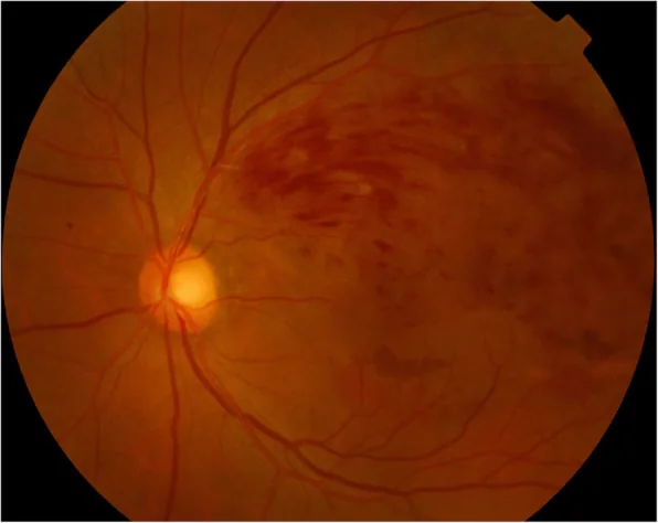
Lee JH, et al. Rapid progression of cataract to mature stage after intravitreal dexamethasone implant injection: a case report. BMC Ophthalmol. 2019. Figure 1. PMCID: PMC6318997. License: CC BY.
The fundus photograph shows fan-shaped retinal hemorrhage and yellowish-white lesions centered on the superotemporal area. Findings indicating impaired drainage of the branch vein are observed, showing the clinical picture of branch retinal vein occlusion.
In the acute phase, characteristic fundus findings are observed corresponding to the occluded area.
Fan-shaped (wedge-shaped) retinal hemorrhage: Distributed in the area drained by the occluded vein. Flame-shaped hemorrhages are characteristic.
Bonnet sign: Hemorrhages concentrated at arteriovenous crossings. Useful for identifying the occlusion site.
Cotton-wool spots (CWS): Infarction of the nerve fiber layer due to occlusion of precapillary arterioles. They indicate microcirculatory disturbance within the retina; when increased, FA is used to assess the degree of ischemia and consider the need for photocoagulation.
Hard exudates: Lipid deposits in the chronic phase.
Dilation and tortuosity of veins: Prominent on the peripheral side of the occluded vein.
Chronic phase findings: Absorption of hemorrhage, formation of collateral circulation (communicating vessels between superior and inferior veins), and capillary microaneurysms.
Important complications of BRVO are macular edema and neovascularization.
Macular edema: Present in more than half of cases, it is the most important cause of vision loss due to BRVO. It occurs in about 30% of all BRVO patients 12).
Neovascularization: Occurs on the optic disc and retina. Unlike CRVO, it rarely occurs in the anterior segment. When neovascularization occurs, vitreous hemorrhage is reported to develop in about 60% of cases.
Epiretinal membrane: Often occurs in eyes affected by BRVO and may coexist with macular edema12).
BRVO occurs when a hardened artery compresses a vein at an arteriovenous crossing. At this site, the artery and vein are encased in a common adventitia (shared adventitia), and thickening and hardening of the arterial wall directly compresses the vein. The artery runs anteriorly (superficially) to the vein in 97.6–100% of cases. Compression narrows the venous lumen, leading to Virchow’s triad (hypercoagulability, abnormal blood flow, and endothelial injury) and thrombus formation.
According to meta-analyses, 48% of RVO cases are attributable to hypertension, 20% to dyslipidemia, and 5% to diabetes 5).
Major risk factors
Hypertension: The greatest risk factor. It increases the risk of BRVO through arteriosclerosis 5).
Dyslipidemia: Meta-analyses show a significant association with BRVO 5). Low HDL cholesterol has been reported as an independent risk factor for RVO12).
Diabetes: Contributes to onset via endothelial injury and hypercoagulability 5).
Aging: Incidence increases with progression of arteriosclerosis.
Obesity and smoking: They are involved through vascular endothelial damage and increased blood viscosity5).
Hypercoagulable state: Screening is recommended in patients under 50 years old, with bilateral involvement, or recurrent cases6).
Sleep apnea syndrome: A large-scale cohort study in Taiwan reported an association with the risk of developing RVO7).
Cardiovascular disease: Patients with RVO have an increased risk of cardiovascular events and all-cause mortality8). An increased risk of RVO in depression has also been reported12).
Cases of BRVO after mRNA vaccination have been reported.
Sugihara et al. (2022) reported a 38-year-old man with BRVO that developed 2 days after BNT162b2 (Pfizer) vaccination9). Best-corrected visual acuity improved from 0.9 to 1.2 after two intravitreal injections of aflibercept 2 mg.
Tanaka et al. (2022) reported two women aged 50 and 56 who developed BRVO 3 days after mRNA vaccination10). Both had a history of tamoxifen use, and it was suggested that the hypercoagulable state after vaccination may have overlapped with the venous thrombosis risk from tamoxifen. Visual acuity improved from 20/25 to 20/20 after three ranibizumab injections.
Girioni et al. (2023) reported a 50-year-old man who developed bilateral BRVO 24 hours after an mRNA-SARS-CoV-2 booster dose, recording it as the first report of bilateral BRVO after vaccination11). Over 50% of vaccine-associated RVO cases were due to mRNA vaccines, with a median time to onset of 2 days.
The spike protein is hypothesized to promote thrombus formation and inflammatory reactions as the mechanism of onset10, 11), but the incidence is extremely rare, and the benefits of vaccination are considered to far outweigh the risks.
QCan branch retinal vein occlusion occur after COVID-19 vaccination?
A
Reports of BRVO after mRNA vaccines (Pfizer, Moderna) have accumulated, with a median time to onset of about 2 days11). The incidence is extremely low, and the benefits of vaccination far outweigh the risks. If you experience sudden vision loss or visual field defects after vaccination, it is recommended to see an ophthalmologist immediately.
The diagnosis of BRVO is based on fundus findings. The characteristic fan-shaped retinal hemorrhage corresponding to the area of the occluded vein makes the diagnosis itself straightforward. However, other tests besides fundus examination are important for determining treatment indications and predicting prognosis.
Fluorescein angiography (FA): This is an essential test for evaluating circulatory dynamics. It shows delayed filling in the affected area, venous dilation, and increased vascular permeability. Early after onset, retinal hemorrhage often makes it difficult to assess areas of capillary nonperfusion, so repeat testing is performed after the hemorrhage resolves. It is also very useful for distinguishing collateral vessels from neovascularization. The definition of ischemic type is capillary nonperfusion of 10 disc areas or more according to the CVOS criteria 12). Wide-field FA allows simultaneous evaluation of peripheral nonperfusion areas, but clinical benefit data are still limited 12). Although the use of FA has decreased in the anti-VEGF era, it remains an important test.
OCT: This is the best test for quantitative evaluation of macular edema. It is useful not only for diagnosis but also for monitoring treatment effects, and in clinical trials, treatment decisions based on OCT measurements have become mainstream 12). The reduction in central subfield thickness (CST) is an indicator of treatment effect. Note that even if retinal thickness decreases, visual acuity may not improve, so thickness and visual acuity do not always correlate 12).
OCTA (OCT angiography): This is a noninvasive method for visualizing blood vessels without contrast agents. It is useful for evaluating capillary nonperfusion areas and quantifying the foveal avascular zone (FAZ) area, but image artifacts and limited field of view remain challenges 12).
Ultrasound: This is used to evaluate the vitreoretinal relationship when there is media opacity such as vitreous hemorrhage12).
Screening for thrombophilia is recommended in patients under 50 years of age, bilateral cases, and recurrent cases 6). It has been reported that nontraditional risk factors were detected in 58% of CRVO cases with onset before age 50. The tests to be performed are as follows:
Protein C and protein S deficiency
Antiphospholipid antibody syndrome
Homocysteine level
Factor V Leiden mutation
All patients should be evaluated for hypertension, dyslipidemia, and diabetes. Since RVO patients have a high risk of cardiovascular disease and stroke, collaboration with internists and primary care physicians is recommended 12).
The following diseases are included in the differential diagnosis. Differentiation is performed using fundus examination and FA. In young-onset cases, collaboration with internal medicine is important to investigate underlying diseases.
Intravitreal injection of anti-VEGF agents is the first-line treatment for macular edema associated with BRVO 12). Efficacy has been established in multiple large-scale RCTs, and it is recommended as initial therapy due to its favorable risk-benefit profile.
Anti-VEGF Therapy
Ranibizumab (BRAVO trial): 0.5 mg monthly for 6 months achieved a mean visual acuity improvement of +18.3 letters, with 61.1% showing improvement of 15 letters or more 13). Vision was maintained at 12 months (HORIZON trial: -0.7 letters) and improvement persisted at 48 months (RETAIN study: mean 53 months, 14.8 injections; ME resolved in 50% of cases even after more than 6 months without retreatment).
Aflibercept (VIBRANT trial): 2 mg monthly achieved 52.7% of patients gaining 15 letters or more at 24 weeks, demonstrating superiority over the grid laser group (26.7%) 14).
Faricimab (BALATON trial): 6 mg monthly. At 24 weeks, showed +16.9 letters improvement, confirming non-inferiority to the aflibercept group (+17.5 letters). 56.1% vs 60.4% gained 15 letters or more. IOI incidence in BALATON trial: faricimab 0.4% vs aflibercept 0% 15).
Bevacizumab: Not covered by insurance, but has clinical use experience. In the SCORE2 trial, it was equivalent to aflibercept in visual acuity at 6 months for CRVO/HRVO 21). The LEAVO trial at 100 weeks reported RBZ +12.5 letters, AFL +15.1 letters, BEV +9.8 letters 12).
Steroid Therapy
Dexamethasone implant (GENEVA trial): A single 0.7 mg dose achieved 41% gaining 15 letters or more (at 6 months) 18). Effect peaks at 90 days and disappears by 6 months. Risks include cataract and increased intraocular pressure (≥25 mmHg in 16%). The COBALT trial showed retreatment every 4 months resulted in 15.3 letters improvement at 12 months, reaching about 70% of maximum effect within 1 week after the first injection 19).
Triamcinolone (SCORE trial): Compared 1 mg and 4 mg with grid laser. Visual acuity improvement at 12 months was similar across groups (about 1/3 gained 15 letters or more), with no superiority of TA. The 4 mg group had significantly higher cataract incidence, and TA for BRVO is indicated only in limited cases.
Indications: Used in cases resistant to anti-VEGF agents, or to extend the dosing interval.
In addition to fixed monthly dosing (loading phase), the following regimens are available for anti-VEGF administration:
PRN (pro re nata; as needed): Retreatment is determined based on OCT findings and visual acuity.
Treat-and-extend (T&E): A regimen that individually extends the treatment interval. It has been reported that T&E and PRN have comparable short-term outcomes. In the SCORE2 trial comparing T&E vs monthly, T&E required 1–2 fewer injections with similar visual outcomes, but the confidence interval was wide, requiring cautious interpretation 16). The 24-month results of the BRIGHTER trial showed no difference in visual outcomes between ranibizumab alone and ranibizumab plus laser, indicating no benefit of adding laser 17). The RETAIN 4-year data similarly showed no effect of adding laser 12).
Approximately half of patients with BRVO require continued anti-VEGF therapy for more than 5 years 12).
Grid laser (BVOS 1984): Grid photocoagulation was applied to the macula in patients with corrected visual acuity of 0.5 or worse at 3–18 months after onset. Visual improvement of 2 or more lines was achieved in 63% of the treated group, compared to 37% in the untreated group 20). At 3-year follow-up, final visual acuity was 20/40 or better in 34% and 20/200 or worse in 23%. However, absolute scotomas can occur due to laser scars, and with the availability of anti-VEGF agents, this is no longer a first-line treatment.
Scatter photocoagulation: Performed for ischemic type. Photocoagulation of capillary non-perfusion areas on FA can reduce the incidence of neovascularization from about 40% to about 20%. In ischemic BRVO with optic discneovascularization (NVD) or retinal neovascularization (NVE), sectoral PRP is recommended 12).
Refractory macular edema: Creation of a posterior vitreous detachment to relieve vitreous traction and remove inflammatory cytokines. Internal limiting membrane peeling and A/V sheathotomy have also been reported. However, the effect of VEGF inhibitors is reduced in vitrectomized eyes, so surgical indications should be carefully considered.
According to the BVOS study, even without treatment, 37% of patients show spontaneous improvement of 2 or more lines in visual acuity, while 23% have a final visual acuity of 20/200 or worse, and 34% achieve 20/40 or better 20). At 3-year follow-up, an average improvement of 2.3 lines was observed. The formation of collateral vessels improves venous drainage, reducing edema and ischemia 12). Given the wide variability in natural course, quantitative assessment of macular edema by OCT is important for deciding on treatment intervention.
QHow long does anti-VEGF injection need to continue?
A
The RETAIN study reported that an average of 14.8 injections over 53 months were needed, and about half of BRVO patients continued treatment for more than 5 years 12). It is common to adjust treatment using PRN (as needed) or T&E (treat-and-extend) based on response. See the “Standard Treatment” section for details.
QCan it heal on its own?
A
According to the BVOS study, 37% of untreated patients show spontaneous improvement of 2 or more lines of vision, but 23% have final visual acuity of 20/200 or worse 20). Collateral circulation may reduce edema, but the natural course is unpredictable, and treatment is recommended if macular edema persists.
At arteriovenous crossings, the artery and vein share a common adventitia. Arteriosclerosis causes thickening and hardening of the arterial wall, compressing the vein from outside and narrowing the venous lumen. OCT studies have confirmed deformation (narrowing rather than flattening) of the venous lumen at crossings. This narrowing creates turbulent flow, leading to chronic endothelial damage, intimal remodeling, and thrombus formation.
Venous occlusion increases venous pressure, causing blood stasis and retinal ischemia. At the same time, leakage from the foveal capillary network due to increased pressure and increased vascular permeability leads to macular edema.
In the vitreous humor of BRVO patients, elevations of the following cytokines have been reported.
VEGF: A major mediator of increased vascular permeability and macular edema formation. It plays a central role as a hypoxia-related factor released from the ischemic retina.
Angiopoietin-2 (Ang-2): Reaches the highest levels among all retinal diseases in RVO patients15). Ang-2 competitively inhibits the binding of Angiopoietin-1 to the Tie2 receptor, preventing vascular stabilization. The dual action of VEGF and Ang-2 amplifies vascular instability, promoting vascular leakage, inflammation, and neovascularization. Dual inhibition of Ang-2/VEGF with faricimab is expected to achieve more sustained retinal vascular stabilization than VEGF inhibition alone.
IL-6, IL-8: Involved in amplifying inflammation.
MCP-1 (Monocyte Chemoattractant Protein-1): Induces accumulation of monocytes and macrophages.
Regarding the mechanism of vaccine-associated BRVO, hypotheses have been proposed that spike protein promotes thrombus formation and inflammatory reactions10, 11). There is also a theory that spike protein directly damages vascular endothelial cells and promotes the release of von Willebrand factor.
7. Latest Research and Future Perspectives (Investigational Reports)
Faricimab is a bispecific antibody that simultaneously inhibits Ang-2 and VEGF-A, and its prefilled syringe for RVO indication was FDA-approved in 202412). In Part 1 (fixed monthly dosing for 24 weeks) of the BALATON/COMINO trials, it showed comparable efficacy to aflibercept in visual improvement and CST reduction15).
In the BALATON trial, comparing faricimab 6 mg and aflibercept 2 mg over 24 weeks, CST reductions were similar: -311.4 μm and -304.4 μm, respectively. Notably, the rate of macular leakage disappearance on FA was significantly higher in the faricimab group (33.6% vs 21.0%, P=0.0023)15). Similarly, in the COMINO trial, rates were 44.4% vs 30.0%, higher in the faricimab group, suggesting a vascular stabilizing effect via Ang-2 inhibition.
In Part 2 (weeks 24-72), extension to up to 16-week intervals using a modified T&E regimen is being investigated15). Data on long-term durability and dosing intervals are awaited.
Biosimilars of anti-VEGF drugs have been successively approved by the FDA, and are expected to contribute to improved treatment access12).
Ranibizumab biosimilars: ranibizumab-nuna (Byooviz, FDA approved in 2021) and ranibizumab-eqrn (Cimerli, FDA approved in 2022) have been approved for RVOmacular edema.
Aflibercept biosimilars: Four products were FDA approved in 2024 (aflibercept-jbvf [Yesafili], aflibercept-yszy [Opuviz], aflibercept-mrbb [Ahzantive], aflibercept-ayyh [Pavblu]). Equivalence to the reference product has been confirmed, but long-term clinical data are still limited.
Accumulating knowledge on COVID-19 vaccine-related RVO
Case reports of RVO after vaccination have been accumulating worldwide.
Girioni et al. (2023) reported the first case of bilateral BRVO after mRNA-SARS-CoV-2 booster administration11). Over 50% of vaccine-related RVO cases were associated with mRNA vaccines, with a median time to onset of 2 days. It has been suggested that direct vascular endothelial damage and procoagulant response to the spike protein may be involved in the pathogenesis.
Song P, Xu Y, Zha M, Zhang Y, Rudan I. Global epidemiology of retinal vein occlusion: a systematic review and meta-analysis of prevalence, incidence, and risk factors. Journal of global health. 2019;9(1):010427. doi:10.7189/jogh.09.010427. PMID:31131101; PMCID:PMC6513508.
Arakawa S, Yasuda M, Nagata M, et al. Nine-year incidence and risk factors for retinal vein occlusion in a general Japanese population: the Hisayama Study. Invest Ophthalmol Vis Sci. 2011;52:5905-9. doi:10.1167/iovs.11-7775. PMID:21693603.
Jaulim A, Ahmed B, Khanam T, Chatziralli IP. Branch retinal vein occlusion: epidemiology, pathogenesis, risk factors, clinical features, diagnosis, and complications. An update of the literature. Retina (Philadelphia, Pa.). 2013;33(5):901-10. doi:10.1097/IAE.0b013e3182870c15. PMID:23609064.
Rogers S, McIntosh RL, Cheung N, et al. The prevalence of retinal vein occlusion: pooled data from population studies from the United States, Europe, Asia, and Australia. Ophthalmology. 2010;117:313-9. doi:10.1016/j.ophtha.2009.07.017. PMID:20022117; PMCID:PMC2945292.
O’Mahoney PR, Wong DT, Ray JG. Retinal vein occlusion and traditional risk factors for atherosclerosis. Archives of ophthalmology (Chicago, Ill. : 1960). 2008;126(5):692-9. doi:10.1001/archopht.126.5.692. PMID:18474782.
Rothman AL, Thomas AS, Khan K, Fekrat S. Central retinal vein occlusion in young individuals: a comparison of risk factors and clinical outcomes. Retina. 2019;39:2205-16.
Chou KT, Huang CC, Tsai DC, et al. Sleep apnea and risk of retinal vein occlusion: a nationwide population-based study of Taiwanese. Am J Ophthalmol. 2012;154:200-5. doi:10.1016/j.ajo.2012.01.011. PMID:22464364.
Wu CY, Riangwiwat T, Limpruttidham N, et al. Association of retinal vein occlusion with cardiovascular events and mortality: a systematic review and meta-analysis. Retina. 2019;39:1635-45. doi:10.1097/IAE.0000000000002472. PMID:30829987.
Sugihara K, et al. Branch Retinal Vein Occlusion after Messenger RNA-Based COVID-19 Vaccine. Case Rep Ophthalmol. 2022;13:28-32. doi:10.1159/000521838. PMID:35221977; PMCID:PMC8832183.
Tanaka H, et al. Branch retinal vein occlusion post severe acute respiratory syndrome coronavirus 2 vaccination. Taiwan J Ophthalmol. 2022;12:202-5. doi:10.4103/tjo.tjo_24_22. PMID:35813793; PMCID:PMC9262010.
Girioni M, et al. Bilateral BRVO after mRNA-SARS-CoV-2 booster dose vaccination. J Clin Med. 2023;12:1325.
Flaxel CJ, Adelman RA, Bailey ST, et al. Retinal Vein Occlusions Preferred Practice Pattern. Ophthalmology. 2024;131:P1-P48.
Campochiaro PA, Heier JS, Feiner L, et al. Ranibizumab for macular edema following branch retinal vein occlusion: six-month primary end point results of a phase III study (BRAVO). Ophthalmology. 2010;117:1102-12. doi:10.1016/j.ophtha.2010.02.021. PMID:20398941.
Clark WL, Boyer DS, Heier JS, Brown DM, Haller JA, Vitti R, et al. Intravitreal Aflibercept for Macular Edema Following Branch Retinal Vein Occlusion: 52-Week Results of the VIBRANT Study. Ophthalmology. 2016;123(2):330-336. doi:10.1016/j.ophtha.2015.09.035. PMID:26522708.
Tadayoni R, Paris LP, Danzig CJ, et al. Efficacy and safety of faricimab for macular edema due to retinal vein occlusion: 24-week results from the BALATON and COMINO trials. Ophthalmology. 2024;131:950-60. doi:10.1016/j.ophtha.2024.01.029. PMID:38280653.
Scott IU, VanVeldhuisen PC, Ip MS, Blodi BA, Oden NL, Altaweel M, Berinstein DM, SCORE2 Investigator Group. Comparison of Monthly vs Treat-and-Extend Regimens for Individuals With Macular Edema Who Respond Well to Anti-Vascular Endothelial Growth Factor Medications: Secondary Outcomes From the SCORE2 Randomized Clinical Trial. JAMA ophthalmology. 2018;136(4):337-345. doi:10.1001/jamaophthalmol.2017.6843. PMID:29476687; PMCID:PMC5876862.
Tadayoni R, Waldstein SM, Boscia F, et al. Sustained benefits of ranibizumab with or without laser in branch retinal vein occlusion: 24-month results of the BRIGHTER study. Ophthalmology. 2017;124:1778-87. doi:10.1016/j.ophtha.2017.06.027. PMID:28807635.
Haller JA, Bandello F, Belfort R Jr, et al. Randomized, sham-controlled trial of dexamethasone intravitreal implant in patients with macular edema due to retinal vein occlusion (GENEVA). Ophthalmology. 2010;117:1134-46. doi:10.1016/j.ophtha.2010.03.032. PMID:20417567.
Branch Vein Occlusion Study Group. Argon laser photocoagulation for macular edema in branch vein occlusion. The Branch Vein Occlusion Study Group. Am J Ophthalmol. 1984;98:271-82. doi:10.1016/0002-9394(84)90316-7. PMID:6383055.
Scott IU, VanVeldhuisen PC, Ip MS, et al. Effect of bevacizumab vs aflibercept on visual acuity among patients with macular edema due to central retinal vein occlusion: the SCORE2 randomized clinical trial. JAMA. 2017;317(20):2072-2087. doi:10.1001/jama.2017.4568. PMID:28492910; PMCID:PMC5710547.
Copy the article text and paste it into your preferred AI assistant.
Article copied to clipboard
Open an AI assistant below and paste the copied text into the chat box.