Intravitreal injection of anti-VEGF drugs is a treatment that inhibits the action of VEGF (angiogenesis and increased vascular permeability) by directly injecting the drug solution into the vitreous cavity. The main indications are retinal vascular diseases such as age-related macular degeneration, diabetic macular edema, and retinal vein occlusion.
Retinopathy of prematurity (ROP): Ranibizumab 0.2 mg (approved 2019), aflibercept 0.4 mg (approved September 2022). Both are low-dose formulations equivalent to 20–40% of the adult dose.3)
Geographic atrophy (GA): Complement inhibitors
Endophthalmitis: Direct administration of antibiotics, antifungals, or antivirals
Age-related macular degeneration, diabetic macular edema, and retinal vein occlusion are the three major indications. In all these conditions, VEGF is overproduced, leading to neovascularization and increased vascular permeability that cause vision loss in the macula. By directly injecting anti-VEGF drugs into the vitreous cavity, these pathological processes are inhibited, aiming to improve or maintain vision. In recent years, indications have expanded to include PDR, mCNV, ROP, and polypoidal choroidal vasculopathy.
Mechanism of action: Simultaneous inhibition of VEGF-A and Ang-2 (angiopoietin-2). World’s first dual-target drug.
Key trials: TENAYA/LUCERNE (nAMD) up to Q16W. 63% of nAMD patients achieved Q16W in the second year. 8)
Features: In RVO (BALATON/COMINO), superior FA leakage resolution rate compared to aflibercept. 8)
Bevacizumab (Avastin®)
Molecular weight: 148 kDa (full-length IgG)
Mechanism of action: VEGF-A inhibition (off-label use in ophthalmology)
Key trials: CATT trial confirmed efficacy equivalent to ranibizumab. 12)
Usage: Significantly lower cost. The intravenous infusion preparation is aseptically dispensed and used.
Pegaptanib (Macugen®)
Mechanism of action: VEGF165-specific aptamer
Current status: First-generation drug approved in 2008. Newer generation drugs are now mainstream. Contributed to establishing the concept of early anti-VEGF therapy.
QWhich drug should be chosen?
A
The choice depends on the indicated disease, desired dosing interval, and risk of complications. Generally, for nAMD, aflibercept, faricimab, and brolucizumab are superior in terms of extended dosing intervals. For DME, aflibercept and faricimab are standard. Brolucizumab has the advantage of a high polyp regression rate in PCV patients, considering the risk of IOI. Faricimab allows dosing up to Q16W (every 16 weeks), minimizing the burden of hospital visits. Ultimately, the attending physician makes a comprehensive decision.
QCan bevacizumab (Avastin) be used?
A
In ophthalmology, it is used off-label. The intravenous infusion preparation is aseptically prepared for ophthalmic use. The CATT study confirmed visual improvement equivalent to ranibizumab, and due to its significantly lower cost, it is widely used worldwide. However, in Japan, it is not covered by insurance, and its use depends on the facility’s discretion.
Anti-VEGF drug administration is performed in two phases: induction and maintenance.
Induction phase: To strongly suppress disease activity, fixed monthly doses are given 3 to 6 times (varies by disease and drug).
Maintenance phase has the following three methods:
PRN (pro re nata): Monthly visits, with injection only if recurrence findings are present
Fixed dosing: Regular injections at fixed intervals, e.g., every 2 months or every 3 months
Treat and Extend (T&E): If no activity findings, the interval is extended by 2 weeks; if recurrence occurs, the interval is shortened.
For ranibizumab, the basic recommended regimen is 3 monthly loading doses followed by PRN maintenance; for aflibercept, it is 3 monthly loading doses followed by fixed dosing every 2 months or a treat-and-extend (T&E) regimen. In recent years, the T&E method has been adopted at many institutions.
Additional at recurrence (interval of at least 1 month) 3)
Management of PCV (polypoidal choroidal vasculopathy): Brolucizumab achieves a polyp regression rate of approximately 79%, superior to other agents, and maintains 12-week intervals in 76% of cases (48 weeks). 14)Faricimab has been reported to be effective even in ranibizumab-resistant PCV cases. 15)
BALATON trial (BRVO, n=553): Faricimab 6.0 mg vs. aflibercept 2.0 mg Q4W; 24-week BCVA change was +16.9 letters and +17.5 letters, respectively (non-inferiority achieved). FA leakage resolution rate was significantly higher with faricimab (33.6% vs. 21.0%, nominal p=0.0023). 8)
COMINO trial (CRVO/HRVO, n=729): Same regimen; 24-week BCVA change was +16.9 letters and +17.3 letters (non-inferiority achieved). CST change was −461.6 μm vs. −448.8 μm. FA leakage resolution rate was significantly higher with faricimab (44.4% vs. 30.0%, nominal p=0.0002), demonstrating vascular stabilization via Ang-2 inhibition. 8)
STTA combination (RVO): Combination of ranibizumab plus suprachoroidal triamcinolone 4 mg significantly reduces the number of injections compared to ranibizumab alone (4.4 vs. 2.47, p<0.001). 11)
3-8. Anti-VEGF therapy for retinopathy of prematurity (ROP)
Aggressive ROP (A-ROP): Perform as soon as possible
Recurrence rate and follow-up3):
The recurrence rate of aflibercept is 13.9–28% (mean recurrence time 11–14.2 weeks), while that of ranibizumab is 20.8–83.0% (recurrence time 5.9–9.3 weeks, earlier). When using ranibizumab, careful observation from early after injection is necessary. If retinal vessels have not extended to Zone III, weekly fundus examination is recommended until 17 weeks after injection.
In A-ROP, anti-VEGF monotherapy requires additional treatment in 75.0–87.5% of cases. Early recurrence within 1–3 weeks after injection may occur, and if fibrous proliferation is extensive, anti-VEGF monotherapy is contraindicated (risk of tractional retinal detachment due to contraction).
Injection technique for ROP (differences from adults)3):
Insertion site: 1.0–1.5 mm posterior to the limbus (significantly different from 3.5–4 mm in adults).
Needle direction: Insert downward (since the lens is relatively large, aiming centrally risks lens penetration).
Use a 30-gauge or smaller needle.
Confirm dosage: ranibizumab 0.02 mL, aflibercept 0.01 mL (carefully verify to prevent overdose).
QWhich drug is most effective for diabetic macular edema?
A
In the Protocol T study, aflibercept showed the greatest visual improvement at 1 year. However, in the mild group (BCVA ≥20/40), there was no statistically significant difference among the three drugs. 13)Faricimab showed visual improvement equivalent to aflibercept in the YOSEMITE/RHINE trials while allowing longer injection intervals during the maintenance phase (Q16W achieved in 60–64% at year 2). 8) Drug selection should be based on patient background and desired visit frequency.
Anesthesia: Considering the possibility of disinfectant splashing into the fellow eye, administer Benoxil® eye drops to both eyes, then apply 4% Xylocaine® eye drops to the treated eye twice.
Points to note for PA iodine disinfection:
PA iodine immediately after removal from the refrigerator has reduced antibacterial and antifungal inactivation effects, so always return it to room temperature.
When stored in a non-airtight container at 25°C, the residual active ingredient decreases to 60% in 5 hours. Do not use PA iodine that has been left for a long time.
Since a contact time of about 1 minute is required for inactivation of bacteria and fungi, keep the eyelids closed after irrigation to ensure sufficient contact time with the conjunctiva.
Oral bacteria control: Use a fenestrated drape and have the surgeon, assistants, and patient all wear masks to prevent droplet spread.
The distance from the surgical limbus for insertion is as follows. Maintaining the correct insertion site allows entry through the pars plana (behind the pars plicata), preventing lens damage and vitreous hemorrhage.
The needle is inserted toward the center of the vitreous cavity. Insertion too close to the limbus may damage the pars plicata, causing vitreous hemorrhage, and increases the risk of lens injury due to proximity to the lens.
Apply a fenestrated drape and open the eyelids with a lid speculum.
Measure the insertion distance from the limbus using calipers.
The injection site should be the superotemporal or inferotemporal quadrant (to prevent damage to the horizontal rectus muscles).
Fix the eyeball with forceps and slightly displace the conjunctiva anteriorly before injection to offset the needle hole after withdrawal and prevent fluid leakage.
Insert a 30G short needle almost perpendicular to the sclera and inject the medication slowly (rapid injection can cause sustained intraocular pressure elevation).
After needle withdrawal, apply pressure to the injection site with a cotton swab.
Immediately after the procedure, check visual acuity (counting fingers). If counting fingers is not perceived, perform anterior chamber paracentesis.
Continue broad-spectrum antibiotic eye drops for 3 days postoperatively.
QDoes the injection hurt?
A
Since the procedure is performed after topical anesthesia (Benoxil® or 4% Xylocaine®), the pain during injection is minimal. You may feel irritation from the disinfectant (PA iodine), but sodium hyaluronate eye drops are effective for early postoperative discomfort.
Infectious endophthalmitis is the most serious complication, with an incidence of approximately 0.027–0.065%. When it occurs, emergency treatment with intravitreal injection of vancomycin 1.0 mg + ceftazidime 2.0 mg is required.
Most important preventive measure:
Proper disinfection with povidone-iodine (allow to reach room temperature, contact time of at least 1 minute)
Use of a fenestrated drape (to prevent droplet contamination from oral bacteria)
Regarding postoperative prophylactic antibiotic eye drops, multiple studies have shown that they do not reduce the incidence of endophthalmitis, and the evidence for their efficacy is inconsistent.
This is an intraocular inflammatory reaction without bacterial infection, with an incidence ranging from 0.005% to 4.4% depending on the drug. 7)
Onset timing: Usually within 24–48 hours after injection (infectious endophthalmitis occurs 2–7 days later) 7)
Main findings: Vitreous opacity (approximately 80%), hypopyon (approximately 5%) 7)
Definitive diagnosis: Negative culture (PCR to rule out pathogens is also useful)
Incidence by drug (‰): Bevacizumab 3.64 / Ranibizumab 1.39 / Aflibercept 0.76 7)
Treatment: Mild cases are managed conservatively (steroid eye drops, subconjunctival injection). The CEVE protocol (immediate complete vitrectomy) has been reported to achieve visual recovery in an average of 17.8 days. 7)
In a matched cohort analysis from the IRIS Registry, there was no significant difference in visual outcomes between injection management alone and early vitrectomy. 21)
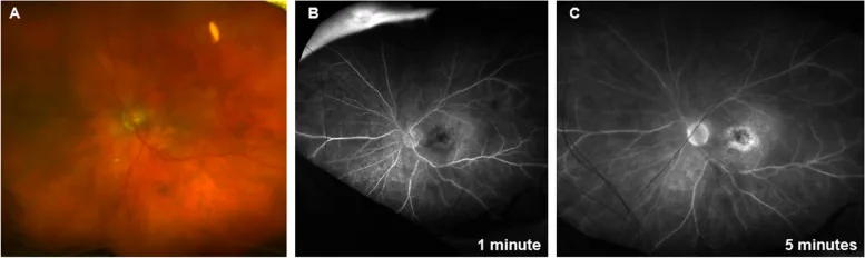
Bahram Bodaghi; Arshad M Khanani; Ramin Khoramnia; Carlos Pavesio; Quan Dong Nguyen. Gains in the current understanding of managing neovascular AMD with brolucizumab. J Ophthalmic Inflamm Infect. 2023 Nov 23; 13:51 Figure 2. PMCID: PMC10667168. License: CC BY.
Panuveitis with nonocclusive vasculitis in the left eye following injection with brolucizumab. A Fundus examination showed vitreous haze, hyperemia of the optic nerve, and sheathing around some of the retinal vessels. B, C Fluorescein angiography showed optic nerve leakage and perivascular leakage in the posterior pole and peripheral retina
Brolucizumab is known to cause intraocular inflammation (IOI) more frequently than other anti-VEGF drugs.
The majority of IOI occurs within 6 months after the first dose and within 4 injections. 10)
Mechanism: The positivity rate of ADA (anti-drug antibodies) is high with brolucizumab at 35–52% (in contrast to <5% for ranibizumab and aflibercept), and it is thought to be a type III hypersensitivity reaction due to immune complex deposition. 10)
Scleritis (first report worldwide): Posterior scleritis after brolucizumab injection has been reported in 3 Japanese patients, accompanied by an increase in intraocular pressure to 24–49 mmHg, and one case progressed to retinal artery occlusion and vasculitis. 9)
Treatment: Subconjunctival or sub-Tenon injection of triamcinolone acetonide (STTA) 5–20 mg is effective. Combination with prophylactic STTA administration has also been reported. 18, 19)
The incidence of IOI is 2.0% in nAMD, 1.3% in DME, and 1.4% in RVO, and was observed in 8.5% of bilateral injection cases. 8) In post-marketing surveillance, retinal vasculitis is rare at 0.17/10,000 injections, but hemorrhagic occlusive retinal vasculitis (HORV) can lead to severe outcomes. 8)HORV is pathologically suggested to be associated with a type IV (delayed-type) hypersensitivity reaction. 16)
RPE tears occurred in the faricimab group in 2.7% (TENAYA) and 3.0% (LUCERNE), with PED height >550 μm being a risk factor. 17)
In ROP patients, after anti-VEGF therapy, the fibrovascular membrane rapidly contracts, leading to tractional retinal detachment (TRD), a complication called crunch syndrome. 3) When extensive fibroproliferation is present, anti-VEGF monotherapy is contraindicated, and vitrectomy is required. It is important to examine changes in proliferative tissue on fundus examination early after administration.
A transient intraocular pressure elevation immediately after injection occurs in all patients receiving the injection. Injection of 0.05 mL raises intraocular pressure to 50 mmHg instantly, but it is usually reversible. In patients with a history of glaucoma, attention should be paid to sustained intraocular pressure elevation, and if necessary, decompression by anterior chamber paracentesis should be performed.
There is a theoretical risk of stroke and myocardial infarction. In the HAWK trial of brolucizumab, ATE was observed in 1.1-1.4%. 10) Caution is required in patients with a history of such events.
QWhat symptoms after injection should prompt a visit to the doctor?
A
If the following symptoms appear, promptly consult an ophthalmologist: ① sudden vision loss, ② worsening eye pain or redness, ③ marked increase in floaters, ④ discharge. These may indicate infectious endophthalmitis or intraocular inflammation (IOI). Particular caution is needed within 24-72 hours after injection.
QWhat should be done if IOI occurs with brolucizumab?
A
If sudden vision loss, worsening floaters, redness, or eye pain occur, promptly consult an ophthalmologist. After diagnosis, subconjunctival or sub-Tenon injection of triamcinolone acetonide (STTA) is effective, and inflammation improves in most cases. 18, 19) Re-administration should be carefully considered after confirming resolution of inflammation using LFP (laser flare cell photometer) or other methods. In cases with severe vascular occlusion, re-administration may be contraindicated, so switching to an alternative drug should be considered.
6. Pathophysiology (Role of VEGF and mechanism of action of the drug)
VEGF binds to VEGFR-1 and VEGFR-2 on vascular endothelial cells, promoting endothelial cell proliferation, increased vascular permeability, and neovascularization.
DME: Breakdown of the blood-retinal barrier (BRB) → macular edema formation
RVO: Ischemia → VEGF overexpression → macular edema and neovascularization
When VEGF levels decrease after intravitreal injection, vascular permeability decreases and macular edema improves. The effect of anti-VEGF drugs is temporary, so regular re-administration is necessary.
Ang-2 acts as an antagonist of the Tie-2 receptor and is involved in vascular destabilization. By inhibiting Ang-2, faricimab normalizes the Tie-2 pathway, improving vascular stability and reducing VEGF sensitivity. This dual inhibition effect is the pharmacological basis for extending the dosing interval.
The reason for administering 3 to 5 fixed monthly doses during the loading phase is to achieve early and strong suppression of disease activity. The treat-and-extend (T&E) method provides a framework that maintains intravitreal drug concentrations within the therapeutic range without allowing recurrence.
7. Latest Research and Future Perspectives (Research-stage Reports)
With the ranibizumabPDS (Port Delivery System), 98% of patients did not require monthly injections after refill administration every 6 months. 12) In 2025, expansion of the indication to DME is being considered. 13) A significant reduction in injection burden is expected.
In the PULSAR trial (AMD), at 48 weeks, 79% maintained 12-week intervals and 77% maintained 16-week intervals. 12) In the PHOTON trial (DME), 93% achieved intervals of 12 weeks or longer. 13)
The PROUD trial (Korea) targeting PCV and macular telangiectasia type 1 (AT1) is ongoing, and accumulation of evidence for Asian-specific diseases is expected. 14)
The SALWEEN trial investigating the effect of faricimab in PCV patients is ongoing. 20) High Ang-2 levels have been confirmed in the aqueous humor of PCV patients, and an additional benefit of Ang-2 inhibition is expected.
Combined administration of subconjunctival/Tenon’s capsule triamcinolone acetonide (STTA) and brolucizumab may be effective for both IOI prevention and exudation control. 18, 19) Research is underway to standardize the optimal dose and timing of administration.
Measurement of cytokine profiles (IL-6, IL-8, IL-10) in vitreous fluid is expected to be a useful adjunct tool for early differentiation between sterile and infectious endophthalmitis. 7)
Biosimilars of ranibizumab and bevacizumab are emerging. Significant cost reductions are expected to improve treatment continuation rates and healthcare access. 12)
Lim JI, Kim SJ, Bailey ST, et al. Diabetic Retinopathy Preferred Practice Pattern. Ophthalmology. 2025 Apr;132(4):P75-P162. doi:10.1016/j.ophtha.2024.12.020. PMID:39918521.
American Academy of Ophthalmology. Retinal Vein Occlusion Preferred Practice Pattern. Ophthalmology. 2024. [BRAVO data]
Kudasiewicz-Kardaszewska A, Ozimek MA, Kardaszewska A, et al. Complete and Early Vitrectomy for Sterile Endophthalmitis After Bevacizumab: A Case Series. Cureus. 2025;17(10):e93996. doi:10.7759/cureus.93996.
Tadayoni R, Paris LP, Danzig CJ, Abreu F, Khanani AM, Brittain C, et al. Efficacy and Safety of Faricimab for Macular Edema due to Retinal Vein Occlusion: 24-Week Results from the BALATON and COMINO Trials. Ophthalmology. 2024;131(8):950-960. doi:10.1016/j.ophtha.2024.01.029. PMID:38280653.
Takayama T, Inoda S, Takahashi H, Tsukii R, Yoshida H, Kasuya Y, et al. Scleritis following intravitreal brolucizumab injection: a case series. Journal of medical case reports. 2024;18(1):80. doi:10.1186/s13256-024-04402-9. PMID:38419100; PMCID:PMC10902930.
Sharma A, et al. Understanding retinal vasculitis associated with brolucizumab. Ocul Immunol Inflamm. 2022;30(6):1508-1510.
Terashima H, Hasebe H, Okamoto F, Matsuoka N, Sato Y, Fukuchi T. COMBINATION THERAPY OF INTRAVITREAL RANIBIZUMAB AND SUBTHRESHOLD MICROPULSE PHOTOCOAGULATION FOR MACULAR EDEMA SECONDARY TO BRANCH RETINAL VEIN OCCLUSION: 6-MONTH RESULT. Retina. 2019;39(7):1377-1384. doi:10.1097/IAE.0000000000002165. PMID:29689025. PMCID:PMC6613833.
Flaxel CJ, Adelman RA, Bailey ST, et al. Age-related macular degeneration preferred practice pattern. Ophthalmology. 2024;131(1):P1-P68.
Flaxel CJ, Adelman RA, Bailey ST, et al. Diabetic retinopathy preferred practice pattern. Ophthalmology. 2024;131(1):P99-P168.
Parveen Sen, George Manayath, Daraius Shroff, Vineeth Salloju, Priyanka Dhar. Polypoidal Choroidal Vasculopathy: An Update on Diagnosis and Treatment. OPTH. 2023;Volume 17:53-70. doi:10.2147/opth.s385827.
Bloom J, Madani R, Haidar AJ, Alasil T. Faricimab treatment of polypoidal choroidal vasculopathy resistant to intravitreal ranibizumab. J VitreoRetin Dis. 2024;8(6):731-734.
Christoph R Clemens, Florian Alten, Julian A Zimmermann, Nicole Eter. Old Problem in a New Guise: Retinal Pigment Epithelium Tear after Intravitreal Faricimab (Vabysmo®) Injection. Case Rep Ophthalmol. 2023;241-244. doi:10.1159/000529930.
The combination therapy of subtenon triamcinolone acetonide injection and intravitreal brolucizumab for brolucizumab-related intraocular inflammation. Medicine (Baltimore). 2021;100(42):e27580. doi:10.1097/MD.0000000000027580.
Saito M, et al. A case of intraocular inflammation after intravitreal brolucizumab injection monitored by laser flare-cell photometer. Am J Ophthalmol Case Rep. 2022;28:101727. doi:10.1016/j.ajoc.2022.101727. PMID:36281264; PMCID:PMC9587367.
Cheung CMG, Lai TYY, Teo K, et al. Polypoidal choroidal vasculopathy: consensus nomenclature and non-indocyanine green angiograph diagnostic criteria. Eye. 2024;39:819-834.