Traumatic retinal detachment is a rhegmatogenous retinal detachment caused by ocular trauma. The mechanism and pathology differ between open globe injury and closed (blunt) globe injury, and the treatment strategy also varies greatly. Open globe injuries often involve significant blunt components, requiring careful fundus examination and imaging to understand the pathology.
Traumatic retinal tears are classified into the following three types based on the location of formation:
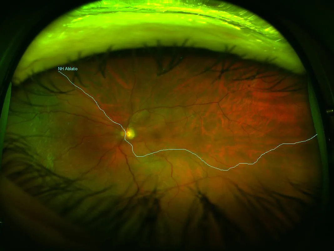
Posterior pole tear: a tear occurring near the vascular arcades.
Dialysis at the ora serrata: an arcuate tear along the ora serrata. Common in young individuals and occurs in sports with repeated ocular contusions such as boxing.
Ciliary epithelial tear: A tear occurring in the ciliary epithelium.
Traumatic retinal detachment is more common in young people and males. The main causes are sports injuries (boxing, ball games, etc.), occupational accidents, and traffic accidents. In sports like boxing, where the eye receives repeated strong blunt trauma, traumatic retinal detachment with ora serrata tears or giant tears can occur. In children, trauma is the leading cause of unilateral blindness, making early diagnosis of traumatic retinal detachment particularly important 1).
QHow is traumatic retinal detachment different from ordinary (non-traumatic) retinal detachment?
A
Non-traumatic rhegmatogenous retinal detachment is mainly caused by traction from vitreous liquefaction and posterior vitreous detachment, leading to peripheral tears, and is more common in middle-aged and elderly people. In traumatic retinal detachment, blunt impact causes eye deformation, leading to large tears at the vitreous base, or in open injuries, incarcerated vitreous gel directly pulls on the opposite retina. The mechanism, tear morphology, and age of onset differ. In young people, even with minimal vitreous liquefaction, shallow retinal detachment can occur, and detection is often delayed.
Visual field defect: A visual field defect corresponding to the area of retinal detachment occurs. If the lower retina detaches, the upper visual field is lost.
Decreased visual acuity: Significant visual loss occurs when the macula detaches.
Slow progression (young people): Because vitreous liquefaction is minimal, the detachment tends to remain shallow, progression is slow, and subjective symptoms may be scarce. Be aware of delayed detection.
Incarcerated vitreous in the corneoscleral laceration: Vitreous becomes incarcerated in the wound, serving as a starting point for traction.
Vitreous hemorrhage: Bleeding associated with the laceration often makes fundus visualization difficult.
Direct retinal break: The external force directly causes a break in the retina.
Retinal tear on the opposite side: Secondary traction from the incarcerated gel may form a tear on the opposite side.
Findings in Non-Open Ocular Trauma
Dialysis of the ora serrata / peripheral retinal tear: Blunt impact causes vitreous base traction, resulting in relatively large peripheral tears.
Irregular retinal hole in necrotic retina: Irregular holes form in areas of retinal contusion necrosis.
Shallow retinal detachment: In young patients, due to less liquefied vitreous, the detachment often appears shallow.
Retinal transparency (good clarity cases): If the media are clear, detailed observation with indirect ophthalmoscopy is possible.
QHow soon after injury does retinal detachment develop?
A
In open ocular trauma, retinal detachment often occurs immediately or within a few days. In non-open (blunt) trauma, dialysis of the ora serrata is common, and shallow retinal detachment progresses slowly, so diagnosis may be made weeks to months after injury. In young patients with blunt ocular trauma and few subjective symptoms, continued ophthalmologic follow-up after injury is necessary2).
In children, the vitreous and retina are strongly adherent, so traction from blunt impact is easily transmitted directly to the entire retina. Additionally, delayed appropriate ophthalmologic examination after trauma is also a risk.
QWhat is the approach when the fundus is not visible due to vitreous hemorrhage?
A
Perform B-mode ultrasonography. Retinal detachment is detected as a characteristic hyperechoic band. In open globe injuries where rupture or intraocular foreign body is suspected, CT is also performed. As soon as the fundus becomes visible, perform a detailed fundus examination using indirect ophthalmoscopy to identify the type and location of the tear.
Cases with PVR (proliferative vitreoretinopathy): Excision and peeling of contractile membranes (membrane peeling) + silicone oil tamponade is required. Removal of silicone oil is considered after the retina stabilizes.
Giant retinal tear: Vitrectomy + perfluorocarbon liquid (PFCL) to unfold the retina → Tamponade with gas (SF6 or C3F8) or silicone oil.
Pediatric cases: Postoperative refractive management and occlusion therapy for amblyopia are essential. Long-term management considering the impact on visual development is necessary.
QHow much does vision recover after surgery?
A
In traumatic retinal detachment associated with closed globe injury, the reattachment rate after scleral buckling surgery is relatively high, and visual prognosis is often good. On the other hand, in open globe injury, there is a risk of PVR progression even after vitrectomy, and many cases have poor visual prognosis. Additionally, if the macula has been detached for a long time, visual function recovery may be insufficient even after anatomical reattachment 3). Associated disorders such as lens damage and traumatic glaucoma also affect final visual acuity.
The mechanism of traumatic retinal detachment varies depending on the type of trauma.
Mechanism of Blunt (Non-Penetrating) Trauma
Traction due to ocular deformation:
Blunt impact → shortening of anteroposterior diameter and expansion of equatorial diameter (ocular deformation) → concentration of traction force on the vitreous base → formation of peripheral retinal tears → subretinal fluid accumulation
Special considerations in young patients:
Because the adhesion between the vitreous and retina is strong, traction forces are directly transmitted to the entire retina. This often results in large peripheral tears (dialysis of the ora serrata, giant retinal tears).
Mechanism of Open-Globe Injury
Direct traction by incarcerated vitreous gel:
Vitreous gel becomes incarcerated in the corneoscleral wound → with eye movement, the incarcerated gel directly pulls on the retina → formation of tears in the opposite or peripheral retina → progression of retinal detachment
Direct retinal laceration:
The external force directly affects the retina, creating a laceration. Retinal detachment progresses from that site.
Progression to Proliferative Vitreoretinopathy (PVR)
Trauma disrupts retinal pigment epithelial cells, glial cells, and macrophages, which then proliferate and form contractile fibrocellular membranes on the surface and underside of the retina. Contraction of these membranes pulls the retina, leading to complex tractional retinal detachment. In open-globe injuries, blood and inflammatory cells often enter the vitreous cavity, resulting in a particularly high risk of PVR progression. PVR is a major cause of postoperative redetachment, so management of the intraocular environment after trauma is important.
Kuhn F, Maisiak R, Mann L, Mester V, Morris R, Witherspoon CD. The Ocular Trauma Score (OTS). Ophthalmology clinics of North America. 2002;15(2):163-5, vi. doi:10.1016/s0896-1549(02)00007-x. PMID:12229231.
Mitry D, Charteris DG, Fleck BW, Campbell H, Singh J.. The epidemiology of rhegmatogenous retinal detachment: geographical variation and clinical associations. Br J Ophthalmol. 2010;94(6):678-684. doi:10.1136/bjo.2009.157727. PMID:19515646.
Soni NG, Bauza AM, Son JH, Langer PD, Zarbin MA, Bhagat N.. Open globe ocular trauma: functional outcome of eyes with no light perception at initial presentation. Retina. 2013;33(2):380-386. doi:10.1097/iae.0b013e318263cefb. PMID:23026847.
Copy the article text and paste it into your preferred AI assistant.
Article copied to clipboard
Open an AI assistant below and paste the copied text into the chat box.
