B-mode ultrasound (ocular B-scan) is a diagnostic imaging test that displays the reflection intensity of ultrasound as a two-dimensional image. Ultrasound waves emitted from the probe reflect at tissue interfaces, and the reflection intensity (brightness) and reflection time (tissue depth) are mapped in two dimensions to obtain cross-sectional images of the intraocular and orbital structures.
In ophthalmology, it is established as a test used when the interior of the eye cannot be observed due to opaque media and for diagnosing orbital lesions. When there is corneal opacity, advanced cataract, severe vitreous hemorrhage, or other opacities of the transparent media, observation of the interior of the eye with a slit lamp or ophthalmoscope becomes impossible. In such situations, B-scan is the only imaging test that can provide structural information about the inside of the eye.
In 1956, Mundt & Hughes reported the application of ultrasound to ophthalmology, which later developed into A-mode and B-mode. Today, it is widely used to evaluate the vitreous, retina, choroid, and orbital structures, and is an indispensable examination tool in outpatient ophthalmology and pre- and postoperative management.
Evaluation of the intraocular structures and fundus in severe vitreous hemorrhage (preoperative confirmation of retinal detachment or intraocular tumor)
Preoperative fundus evaluation in advanced cataract (assessment of retinal detachment and vitreous opacity)
Localization and material estimation of intraocular foreign bodies (differentiation of metal, non-metal, and wood)
Diagnosis and follow-up of intraocular tumors (choroidal melanoma, metastatic tumors, choroidal hemangioma, etc.)
Screening for orbital tumors and orbital inflammation
Preoperative planning for vitreous surgery (assessment of the extent and shape of traction membranes)
Monitoring of postoperative vitreous and retinal changes
QWhen is B-mode ultrasound necessary?
A
This is mainly necessary when the transparent media (cornea, lens, vitreous) are opaque and the interior of the eye cannot be directly visualized, or when there are lesions that can only be evaluated with B-scan. Typical indications include confirming the presence or absence of retinal detachment or tumors in cases of severe vitreous hemorrhage, mature cataract, or corneal opacity, localizing intraocular foreign bodies, and diagnosing orbital tumors. Even when the interior of the eye can be directly visualized, it is useful for detailed evaluation of orbital lesions.
The examination is generally performed according to the following steps.
Transpalpebral approach: Apply sufficient coupling gel (e.g., Scopisol) to the closed upper eyelid (no topical anesthesia required).
Only when choosing the direct corneal approach, open the eyelid and use topical anesthesia (e.g., 0.4% oxybuprocaine).
Gently place the probe on the eyelid without pressing on the eyeball.
Fix the head and perform a full circumferential scan while changing the probe position and angle.
Observe the dynamics of the vitreous membranes and retina during eye movements (kinetic echography).
Start with high gain (amplification sensitivity) and gradually decrease it to check changes in lesion brightness1).
The transpalpebral approach is standard, with less patient invasiveness and is the first choice in many situations. It is essential not to press on the eyeball with the probe, and head fixation is important. In the supine position, it is desirable to stabilize the head with a pillow; when examining in a sitting position, head fixation by an assistant from behind is indispensable.
Electronically switches multiple transducers to move the ultrasound beam
Provides uniform images without defects
Probe is wide and expensive
Sector scan (mechanical scanning)
Mechanically scans the transducer at high speed
Probe tip is small, good maneuverability, and inexpensive
Image defects occur near the periphery of the anterior segment
For evaluation of the posterior segment and orbit, sector scan is commonly used in daily practice, while linear scan is suitable for detailed evaluation of the anterior segment.
QIs the examination painful?
A
With the standard transpalpebral approach (placing the probe over the closed eyelid), topical anesthesia is not required and there is almost no pain. Only when the probe is placed directly on the cornea, topical anesthesia such as 0.4% oxybuprocaine is administered. The examination takes only a few minutes. Coupling gel is used and can be wiped off after the examination.
3. Normal values and interpretation of imaging findings
In a normal eye, the lens, retina, choroid, and sclera are visualized as a single layer within the eye. Outside the eye, relatively uniform tissue patterns are seen, and the optic nerve is identified as a low-echoic tubular structure.
Ultrasound brightness reflects differences in acoustic impedance of tissues. High-echoic echoes originate from interfaces with large acoustic impedance differences (e.g., sclera, calcifications, intraocular foreign bodies), while low-echoic echoes originate from fluids (e.g., normal vitreous, aqueous humor, cyst fluid).
When interpreting ocular B-mode ultrasound, focus on the following five points.
Presence of intraorbital space: The presence of hypoechoic or hyperechoic areas in the orbit may indicate tumor or inflammation.
Visualization of the optic disc: Confirm the position of the optic disc and understand its relationship with lesions.
Characteristics of elevated lesions: Determine whether the lesion is solid or cystic based on echogenicity and shape (important for tumor differentiation).
Presence of hyperechoic lesions in the vitreous: Check for hemorrhage, inflammation, or proliferative membranes.
Presence of retinal detachment: Identify detached retina based on the shape, continuity, and dynamics of membranous echoes.
Differentiation between Retinal Detachment and Vitreous Membrane
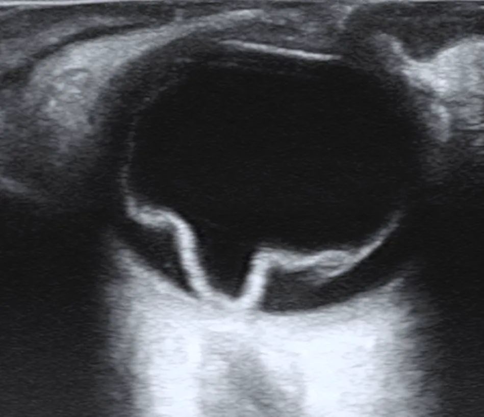
CheckDO. Ultrasound of a retinal detachment in a patient presenting with complete vision loss and light perception only. Wikimedia Commons. 2022. Figure 1. Source ID: commons.wikimedia.org/wiki/File:Retinal_Detachment.jpg. License: CC BY-SA 4.0.
A V-shaped hyperechoic membranous echo with the optic disc as the apex is observed in the vitreous cavity, showing a characteristic morphology of rhegmatogenous retinal detachment. This corresponds to the morphological diagnosis (continuity with the optic disc) discussed in the section “Differentiation between Retinal Detachment and Vitreous Membrane.”
In eyes with vitreous hemorrhage, B-mode ultrasound may show membranous hyperechoic echoes. Differentiating whether this is a detached retina or a vitreous membrane (e.g., fibrous membrane associated with posterior vitreous detachment) greatly influences treatment strategy. Dynamic B-scan has been reported to have a sensitivity of approximately 96% and specificity of approximately 98% for detecting retinal tears in acute posterior vitreous detachment cases 2). Evaluate using a combination of the following three diagnostic methods.
Morphological Diagnosis
Evaluation: Check whether the membranous echo is connected to the optic disc.
Features of detached retina: Continuous with the optic disc. The membranous echo is continuous, smooth, and curved. The membrane thickness is uniform.
Features of vitreous membrane: Connection with the optic disc is unclear. May show intermittent and irregular morphology.
Quantitative Diagnosis (Gain Reduction Method)
Evaluation: Observe the order of disappearance of membrane echoes while gradually decreasing the amplification sensitivity dial (gain).
Characteristics of detached retina: Shows strong reflection, so echoes persist even when gain is reduced.
Characteristics of vitreous membrane: Reflection is weak, so when gain is reduced, it disappears before retinal echoes.
Dynamic Diagnosis (Eye Movement)
Evaluation: Observe the movement of membrane echoes while the patient moves their eyes.
Characteristics of detached retina: Shows regular, smooth, and continuous movement with eye movement. Movement stops when eye movement ceases.
Characteristics of vitreous membrane: Shows irregular, coarse, and discontinuous movement with eye movement. After eye movement stops, a slow undulating movement remains (after-motion).
QHow do you distinguish between retinal detachment and vitreous membrane?
A
Differentiate using a combination of three methods. ① Morphological diagnosis: Detached retina appears as a continuous membrane connected to the optic disc, smooth and uniform in thickness. ② Gain reduction method: When gain is gradually reduced, the vitreous membrane disappears first, so the echo that remains until the end corresponds to the retina. ③ Eye movement: Detached retina moves regularly and smoothly, stopping when the eye is still. Vitreous membrane shows residual undulating movement (after-motion) even after eye movement stops.
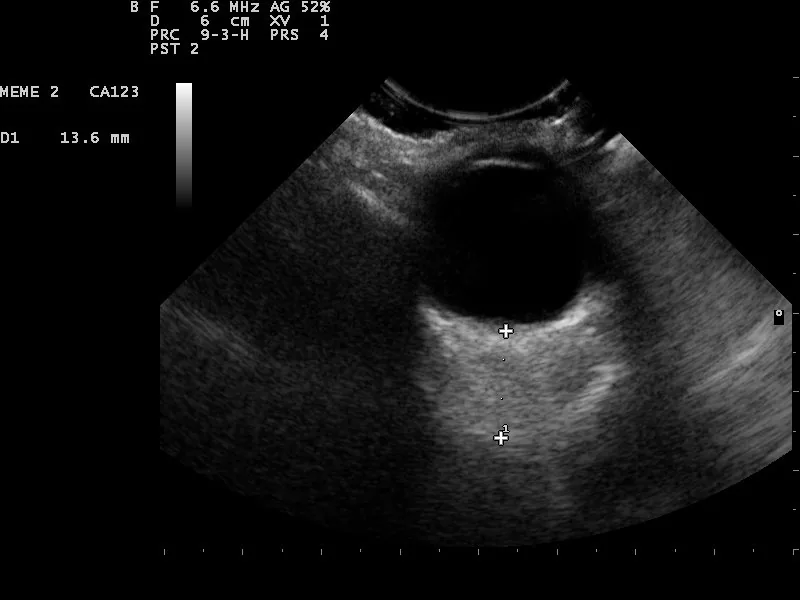
Nevit Dilmen. Eye ultrasound — orbital mass with caliper measurement (6.6 MHz, D1 13.6 mm). Wikimedia Commons. 2010. Figure 2. Source ID: commons.wikimedia.org/wiki/File:Eye_ultrasound_110318153108_1539230.jpg. License: CC BY-SA 3.0.
Retrobulbar orbital space-occupying lesion imaged with a 6.6 MHz sector scan. Caliper measurement D1 13.6 mm is displayed, assessing the size of a solid mass. This corresponds to orbital tumor screening and intraocular tumor detailed examination discussed in the section “Clinical Significance and Indicated Diseases.”
B-scan is an important examination method that provides structural information of the intraocular and orbital regions not only in eyes with opaque media but also in situations where ophthalmoscopy is difficult, and is used in the following clinical scenarios.
Preoperative planning before vitreous surgery: Understand the extent, shape, and positional relationship of traction membranes with the retina to plan the surgical approach. Confirming the presence or absence of retinal detachment in cases with vitreous hemorrhage is particularly important.
Preoperative evaluation of rhegmatogenous retinal detachment: Assess the extent, height, freshness, and presence of traction to help decide between buckling and vitrectomy.
Detailed examination of intraocular tumors: Choroidal melanoma shows high reflectivity at the anterior edge, low internal reflectivity (acoustic hollowness), choroidal excavation, and often a mushroom (collar-stud) shape. Combined A- and B-scan diagnostic accuracy is reported to be 95% for tumors ≥3 mm3).
Screening for orbital tumors: Confirm the presence of an orbital mass and determine the need for further evaluation with MRI or CT1).
Detection of intraocular foreign bodies: Foreign bodies such as metal, glass, or wood show different ultrasound reflection patterns. Metal produces strong high-intensity echoes with posterior acoustic shadowing; wood may appear hypoechoic initially and become hyperechoic after inflammation. In open globe injuries, sensitivity for detecting retinal detachment and vitreous hemorrhage is generally high, but detection of retinal breaks is limited4).
Postoperative follow-up: Assess the status of gas or silicone oil tamponade, retinal reattachment, and monitor for proliferative changes after vitrectomy.
5. Related treatment guidelines (examination findings and management)
The principle of ultrasound B-mode examination is as follows.
An ultrasound pulse is emitted from a piezoelectric element built into the probe, and the ultrasound reflected at the interface of ocular tissues (discontinuity of acoustic impedance) is received.
The higher the intensity (brightness) of the received reflected wave, the higher the brightness of the point, and the depth is calculated from the reflection time (propagation time) to construct a two-dimensional image.
High brightness: interfaces with large acoustic impedance difference (sclera, calcification, intraocular foreign body, etc.)
Detailed observation of the anterior segment (cornea, iris, lens)
10–20 MHz
Moderate
Moderate
Standard observation of the posterior segment (vitreous, retina, choroid)
5–10 MHz
Somewhat low
Deep
Evaluation of orbital lesions
The higher the frequency, the shorter the wavelength, which improves resolution, but attenuation in tissue increases, reducing ultrasound penetration (depth). A 10 MHz probe has a calculated resolution of approximately 0.2 mm. For standard evaluation of the posterior segment, probes around 10 MHz are widely used, while high-frequency probes in the 50–80 MHz range are used for ultrasound biomicroscopy (UBM) of the anterior segment.
Color Doppler ultrasound is a technique that superimposes color-coded blood flow velocity information on a conventional B-mode image. It allows quantitative evaluation of blood flow velocity and resistive index (RI) in orbital vessels (ophthalmic artery, central retinal artery, short posterior ciliary arteries, and ophthalmic veins). In glaucoma, changes in ophthalmic artery Doppler waveform parameters have been reported to correlate with the severity of glaucomatous optic neuropathy, and its application in evaluating ischemic optic neuropathy and orbital vascular lesions is being studied.
Using real-time B-mode ultrasound during vitreous surgery can help assess the status of traction membrane peeling, silicone oil fill, and track intraocular foreign bodies. It is gaining attention as an auxiliary tool in cases such as open globe injuries where fundus observation under a microscope is difficult.
Advances in AI Automated Analysis and Quantitative Evaluation
Deep learning is increasingly applied to automated analysis of B-mode ultrasound images. An InceptionV3-Xception fusion model has been reported to achieve an accuracy of 0.97 and AUC of 0.999 for classifying retinal detachment, vitreous hemorrhage, and intraocular tumors 5). In the future, it is expected to reduce operator dependency and standardize diagnosis.
Aironi VD, Gandage SG. Pictorial essay: B-scan ultrasonography in ocular abnormalities. Indian J Radiol Imaging. 2009;19(2):109-115.
Lorenzo-Carrero J, Perez-Flores I, Cid-Galano M, et al. B-scan ultrasonography to screen for retinal tears in acute symptomatic age-related posterior vitreous detachment. Ophthalmology. 2009;116(1):94-99.
Soliman N, Mamdouh D, Elkordi A. Choroidal melanoma: a mini review. Medicines (Basel). 2023;10(1):11.
Mansoor M, Hunt MS, Binkley EM, et al. Diagnostic accuracy of B-scan ultrasonography in detecting vitreoretinal pathology after open-globe injury. Ophthalmol Retina. 2025;9(5):453-459.
Li Z, Yang J, Wang X, Zhou S. Establishment and evaluation of intelligent diagnostic model for ophthalmic ultrasound images based on deep learning. Ultrasound Med Biol. 2023;49(8):1760-1767.
Copy the article text and paste it into your preferred AI assistant.
Article copied to clipboard
Open an AI assistant below and paste the copied text into the chat box.