Orbital compartment syndrome (OCS) is an emergency condition in which intraorbital pressure rises to exceed the perfusion pressure of the ophthalmic artery and the nutrient vessels of the optic nerve, leading to ischemia of the retina and optic nerve.
The orbit is a closed space bounded by four bony walls and the anterior eyelids and orbital septum, with a volume usually less than 30 mL. The globe and orbital soft tissues occupy up to 26.5 mL, leaving little room to accommodate volume increases from hemorrhage, edema, or masses. Because the bony walls and orbital septum are nearly non-distensible, an increase in contents leads to rapid pressure elevation.
The most common cause is retrobulbar hemorrhage after trauma. Other causes include local injections such as retrobulbar or peribulbar anesthesia, orbital infections, orbital emphysema, rapid enlargement of orbital tumors, thyroid eye disease, spontaneous hemorrhage related to Valsalva maneuvers, and fluid resuscitation after severe burns.
The time window for irreversible damage is extremely short. Experiments in rhesus monkeys have shown that irreversible retinal damage may occur 75 minutes after perfusion arrest, and in clinical cases, irreversible damage has been reported with sustained compression for 105 minutes or more 2). Decompression within 2 hours of onset offers the best prognosis. The incidence of this syndrome in the average patient population is reported to be approximately 0.88% 6).
Orbital compartment syndrome related to the Valsalva maneuver is rare but easily overlooked. Cases have been reported where increased intrathoracic and intra-abdominal pressure due to vomiting caused congestion and rupture of the orbital veins, leading to retrobulbar hemorrhage even in patients not using anticoagulants 1).
QHow quickly can orbital compartment syndrome lead to vision loss after onset?
A
Animal studies have shown that irreversible retinal damage can occur 75 minutes after perfusion cessation, and clinical cases report irreversible damage after sustained compression for 105 minutes or more 2). Decompression within 2 hours of onset offers the best prognosis, so immediate treatment is required when orbital compartment syndrome is suspected.
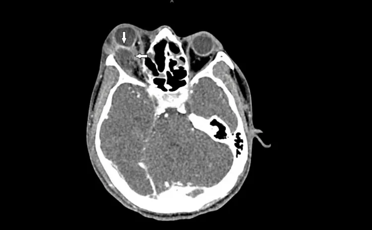
Emard A, et al. A 19-Year-Old Male With Orbital Cellulitis and Abscess Due to Fusobacterium necrophorum With Chronic Aspergillosis Resulting in Orbital Compartment Syndrome. Cureus. 2023. Figure 2. PMCID: PMC10644790. License: CC BY.
Axial CT image of a convex orbital abscess arising from the right lateral orbital wall, obscuring the lateral rectus muscle and causing globe deformity and optic nerve compression. This corresponds to the orbital abscess discussed in the section “2. Main Symptoms and Clinical Findings.”
Findings to be confirmed during examination are as follows.
Elevated intraocular pressure: IOP >40 mmHg is an indication for lateral canthotomy. In reported cases, IOP has been observed in the range of 35–80 mmHg 2, 4, 5, 6, 7, 9).
Proptosis (exophthalmos): Forward displacement of the eye due to increased pressure in the posterior orbit. Also accompanied by resistance to retropulsion.
Relative afferent pupillary defect (RAPD): The pupillary light reflex on the affected side is diminished or absent. This is an important sign of optic nerve damage.
Restricted eye movement: Compression of the extraocular muscles limits eye movement and may lead to complete ophthalmoplegia.
Eyelid and conjunctival findings: Chemosis and ecchymosis are prominent in traumatic and infectious cases. May be accompanied by eyelid subcutaneous hemorrhage and tightness of the eyelid skin.
Fundoscopic findings: Dilated fundus examination may reveal optic disc edema and vascular engorgement. Choroidal folds are a sign that the hematoma is adjacent to the globe.
Crepitus: A characteristic finding felt when palpating the periorbital area in cases of orbital emphysema.
Globe tenting: A CT finding that indicates a poor prognosis.
Post-traumatic retrobulbar hemorrhage: Orbital and facial trauma (including iatrogenic surgical trauma) is the most common cause.
Retrobulbar and peribulbar anesthesia: Can occur due to bleeding associated with local anesthesia for ophthalmic surgery.
Valsalva maneuver: Vomiting or severe straining increases intra-abdominal and intrathoracic pressure, causing rupture of orbital veins. Cases have been reported even in patients not using anticoagulants1).
Infectious
Orbital cellulitis/abscess: Increased intraorbital pressure due to mixed infection with anaerobic bacteria (e.g., Fusobacterium necrophorum) and Aspergillus2).
Mucormycosis: Can develop in the setting of COVID-19 or diabetic ketoacidosis. Delayed treatment initiation leads to a rapid increase in mortality4).
Subperiosteal abscess: Cases developing from sinusitis triggered by barotrauma have also been reported 8).
Orbital plasmacytoma: A case of rapid enlargement of an orbital mass as extramedullary extension of multiple myeloma leading to orbital compartment syndrome has been reported 9).
Intraorbital vascular lesions: Varices, hemangiomas, and arteriovenous malformations can also be sources of bleeding.
Other
Orbital emphysema: Develops due to communication with the paranasal sinuses after orbital wall fracture, or from subcutaneous emphysema spreading from pneumothorax 3).
Fluid resuscitation after severe burns: Orbital compartment syndrome has been reported even with non-aggressive fluid resuscitation (3.5 mL/kg/%TBSA) 5).
Post-cardiac arrest: Cases of orbital compartment syndrome after CPR due to capillary leakage and hypoxia have been reported 6).
Risk factors:
History of orbital or periorbital surgery: Postoperative bleeding or edema can trigger this syndrome.
Anticoagulant therapy / coagulation disorders: Increased bleeding and difficulty in hemostasis raise the risk.
Graves’ disease (thyroid eye disease): Chronic increase in orbital content volume serves as a background.
Diabetes / immunocompromised state: Risk factors for invasive fungal infections such as mucormycosis4).
Severe facial and periorbital full-thickness burns with fluid resuscitation: Burn area and depth are independent risk factors 5).
QCan orbital compartment syndrome occur without trauma?
A
It can develop from various causes including infection (orbital cellulitis, mucormycosis), retrobulbar hemorrhage due to Valsalva maneuver, carotid-cavernous fistula, orbital tumor, fluid resuscitation after severe burns, and post-cardiac arrest 1, 2, 4, 5, 6, 7, 9). It is important to consider the possibility of orbital compartment syndrome even without a history of trauma.
Intraocular pressure measurement is the most important diagnostic aid. Suspect this syndrome when IOP is 30 mmHg or higher, and IOP exceeding 40 mmHg is an indication for lateral canthotomy2). If the following triad is present, prioritize immediate treatment.
The assumption that “because an orbital wall fracture exists, decompression has occurred” is dangerous; orbital compartment syndrome can coexist with orbital fractures.
Auxiliary assessment in emergencies. Used to differentiate subperiosteal hematoma from soft tissue hematoma.
On CT, hematoma appears as a hyperdense area; in subperiosteal hematoma, a well-defined hyperdense area is seen between the orbital bone and periosteum. Tent-like deformation of the posterior globe (globe tenting) is an indicator of poor prognosis.
Orbital compartment syndrome is a clinical diagnosis; treatment should not be delayed for imaging. If elevated intraocular pressure, RAPD, and acute proptosis are present, lateral canthotomy should be performed immediately without waiting for CT results. CT is used in parallel with treatment to investigate the cause and assess the condition.
The goal of treatment for orbital compartment syndrome is to protect visual function by immediately relieving intraorbital pressure. Medical therapy should not delay necessary surgical decompression.
This is the first-line procedure for this syndrome. It can be performed at the bedside and is widely used as an emergency intervention in the emergency department.
Indications: IOP >40 mmHg. If orbital compartment syndrome is strongly suspected based on symptoms and findings, perform immediately.
Procedure:
Disinfect and drape the surgical site
Inject approximately 2 mL of 1-2% lidocaine with epinephrine for local infiltration anesthesia at the lateral canthus (using a 25G needle)
Remove and irrigate any intraocular foreign bodies or debris
Compress the lateral canthus to the lateral orbital rim with sterile hemostatic forceps (for hemostasis and tissue identification)
Perform lateral canthotomy from the lateral canthus to the lateral orbital rim using blunt scissors or a scalpel
Identify and cut the inferior crus of the inferior canthal ligament
When successful: eyelid tension relief, proptosis, immediate IOP reduction and visual improvement
If pressure remains high, also cut the superior crus of the superior canthal ligament (superior cantholysis)
The wound usually heals without additional intervention
About superior cantholysis: Because of increased risk of bleeding from the lacrimal artery, it should be considered only when adequate decompression is not achieved with proper inferior cantholysis.
Reported outcomes: IOP 35→11 mmHg (pupillary reflex also recovered) 6), IOP 60→25 mmHg (visual acuity 20/20 at 9 months) 7), IOP 34→15 mmHg (AION prevention) 5), canthotomy performed for IOP 80 mmHg 4).
If visual improvement is not achieved with cantholysis, orbital decompression via subciliary or transconjunctival approach under general anesthesia is performed. Consultation with an orbital surgeon is necessary. In infectious orbital compartment syndrome with abscess, incision and drainage (I&D) and sinus debridement are indicated 2, 4, 8).
If medical therapy is insufficient and there is elevated IOP with proptosis and globe tenting, orbital decompression via lateral canthotomy is performed as the most urgent measure, along with treatment of the bleeding source and hematoma evacuation.
For infectious orbital compartment syndrome, intravenous antibiotics (including anaerobic coverage) according to the causative organism are required; for mucormycosis, amphotericin B administration and surgical debridement are essential 4).
When orbital emphysema is present, use antiemetics, antitussives, and nasal decongestants to suppress orbital pressure fluctuations.
QWhere is lateral canthotomy performed?
A
Lateral canthotomy and inferior lateral cantholysis can be performed at the bedside, and there are many reports of emergency management in the emergency department 5, 6, 7). No dedicated operating room is required; it can be performed under local anesthesia with sterile scissors and forceps. However, concurrent consultation with an orbital surgeon is desirable.
The orbit is a closed space surrounded by four bony walls (superior, inferior, medial, lateral) and the anterior eyelids and orbital septum. Its volume is less than 30 mL, with the globe and orbital soft tissues occupying up to 26.5 mL. Because the bony walls and orbital septum are non-distensible, an increase in content causes a rapid rise in pressure. Since the anterior boundary (eyelids and orbital septum) limits proptosis, releasing this anterior boundary via lateral canthotomy and cantholysis reduces compartment pressure.
The optic nerve has natural redundancy within the orbit, allowing some degree of proptosis without axonal damage. However, when intraorbital pressure exceeds the perfusion pressure of the vasa nervorum or the central retinal artery, ischemia occurs, establishing the pathophysiology of this syndrome.
Capillary leakage and inflammatory response due to endothelial damage cause leakage of fluid and protein into the orbit. Because hardening of the skin from full-thickness eyelid burns inhibits elastic expansion, this syndrome can occur with a smaller increase in volume than usual 5). Blood flow to the optic nerve is determined by “perfusion pressure/blood flow resistance”; interstitial edema increases blood flow resistance, and decreased intravascular volume reduces perfusion pressure, leading to ischemia.
A burn case has been reported in which AION was avoided on the side where lateral canthotomy was performed (left eye), but AION developed on the untreated side (right eye), suggesting that early lateral canthotomy may prevent AION in severe burn patients 5).
Subperiosteal hematomas are not easily absorbed spontaneously, and organization and fibrosis of the hematoma can cause permanent tissue damage. Intramuscular hemorrhage within the extraocular muscles can cause diplopia, and if a subperiosteal hematoma extends to the orbital apex, it can lead to optic neuropathy.
7. Latest research and future perspectives (reports under investigation)
Kushwaha et al. (2021) reported a case of orbital compartment syndrome in a 57-year-old male who developed proptosis, IOP 35 mmHg, and loss of pupillary reflex after cardiac arrest and CPR 6). CT showed no retrobulbar hematoma, only soft tissue swelling. Lateral canthotomy and cantholysis reduced IOP to 11 mmHg and pupillary reflex recovered. The cause was presumed to be capillary leakage and prolonged hypoxia after CPR, and recognition as a rare cause of non-traumatic orbital compartment syndrome is needed.
Mathews & Knight (2022) reported a 28-year-old female who developed orbital compartment syndrome with IOP 60 mmHg due to rupture of a carotid-cavernous aneurysm after vomiting and dry heaves, via a carotid-cavernous fistula7). Lateral canthotomy reduced IOP to 25 mmHg, followed by coil embolization. Visual acuity recovered to 20/20 at 9 months. CCA/carotid-cavernous fistula should be considered as a cause of spontaneous orbital compartment syndrome without trauma or surgery.
Toh & Cameron (2022) reported a case where a patient with sinusitis experienced rupture of the lamina papyracea triggered by changes in cabin pressure during air travel, leading to orbital subperiosteal abscess and subsequent orbital compartment syndrome 8). The abscess was drained via lateral canthotomy and transconjunctival approach. Culture was positive for Serratia marcescens. Final visual acuity was 6/120. This case demonstrates that air travel in patients with sinusitis can precipitate this syndrome due to barotrauma.
Clinical Severity Classification of Orbital Emphysema (Hunts Classification)
A classification system evaluating the severity of orbital emphysema in four stages (Stage I to IV) has been reported 3). It ranges from Stage I (no clinical signs, only imaging findings) to Stage IV (central retinal artery occlusion), and may be useful in determining treatment strategy.
Orbital Compartment Syndrome Related to Orbital Plasmacytoma
Pyon et al. (2022) reported a 60-year-old woman who developed orbital compartment syndrome due to bilateral orbital plasmacytoma as extramedullary extension of multiple myeloma 9). Despite triple topical therapy, IOP only decreased to 30–33 mmHg. Improvement was achieved with chemotherapy (DCEP regimen) plus palliative radiotherapy (2000 cGy/10 fractions). The median survival of patients with orbital plasmacytoma is reported to be 28 months, which is worse than that of extramedullary plasmacytoma at other sites (8.3 years).
Direct Measurement of Orbital Pressure Using a Needle Manometer
A needle manometer technique for directly measuring orbital compartment pressure is being developed as a new diagnostic aid. It has the potential to directly quantify the compartment pressure of orbital soft tissues, which cannot be measured with a tonometer, and is expected to improve the accuracy of treatment decisions.
Kumar S, Beketova T, Rosenbaum PS, Amin A.. A Rare Case of Vomiting-Induced Retrobulbar Hemorrhage. Cureus. 2023;15(2):e34839. doi:10.7759/cureus.34839. PMID:36919065; PMCID:PMC10008326.
Emard A, Long B, Birdsong S.. A 19-Year-Old Male With Orbital Cellulitis and Abscess Due to Fusobacterium necrophorum With Chronic Aspergillosis Resulting in Orbital Compartment Syndrome. Cureus. 2023;15(10):e47061. doi:10.7759/cureus.47061. PMID:38022104; PMCID:PMC10644790.
Manata JP, Moniz Ramos M, Baiherych T, Alçada M, Matos Costa J.. Periorbital Emphysema Due to Traumatic Pneumothorax. Cureus. 2024;16(1):e51691. doi:10.7759/cureus.51691. PMID:38187024; PMCID:PMC10767690.
Werthman-Ehrenreich A.. Mucormycosis with orbital compartment syndrome in a patient with COVID-19. Am J Emerg Med. 2021;42:264.e5-264.e8. doi:10.1016/j.ajem.2020.09.032. PMID:32972795; PMCID:PMC7493738.
Pircher A, Holm S, Huss F. Left orbital compartment syndrome and right anterior ischemic optic neuropathy in a patient with severe burns despite non-aggressive fluid resuscitation. Scars Burns Heal. 2021;7:20595131211006659. doi:10.1177/20595131211006659.
Kushwaha R, Agil J, Furiato A, Webb McAdams AL.. Case of Orbital Compartment Syndrome Post Cardiac Arrest. Cureus. 2021;13(7):e16514. doi:10.7759/cureus.16514. PMID:34430128; PMCID:PMC8375638.
Mathews B, Knight OJ.. Carotid cavernous fistula secondary to ruptured carotid cavernous aneurysm causing orbital compartment syndrome. Am J Ophthalmol Case Rep. 2022;25:101310. doi:10.1016/j.ajoc.2022.101310. PMID:35128158; PMCID:PMC8807974.
Toh ZYC, Cameron A.. Orbital compartment syndrome secondary to subperiosteal abscess initiated by barotrauma. BMJ Case Rep. 2022;15(3):e248540. doi:10.1136/bcr-2021-248540. PMID:35332011; PMCID:PMC8948390.
Pyon RE, Wang GC, Chu Y, Tulpule S.. Bilateral Orbital Plasmacytomas With Orbital Compartment Syndrome. Cureus. 2022;14(6):e26269. doi:10.7759/cureus.26269. PMID:35898379; PMCID:PMC9308857.
Copy the article text and paste it into your preferred AI assistant.
Article copied to clipboard
Open an AI assistant below and paste the copied text into the chat box.