Orbital cellulitis is a bacterial infection of the soft tissues within the orbit posterior to the orbital septum (a fibrous membrane at the front of the eyelid). It represents an acute infectious inflammatory process of the orbital tissues and is considered an ophthalmic emergency.
The severity of orbital infection is assessed using the Chandler classification (1970). This classification is also called Chan’s group classification and divides the inflammation into Groups 1 to 5 based on the primary location (see table below).
Chandler I–III
Grade I (Preseptal cellulitis): Edema of the eyelids and surrounding soft tissues. Limited to the anterior orbital septum. No proptosis or ophthalmoplegia.
Grade II (Orbital cellulitis): Infection spreads to orbital fat. Accompanied by proptosis and ophthalmoplegia.
Grade III (Subperiosteal abscess): Abscess formation between the orbital periosteum and orbital wall. Often requires surgical drainage.
Chandler IV–V
Grade IV (Orbital abscess): Abscess formation within orbital fat. Severe proptosis and complete restriction of eye movement. Marked vision loss. Indication for emergency drainage.
Grade V (Cavernous sinus thrombosis): Infection spreads intracranially. Bilateral findings and impaired consciousness; most severe form. Life-threatening.
This disease commonly occurs in children, but also affects young adults. It is closely associated with sinusitis (especially ethmoid sinusitis), with infection spreading to the orbit through the thin lamina papyracea that forms the medial orbital wall. In adults, dental infections and trauma can also be causes. In patients with sinusitis or upper respiratory infection, this disease should be strongly suspected when rapid eyelid swelling, pain, and proptosis appear.
QWhat is the difference between preseptal cellulitis (eyelid cellulitis) and orbital cellulitis?
A
Preseptal cellulitis is an infection limited to the area anterior to the orbital septum (eyelid side), without proptosis, ophthalmoplegia, or vision loss. Orbital cellulitis involves infection extending posterior to the septum into the orbit, adding these findings. CT is useful for differentiation.
In a study of 9 cases of orbital cellulitis caused by MRSA (methicillin-resistant Staphylococcus aureus), eyelid edema (88.9%), pain (88.9%), proptosis (66.7%), restricted eye movement (66.7%), and fever (55.5%) were recorded. Median CRP was 178 mg/L and median WBC was 17.9×10⁹/L. 1)
Finding
Characteristic
Proptosis
Caused by increased orbital contents due to abscess or edema. The greater the degree, the more severe.
Restricted eye movement
Due to inflammatory infiltration of extraocular muscles or nerve involvement. In hematogenous cases, prolonged restriction has been reported. 3)
Eyelid edema and conjunctival edema (chemosis)
Due to impaired venous and lymphatic drainage.
Elevated intraocular pressure and optic disc edema
Findings indicating that visual function is threatened due to increased orbital pressure.
Care must be taken not to confuse with eyelid swelling
Eyelid retraction in newborns has been reported as a finding observed in severe neonatal cases2).
Positive RAPD (relative afferent pupillary defect) is a dangerous sign indicating optic nerve compression, and if confirmed, emergency treatment is required.
QIs emergency surgery necessary if there is vision loss?
A
Vision loss is a dangerous sign indicating optic nerve compression and is highly likely to require emergency treatment. However, surgical indication is not based solely on vision loss; it is determined comprehensively by CT findings of abscess size, location, age, and response to antibiotic therapy. See Section 5 for treatment details.
The routes of onset of orbital cellulitis are mainly classified into three categories.
Direct spread from sinusitis: The most common route. In pediatric orbital infections, 60–91% are reported to be associated with sinusitis7, 8). Infection easily progresses into the orbit through the thin lamina papyracea. Direct hematogenous spread via valveless veins is also involved7).
Hematogenous infection (bacteremia): Infection via the bloodstream can occur in immunocompromised individuals and newborns.
Exogenous infection: Direct spread from periorbital trauma, ophthalmic surgery, or surrounding tissues. In adults, dental infection is also an important route.
In addition, routes of pathogen arrival include infiltration from sinusitis, retrograde propagation from cavernous sinus thrombosis, and spread from endogenous endophthalmitis to the orbit. Orbital infection can also complicate acute retinal necrosis (varicella-zoster virus retinal infection).
Major bacterial species: Staphylococcus aureus, Streptococcus pyogenes, and Streptococcus pneumoniae are representative causative organisms.
MRSA: The incidence has been increasing in recent years. In Taiwan, the proportion of MRSA has risen from 14.5% to 37.5%, and in Australia it has been reported at 28.6%. 1) PVL (Panton-Valentine leukocidin) toxin-producing strains are strongly associated with abscess formation. 1)
Immunocompromised patients (HOC; hematogenous orbital cellulitis): This is a rare form, and various pathogens such as Candida, MRSA, Klebsiella, Enterococcus, and zygomycetes may be involved. 3)
Neonates: MSSA (methicillin-sensitive Staphylococcus aureus) is common, but bacteremia and meningitis are frequent complications. 2)
Fungi (Aspergillus, Mucor): Fungal orbital cellulitis can occur in immunocompromised patients and diabetic patients. Early diagnosis is important because of poor prognosis.
Upper respiratory tract infection, sinusitis, facial trauma, odontogenic infection, and immunocompromised status (including HIV infection) are the main risk factors. 8)
Pott’s puffy tumor is a subperiosteal abscess and osteomyelitis of the frontal bone associated with frontal sinusitis, and it can present as orbital cellulitis. A case of a 12-year-old boy with orbital and temporal subperiosteal abscesses complicated by intracranial epidural abscess has been reported. In cases of orbital infection with frontal swelling, this condition should be considered in the differential diagnosis. 10)
QWhy does infection spread from sinusitis to the eye?
A
The medial wall of the orbit (lamina papyracea) is very thin and adjacent to the ethmoid sinus. Additionally, valveless veins run between the sinuses and the orbit, allowing infection to spread bidirectionally. 7) Therefore, ethmoid sinusitis can easily spread directly into the orbit.
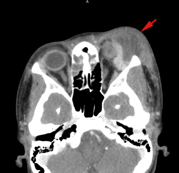
Celebi TB, Shamulzai A, Dahhan H. Worsening preseptal cellulitis with an orbital abscess and intracranial extension in a pediatric patient. Cureus. 2024;16(11):e73772. Figure 1. PMID: 39677106; PMCID: PMC11646562. DOI: 10.7759/cureus.73772. License: CC BY 4.0.
Contrast-enhanced CT axial image (pediatric case) showing a large abscess (red arrow) formed under the periosteum of the left orbit, displacing the globe, superior rectus muscle, and lacrimal gland, with near-complete opacification (purulent retention) of the ethmoid and frontal sinuses. This corresponds to the CT findings of subperiosteal abscess (low-density area, associated sinusitis, mass effect on the globe) discussed in the “Diagnosis and Examination Methods” section.
Examination
Use/Features
CT (contrast-enhanced)
First choice. Evaluates presence, size, and location of subperiosteal abscess. Also assesses concurrent sinusitis.
MRI (STIR sequence)
Detailed evaluation of soft tissues, osteomyelitis, and intracranial lesions. Can detect osteomyelitis that is difficult to identify on CT.
Auxiliary test when radiation exposure is to be avoided.
CT scan is the cornerstone of diagnosis, and imaging with slice thickness of 3 mm or less, including coronal views, is recommended. Contrast-enhanced CT allows identification of subperiosteal and orbital abscesses. Particular attention should be paid to findings of sinusitis near the lesion (mucosal thickening, opacification of the sinus cavity).
MRI (especially STIR sequence) provides excellent soft tissue contrast and is useful for evaluating intraorbital soft tissues, osteomyelitis, and intracranial complications 8). DWI (diffusion-weighted imaging) also helps confirm abscess formation 1).
Blood tests: Evaluate WBC, CRP, and procalcitonin (PCT) to assess systemic inflammation and treatment response.
Blood culture: The positivity rate in typical orbital cellulitis is only about 2–7.9%, but in immunocompromised patients (HOC) it is as high as 75%. 3)
Next-generation sequencing (NGS): Can identify pathogens within 48 hours and is useful even in cases where conventional culture methods are difficult. 3)
Visual acuity and pupillary examination: Confirmation of RAPD (relative afferent pupillary defect) is essential and indispensable for evaluating optic nerve compression.
Ocular motility examination: Used to assess the degree of restriction and evaluate severity.
Orbital tumor/lymphoma: In recurrent treatment-resistant cases, tumor must be ruled out. In culture-negative recurrent cases, consider the possibility of malignant lymphoma. 9)
Orbital cellulitis is treated with intravenous antibiotics upon hospitalization as a rule. Collaboration with otorhinolaryngology is essential, and treatment effect is evaluated by repeated CT observation of the lesion to determine drug dosage and duration. If visual acuity decreases, reduction of intraorbital pressure is important, and surgical drainage is performed as needed.
Transition to outpatient parenteral antibiotic therapy (OPAT) or oral medication should be considered after confirming defervescence, improvement of inflammatory response, and stabilization of ocular findings 4, 8).
Antibiotic failure: When there is worsening or no improvement after appropriate antibiotic therapy.
Intracranial extension: When spread to epidural abscess or brain abscess is observed.
Surgical Procedures
FESS (Functional Endoscopic Sinus Surgery): Drainage of sinusitis. Performed in 88.9% of MRSA cases. 1)
External orbital drainage: Abscess drainage via external incision. Combined approach with endoscopic surgery is also performed. 4)
Multidisciplinary collaboration: Collaboration among otorhinolaryngology, ophthalmology, and neurosurgery is essential in severe cases. 4)
Note that not all subperiosteal abscesses require surgery. If the abscess is small, visual function is preserved, and there is a good response to antibiotic therapy, conservative treatment may be attempted.
The usefulness of dexamethasone as adjunctive therapy has been reported.
AlQahtani et al. reported a case of a 3-year-old with MRSA and Pseudomonas aeruginosa infection (subperiosteal abscess 6.6 mm) treated with ceftazidime plus clindamycin and three courses of dexamethasone 6 mg (q12h, 3 days), achieving dramatic improvement. 6)
Heri-Kovacs et al. reported that in a case of orbital cellulitis following COVID-19 vaccination, intravenous dexamethasone 250 mg/day for 4 days was effective in a patient without sinusitis. 5)
The use of steroids requires judgment based on individual clinical circumstances, and a standard dosing protocol has not been established.
Not all SPAs require surgical drainage. If the abscess is small, visual function is preserved, and there is a good response to antibiotic therapy, conservative treatment may be attempted. However, if visual loss, elevated intraocular pressure, or antibiotic failure occurs, surgical drainage should be considered promptly.
The lamina papyracea, which forms the contact surface between the orbit and the paranasal sinuses, is a bony plate that forms the medial orbital wall and is extremely thin and prone to perforation. This anatomical feature facilitates the spread of infection from ethmoid sinusitis to the orbit.
Between the sinuses and the orbit, there are valveless veins that allow bidirectional hematogenous spread of infection. 7) Frontal sinusitis can also lead to direct extension to the epidural space and intracranially. 4)
Inflammation of the ethmoid and frontal sinuses spreads to the subperiosteal space, progressing from subperiosteal abscess (Chandler stage III) to orbital abscess (stage IV). Direct bone destruction or dissemination via emissary veins are the main routes.
PVL toxin (Panton-Valentine leukocidin): A toxin produced by community-acquired MRSA, strongly associated with leukocyte damage and abscess formation. 1)
Intracranial spread via valveless veins: Infection of the frontal sinus can extend to the orbit and further to the epidural space and brain. 7)
Severe disease in immunocompromised (HOC): In immunocompromised individuals, hematogenous orbital cellulitis (HOC) can develop, involving multiple opportunistic pathogens. 3) Recovery of ocular motor palsy may take up to 18 months. 3)
Fungal pathology: Orbital infections caused by Aspergillus and Mucor occur in immunocompromised and diabetic patients, with rapid tissue invasion and very high mortality.
Pott’s puffy tumor: Frontal sinusitis leads to frontal bone osteomyelitis and subperiosteal abscess, which can spread to the orbit and intracranially. 10)
With early diagnosis and adequate antibiotic therapy, most cases recover. If vision loss occurs, there is a risk of sequelae. Chandler grade V (cavernous sinus thrombosis) has high mortality and morbidity rates, and fungal infections (mucormycosis) have very high mortality, especially in diabetic patients.
In a retrospective study of 9 cases by Ang et al., the median hospital stay for MRSA orbital cellulitis was 13.7 days, and 100% of cases required surgical intervention. 1) The proportion of MRSA in orbital cellulitis varies by region, rising from 14.5% to 37.5% in Taiwan. 1) Optimizing antibiotic selection and establishing initial treatment protocols considering PVL-producing MRSA are future challenges.
Diagnostic Application of Next-Generation Sequencing (NGS)
Tang et al. reported 4 cases of HOC in immunocompromised patients, emphasizing that NGS can identify pathogens within 48 hours. 3) It is useful for identifying diverse pathogens that are difficult to detect by conventional culture methods and can contribute to appropriate antibiotic selection.
Heri-Kovacs et al. reported a 72-year-old man who developed orbital cellulitis (5 mm proptosis, ophthalmoplegia) without sinusitis 9 days after the second dose of VeroCell (inactivated COVID-19 vaccine). 5) He was treated with IV dexamethasone 250 mg/day for 4 days and recovered within 4 days. The pathogenesis remains unclear, and further case accumulation is needed.
Ishak et al. reported a case repeatedly treated as culture-negative “orbital cellulitis” that was ultimately diagnosed as B-cell lymphoma. 9) In treatment-resistant or recurrent orbital cellulitis, early suspicion of tumor or granulomatous disease and thorough investigation including biopsy are essential.
Deng & Shinder reported a 12-year-old boy with Pott’s puffy tumor presenting as orbital cellulitis secondary to frontal sinusitis. He had subperiosteal abscesses in the orbit and temporal region, as well as intracranial epidural abscess. Treatment included IV antibiotics, endoscopic sinus drainage, and craniotomy for epidural abscess drainage, with improvement after 6 weeks of antibiotics. 10) Orbital infection with frontal swelling should raise suspicion for this condition.
Ang T, Smith JEH, Maqsood N, et al. Orbital cellulitis caused by methicillin-resistant Staphylococcus aureus: a case series. Int Ophthalmol. 2023;43:2925-2933.
Kulkarni V, Sundaram V, Sameeksha TH. Overwhelming orbital cellulitis in a neonate. BMJ Case Rep. 2023;16(7):e252390. doi:10.1136/bcr-2022-252390. PMID: 37491125.
Tang X, Li H. A rare ocular complication of septicemia: a case series report and literature review. BMC Infect Dis. 2023;23:522. doi:10.1186/s12879-023-08489-1.
Wong SJ, Levi J. Management of pediatric orbital cellulitis: a systematic review. Int J Pediatr Otorhinolaryngol. 2018;110:123-129. doi:10.1016/j.ijporl.2018.05.006. PMID: 29859573.
Heri-Kovacs A, Eibenberger K, Tausch MK, et al. Orbital cellulitis following SARS-CoV-2 vaccination: a case report. Case Rep Ophthalmol. 2022;13:210-214.
AlQahtani DS, Alshahrani OA, Abu AlOla MA, et al. Refractory orbital cellulitis: a management challenge. Saudi J Ophthalmol. 2021;35:261-262.
Torretta S, Guastella C, Marchisio P, et al. Sinonasal-related orbital infections in children: a clinical and therapeutic overview. J Clin Med. 2019;8(1):101. doi:10.3390/jcm8010101. PMID: 30654566.
Tsirouki T, Dastiridou AI, Ibáñez Flores N, et al. Orbital cellulitis. Surv Ophthalmol. 2018;63(4):534-553. doi:10.1016/j.survophthal.2017.12.001. PMID: 29248536.
Ishak F, Hassan S, Abdul Rahim A, et al. Orbital Lymphoma Presenting As Recurrent Orbital Cellulitis: A Diagnostic Challenge. Cureus. 2024;16(10):e70759. doi:10.7759/cureus.70759.
Deng W, Shinder R. Pott’s puffy tumor presenting as orbital cellulitis. Ophthalmology. (症例報告: 12歳男児、前頭洞炎→骨膜下膿瘍→硬膜外膿瘍合併例)
Copy the article text and paste it into your preferred AI assistant.
Article copied to clipboard
Open an AI assistant below and paste the copied text into the chat box.