Traumatic cataract is an opacity of the lens caused by trauma, and it is more common in younger individuals compared to age-related cataract. When unilateral cataract is found in a young patient without underlying disease, traumatic cataract should be suspected first. The lifetime prevalence of ocular trauma in the general population is about 14%, with a higher incidence in children and young males. 27–65% of ocular trauma cases lead to cataract, and most of these significantly affect visual function and require surgery 3).
Traumatic cataracts often involve damage to other ocular tissues and frequently occur in younger individuals, making them a significant public health burden. Even in the absence of cataracts that severely affect visual function, subluxation of the lens due to damage to the zonules of Zinn may occur, sometimes requiring surgical intervention.
Mechanism of Cataract Formation
Rapid opacification: Aqueous humor enters the lens fibers due to rupture of the lens capsule
Delayed opacification: Even without capsule rupture, trauma can damage lens fibers, leading to cataract formation months to years later.
Typical appearance: Rosette-shaped or stellate opacity
Characteristics of Traumatic Cataract
Commonly affected group: Children and young males
Other ocular injury complications: Iris injury, Zinn zonule injury, vitreous prolapse, etc.
Urgency: Emergency extraction is required in cases of capsule rupture or elevated intraocular pressure.
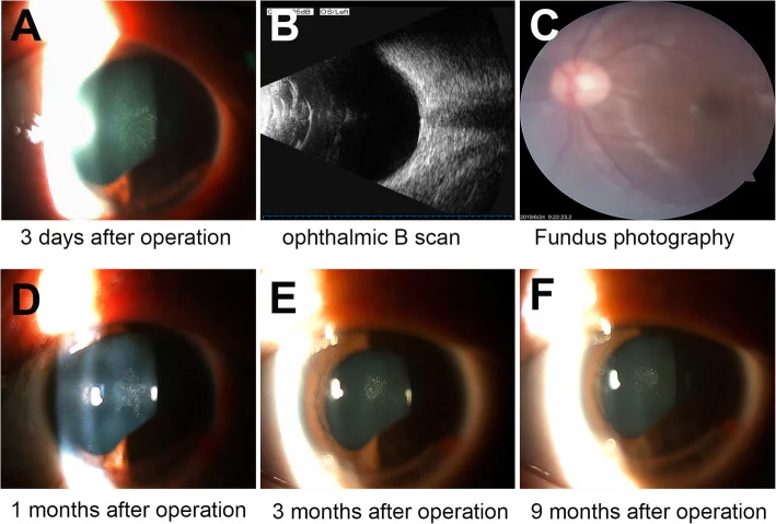
Zhang YT, Du LQ, Liu M, et al. Spontaneous resolution of a traumatic cataract in a patient with an open-globe ocular injury: a case report. BMC Ophthalmol. 2020;20(1):285. Fig. 2. PMID: 32660455; PMCID: PMC7359477; DOI: 10.1186/s12886-020-01555-1. License: CC BY.
Serial slit-lamp and fundus photographs showing a rosette-shaped posterior capsule opacification (PCC) observed on postoperative day 3 (Panel A), which spontaneously regressed and resolved over 9 months. This corresponds to the rosette opacification discussed in the section “What is traumatic cataract?”.
Posterior subcapsular opacity. See separate article “Radiation cataract”.
Drug-induced
Steroids, etc.
See separate article “Effects of Steroids on the Eye”
Blunt trauma object size and opacity pattern:
When an object enters the orbit at high speed, such as a badminton shuttlecock: posterior subcapsular opacity and posterior capsule rupture occur immediately.
Softball/baseball: posterior subcapsular opacity → anterior subcapsular opacity over time
Atopic dermatitis (habit of hitting or rubbing): Radial opacities under the anterior and posterior capsules
Vossius ring: Iris pigment deposits in a ring shape on the anterior lens capsule corresponding to the pupillary margin. It is characteristic of blunt trauma and serves as evidence of trauma to the lens capsule.
If an iron foreign body remains in the eye, it can cause siderosis lentis, presenting with characteristic opacities. Retention of a copper foreign body leads to chalcosis lentis. If a retained foreign body is suspected, CT or other imaging for localization and early removal are necessary.
Opacity location, anterior capsule rupture, subluxation, Vossius ring
In blunt trauma, if findings such as posterior synechiae, anterior capsule opacity, or localized cortical opacity are observed in only one eye, traumatic cataract is strongly suspected. Carefully observe for differences in anterior chamber depth between the eyes, presence of angle recession, zonular rupture, vitreous prolapse into the anterior chamber, and ora serrata tears.
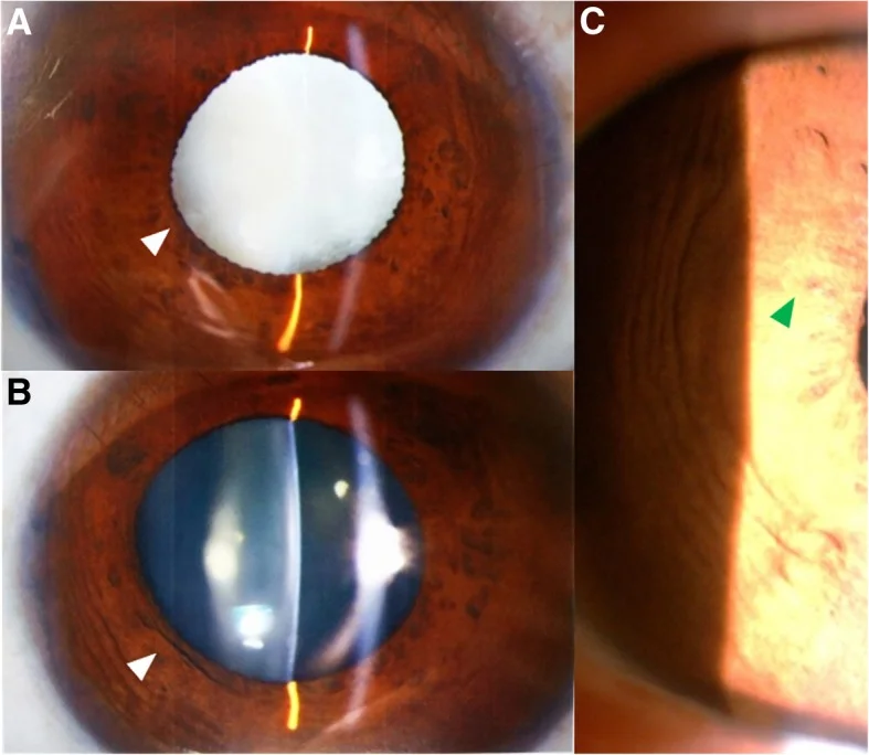
Miao A, Zhang K, Yu J, et al. How many challenges we may encounter in anterior megalophthalmos with white cataract: a case report. BMC Ophthalmol. 2019;19(1):122. Fig. 1. PMID: 31146719; PMCID: PMC6543662; DOI: 10.1186/s12886-019-1133-y. License: CC BY.
Preoperative slit-lamp examination reveals white cataract, corneal enlargement, deep anterior chamber, insufficient mydriasis (Panel A and B, white arrows), and mild iris atrophy (Panel C, green arrow) in the right eye. These findings correspond to the anterior capsule, iris, and pupil findings discussed in the section “Main Symptoms and Clinical Findings.”
Diagnostic System
The Birmingham Eye Trauma Terminology (BETT) system is used to record trauma.
Imaging tests
B-mode ultrasound: evaluation of intraocular foreign bodies, retinal detachment, and vitreous opacities when posterior segment observation is difficult
CT scan: Exclusion of intraocular and orbital foreign bodies and abnormal eye shape (search for foreign bodies in perforating trauma)
Ultrasound biomicroscopy (UBM): evaluation of the posterior capsule, lens position, angle, and zonular integrity
QCan cataracts develop immediately after trauma, or can they take time to appear?
A
Yes. In perforating trauma, the lens capsule is damaged and aqueous humor enters, causing rapid opacification immediately after injury. Small wounds (e.g., from a needle) result in localized anterior subcapsular opacification, while large wounds (e.g., from a cutter) lead to rapid spread of opacification. In contrast, blunt trauma can cause metabolic disturbances and osmotic changes due to external force even without capsular rupture, and opacification often progresses gradually over several months to years after injury.
Unilateral cataract in a young patient without underlying disease → first suspect traumatic cataract
Children and young men are at the center of risk
Sports and occupational injuries are the main mechanisms of injury7)
In blunt trauma, the course is long, so patients may forget their trauma history → actively confirm during history taking
When foreign bodies (iron, copper) remain in the eye: characteristic opacities form as siderosis lentis or chalcosis lentis
Infrared cataract (glassblower’s cataract): Posterior subcapsular opacity commonly seen in furnace and blast furnace workers. Chronic infrared exposure causes opacity in the posterior subcapsular region of the lens.
Electric cataract: Occurs after lightning strike or electric shock. Characterized by cortical and subcapsular opacities. Often forms within a few months after injury.
The diagnosis of traumatic cataract itself is straightforward, but it is important to confirm that the cause is trauma. Since cataract surgery for traumatic cataracts is more likely to be a difficult case compared to routine cataract surgery, thorough preoperative evaluation is essential.
Inquiry about trauma history (patients may forget a history of blunt trauma)
Integrity of the anterior capsule (presence or absence of rupture)
Integrity of the zonule of Zinn (dislocation, phacodonesis)
Intraocular pressure (presence and type of glaucoma)
X-ray/CT (useful for searching for foreign bodies in perforating trauma, as well as head X-ray and CT examinations)
General condition and indications for emergency surgery
Key differentiation points: If posterior synechiae, anterior capsule opacity, and localized cortical opacity are observed in only one eye, suspect traumatic origin. In penetrating trauma, it is essential to assess the condition of the wound, the lens capsule (presence of posterior capsule rupture), the posterior segment, and the presence, nature, and location of any foreign body.
Generally, lens reconstruction is performed after opacification becomes severe and visual impairment progresses. On the other hand, urgent surgery may be necessary in some cases, such as for lens-induced uveitis (due to leakage of lens proteins) upon capsule rupture, or to prevent bacterial endophthalmitis after foreign body or laceration.
Removal of traumatic cataract is broadly divided into “primary removal” immediately after open globe injury and “secondary removal” several weeks to months after the injury.
Primary repair of open globe injuries is ideally performed within 24 hours, and the group repaired within 24 hours has a significantly lower risk of endophthalmitis (OR 0.39, 95% CI 0.19-0.79)1).
Emergency surgery is common. First, suturing of the corneoscleral perforation wound is necessary. If the anterior chamber can be maintained and there is a relatively small perforation only in the anterior capsule with no intraocular foreign body, standard ultrasonic cataract surgery (PEA) is possible. In cases of perforation extending to the posterior capsule, foreign bodies often reach the vitreous, so simultaneous vitrectomy is required. Topical and systemic administration of antibiotics is necessary.
Primary IOL implantation: Consider primary implantation if preoperative axial length measurement is possible and infection risk is low. If difficult, consider secondary implantation.
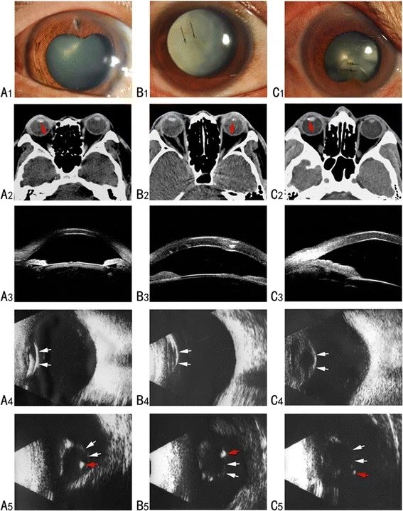
Wang K, Liu J, Chen M. Role of B-scan ultrasonography in the localization of intraocular foreign bodies in the anterior segment: a report of three cases. BMC Ophthalmol. 2015;15:102. Fig. 1. PMID: 26268356; PMCID: PMC4535674; DOI: 10.1186/s12886-015-0076-1. License: CC BY.
Preoperative slit-lamp, CT, UBM, and B-mode ultrasound findings in 3 cases are evaluated using multiple modalities, from corneal laceration and lens opacity (A1–C1) to CT intraocular foreign body (red arrow, A2–C2) and its positional relationship with the posterior capsule (A5–C5). This corresponds to intraocular foreign body-associated trauma discussed in the section “Treatment of Penetrating Trauma.”
Surgical indications are determined in the same way as for ordinary cataracts. Zinn zonules are often fragile or torn, so the ultrasound device settings should be set to low perfusion and low aspiration pressure. Use a capsule expander or capsular tension ring (CTR) as appropriate. If extensive zonular dialysis is present, consider intraocular lens suturing.
Posterior synechiae (small pupil, pupillary distortion): Gently release adhesions with a needle while injecting viscoelastic material
Anterior capsule fibrosis: If fibrosis crosses the planned anterior capsulotomy site, cut the fibrotic portion with scissors and proceed.
Zinn zonule weakness/rupture: Use low perfusion and low aspiration pressure settings, and consider CTR use
Evaluation of anterior capsule integrity: Intraoperative use of trypan blue helps identify anterior capsule tears and allows visualization of the capsule even in white cataracts. If a capsular tear is suspected, perform hydrodissection conservatively and carefully.
If the extent of the tear is 1/4 or more, use a CTR (capsular tension ring) to fix the intraocular lens within the capsule. If fixation within the capsule is not possible, suture the intraocular lens to the ciliary sulcus or fix it within the sclera (e.g., Yamane method)6).
In cases of complete dislocation, liquid perfluorocarbon (LPFC/PFCL) is used during vitreous surgery to float the lens up to the iris plane, and it is removed via a transscleral approach.
Regular checkups are performed on postoperative day 1, week 1, and month 1. Complete the course of topical antibiotics and steroid eye drops. If complications occur, follow up more frequently and adjust steroids or administer intraocular pressure-lowering medications. In cases with zonular rupture, be aware of the risk of intraocular lens dislocation or drop postoperatively, and conduct long-term follow-up.
QHow is traumatic cataract surgery different from regular cataract surgery?
A
Traumatic cataract surgery is more difficult than standard cataract surgery. Many intraoperative difficulties are anticipated, such as possible rupture of the anterior capsule, lens instability due to zonular damage, difficulty in dilating the pupil due to posterior synechiae, anterior capsule fibrosis, and a high risk of posterior capsule rupture. It is important to make full use of auxiliary tools such as trypan blue, CTR, and Malyugin ring, and to carefully plan the surgical strategy according to the cataract morphology and associated injuries. Adequate preparation of instruments before surgery is also essential.
QCataract found after trauma, but is surgery needed immediately?
A
If the opacity is mild and does not significantly affect vision, observation is possible. However, if there is rupture of the lens capsule, increased intraocular pressure, or lens-induced uveitis (a condition where lens proteins leak and cause inflammation), emergency surgery is indicated. In children, due to the risk of amblyopia, more aggressive early intervention is required compared to adults.
QIs it always possible to insert an intraocular lens?
A
If the lens capsule is preserved, primary insertion is possible. If the capsule is damaged, secondary insertion is performed via ciliary sulcus suturing or intrascleral fixation (Yamane method). IOL insertion may be postponed if axial length measurement is difficult or the risk of infection is high.
Children are disproportionately susceptible to the effects of ocular trauma and require special management due to the risk of stimulus deprivation amblyopia, which differs from that in adults.
Preoperative considerations
In children, the threshold for determining a significant impact on visual function is lower than in adults. If there is opacity exceeding 3 mm on the visual axis, extraction should be considered. Since delay increases the risk of amblyopia, primary extraction is recommended as an emergency procedure 5).
In perforating trauma, remove the lens immediately and insert an IOL if possible. In blunt trauma, perform surgery based on the progression of the cataract.
Intraoperative considerations
In children under 2 years of age, pars plana vitrectomy is often performed simultaneously with cataract extraction. In this age group, IOL implantation is deferred and performed as a secondary procedure.
Postoperative considerations
Postoperative amblyopia treatment (optical correction and occlusion of the healthy eye) is essential. Active amblyopia therapy combining optical correction with glasses or contact lenses and occlusion therapy of the healthy eye should be continued. Posterior capsule opacification (PCO) is a common postoperative complication in children and requires early intervention as it can lead to amblyopia if left untreated. Young patients have a strong inflammatory response and are at risk for fibrinous uveitis, so aggressive preoperative and postoperative steroid eye drop management is necessary.
QHow soon does a child with traumatic cataract need surgery?
A
In children, there is a risk of amblyopia, so more aggressive early intervention is required than in adults. If there is opacity exceeding 3 mm on the central visual axis, extraction is indicated, and emergency primary extraction is recommended. Delay in surgery increases the risk of amblyopia and may lead to permanent visual impairment. After surgery, active amblyopia treatment such as occlusion therapy of the healthy eye and optical correction is necessary.
Sharp trauma (perforating): Damage to the lens capsule allows aqueous humor to rapidly enter the lens fibers, causing rapid opacification due to osmotic changes and metabolic disturbances. Small perforations result in localized anterior subcapsular opacities, while large perforations lead to total rapid opacification.
Blunt trauma: Sudden deformation of the eyeball applies mechanical stress to the lens fibers. Even without capsular rupture, metabolic disturbances and osmotic changes are induced, leading to gradual opacification over months to years. Mechanical stress on the zonules of Zinn may also cause rupture.
Lens swelling (intumescent cataract): The lens becomes swollen, causing an increase in intraocular pressure. Leakage of lens proteins into the aqueous humor induces lens-induced uveitis, leading to further elevation of intraocular pressure and worsening of inflammation.
Mechanism of Vossius ring formation: When the eyeball is abruptly deformed by blunt external force, the iris is strongly pressed against the anterior surface of the lens. At this time, pigment from the iris pigment epithelial cells is transferred to the anterior lens capsule surface, forming a circular ring-shaped deposit (Vossius ring) corresponding to the pupillary margin.
The Ocular Trauma Score (OTS) is widely used to predict visual prognosis in traumatic cataract. The OTS calculates prognosis from six factors: initial visual acuity, presence of globe rupture, endophthalmitis, penetrating injury, retinal detachment, and relative afferent pupillary defect2). A retrospective study of over 300 children showed that the OTS reliably predicts visual prognosis in pediatric traumatic cataract 5).
There remains conflicting data regarding the superiority of primary versus secondary enucleation, and no consensus has been reached4). A report indicates that primary repair of open globe injuries within 24 hours is associated with a reduced risk of endophthalmitis (OR 0.39), supporting early intervention1).
For scleral-fixated IOLs, sutureless fixation methods such as the Yamane technique are also options. In cases without capsular support, selecting a fixation method based on associated injuries and surgeon experience is important 6). For prognostic evaluation of traumatic cataracts, individualized assessment combining the OTS and findings from pediatric trauma studies is required 2,5).
McMaster D, Bapty J, Bush L, Serra G, Kempapidis T, McClellan SF, et al. Early versus Delayed Timing of Primary Repair after Open-Globe Injury: A Systematic Review and Meta-analysis. Ophthalmology. 2025;132(4):431-441. doi:10.1016/j.ophtha.2024.08.030. PMID:39218161.
Kuhn F, Maisiak R, Mann L, Mester V, Morris R, Witherspoon CD. The Ocular Trauma Score (OTS). Ophthalmology clinics of North America. 2002;15(2):163-5, vi. doi:10.1016/s0896-1549(02)00007-x. PMID:12229231.
Mehul A Shah, Shreya M Shah, Shashank B Shah, Chintan G Patel, Utsav A Patel. Morphology of traumatic cataract: does it play a role in final visual outcome?. BMJ Open. 2011;1(1):e000060. doi:10.1136/bmjopen-2011-000060.
Rumelt S, Rehany U. The influence of surgery and intraocular lens implantation timing on visual outcome in traumatic cataract. Graefe’s archive for clinical and experimental ophthalmology = Albrecht von Graefes Archiv fur klinische und experimentelle Ophthalmologie. 2010;248(9):1293-7. doi:10.1007/s00417-010-1378-x. PMID:20585800.
Ram J, Verma N, Gupta N, Chaudhary M. Effect of penetrating and blunt ocular trauma on the outcome of traumatic cataract in children in northern India. The journal of trauma and acute care surgery. 2012;73(3):726-30. doi:10.1097/TA.0b013e31825eeac9. PMID:22929502.
Morikawa S, Okamoto F, Okamoto Y, Mitamura Y, Ishikawa H, Harimoto K, et al. Clinical characteristics and visual outcomes of work-related open globe injuries in Japanese patients. Scientific reports. 2020;10(1):1208. doi:10.1038/s41598-020-57568-9. PMID:31988287; PMCID:PMC6985116.
Copy the article text and paste it into your preferred AI assistant.
Article copied to clipboard
Open an AI assistant below and paste the copied text into the chat box.