Horner syndrome is a syndrome that presents with various ocular and systemic findings due to disruption of the ocular sympathetic pathway. The three main signs are miosis, ptosis, and anhidrosis, and the lesion can occur at central, preganglionic, or postganglionic sites.
The condition is named after Claude Bernard (1852) and Johann Friedrich Horner (1869). It is also called Bernard-Horner syndrome. The ocular sympathetic pathway is a long route from the hypothalamus through three neurons to the pupillary dilator muscle, and the causative disease and urgency vary greatly depending on the lesion site.
Miosis is moderate, and anisocoria is most noticeable in dim light. The pupillary light reflex remains normal, which is an important distinguishing feature from Adie pupil and oculomotor nerve palsy. Denervation of Müller’s muscle in the upper eyelid causes mild ptosis of about 2 mm, and involvement of Müller’s muscle in the lower eyelid causes mild elevation (upside-down ptosis). Together, these result in narrowing of the palpebral fissure and apparent enophthalmos.
QIs the ptosis in Horner syndrome severe?
A
It is usually mild, about 2 mm, due to sympathetic denervation of Müller’s muscle in the upper eyelid. The lower eyelid also elevates slightly (upside-down ptosis), causing apparent narrowing of the palpebral fissure and enophthalmos. It is important to distinguish from complete ptosis (oculomotor nerve palsy).
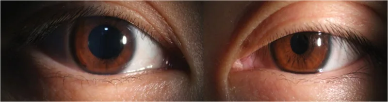
Li XM, et al. Neuro-ophthalmic observation and 16-month follow-up of horner syndrome after thyroidectomy: A case report. Medicine (Baltimore). 2026. Figure 2. PMCID: PMC12826322. License: CC BY.
External eye photographs of both eyes showing mild ptosis and miosis in one eye. This visually demonstrates the typical clinical findings of Horner syndrome and is suitable for explaining the main symptoms and clinical findings.
Miosis: Moderate miosis. Anisocoria is most pronounced in dim light. The pupillary light reflex is preserved.
Ptosis: Mild ptosis of about 2 mm due to dysfunction of the Müller muscle in the upper eyelid. Dysfunction of the Müller muscle in the lower eyelid causes elevation of the lower eyelid (upside-down ptosis) → narrowing of the palpebral fissure.
Anhidrosis: The extent varies depending on the lesion site and is directly linked to localization diagnosis. It may be accompanied by flushing and warmth in the area of decreased sweating.
If congenital or onset within the first year of life, it is accompanied by iris hypopigmentation (heterochromia). Causes include birth trauma such as forceps delivery, but many cases are idiopathic.
Raeder syndrome: Trigeminal neuralgia + postganglionic Horner syndrome. Indicates a lesion near the trigeminal ganglion; requires evaluation for internal carotid artery aneurysm, middle cranial fossa tumor, or nasopharyngeal tumor.
Alternating Horner sign: The affected side alternates with a nearly regular cycle (every 1–3 days). Occurs during nighttime sleep. Reported in association with Shy-Drager syndrome, cervical spinal cord injury, and multiple sclerosis.
GCA is a granulomatous vasculitis of medium and large vessels that occurs in individuals aged 50 years or older, and it has been reported to be associated with Horner syndrome 1). In an analysis of a case series of GCA patients, Horner syndrome was observed in 2 out of 53 cases (approximately 4%) 1).
In a case report, a 67-year-old man presented with right trochlear nerve palsy and left Horner syndrome, with elevated ESR of 70 mm/hr and CRP of 10 mg/L. After starting prednisone 60 mg, diplopia resolved, but Horner syndrome persisted 1). Another 71-year-old man presented with bilateral headache, jaw pain, left Horner syndrome, and skew deviation, with ESR of 68 mm/hr and CRP of 46 mg/L. All symptoms including Horner syndrome resolved 3 days after starting prednisone 60 mg 1). In both cases, temporal artery biopsy confirmed GCA, and MRI/MRA showed no abnormalities 1).
Use 5% cocaine instillation to inhibit norepinephrine reuptake and assess mydriasis. In normal eyes, mydriasis (++) occurs after 90–120 minutes, but in Horner eyes, mydriasis is poor.
Differentiation of lesion site (pharmacological localization)
Tyramine causes the release of norepinephrine from nerve terminals. In preganglionic lesions, norepinephrine remains in the nerve terminals, resulting in mydriasis. In postganglionic lesions, it does not remain, so no mydriasis occurs.
Hydroxyamphetamine (1%) eye drops can also differentiate between preganglionic and postganglionic lesions: preganglionic lesions show mydriasis (+), while postganglionic lesions show no mydriasis (−)2).
QCan the apraclonidine test be performed at any time?
A
Since denervation supersensitivity is not fully acquired until at least 3 days after onset, false negatives may occur in the acute phase (within 3 days of onset). The cocaine eye drop test is recommended in the acute phase, but cocaine eye drops may be difficult to obtain in Japan.
Perform imaging tests according to the lesion site. Imaging tests corresponding to each lesion site are required, but chest imaging is prioritized to rule out lung cancer and mediastinal tumors.
Postganglionic: Neck MRI/MRA (urgent to rule out carotid artery dissection)
Central: Head MRI (search for brainstem lesions)
Pediatric: Abdominal/chest CT/MRI + urinary catecholamines (to rule out neuroblastoma)
Treatment of the underlying disease is the highest priority. If no other systemic findings are present and it is judged to be benign, observation is appropriate.
Carotid artery dissection (acute Horner with neck pain/headache): Immediate referral to neurology or neurosurgery. Consider antithrombotic therapy (anticoagulation or antiplatelet).
Pancoast tumor (apical lung shadow): Refer to oncology.
Children/neuroblastoma (acquired pediatric Horner): Refer to pediatrics or pediatric surgery.
QDoes miosis improve after treatment of Horner syndrome?
A
If the underlying disease is treated, the prognosis for recovery varies depending on the site and cause of the lesion. Iatrogenic (post-surgical) and idiopathic cases often have a benign course. In carotid artery dissection, symptoms may improve with emergency treatment. Neoplastic causes depend on the prognosis of the tumor itself. In GCA cases, there are reports of symptom improvement with prednisone, but Horner syndrome may persist in some cases.
First-order neuron (central): Hypothalamus → descends through the brainstem → synapses at the Budge center (ciliospinal center, C8-T2 intermediolateral column)
Second-order neuron (preganglionic): Budge center → exits the spinal cord and passes through the lung apex → synapses at the stellate ganglion (superior cervical ganglion)
Third-order neuron (postganglionic): Stellate ganglion → ascends along the internal carotid artery → passes through the cavernous sinus → reaches the dilator pupillae muscle and Müller’s muscle as the long ciliary nerve
Because this pathway is long, various causes from the brainstem to the lung apex and neck can be involved.
Sympathetic Innervation of Müller’s Muscle and Mechanism of Symptom Development
The superior tarsal muscle (Müller’s muscle) of the upper eyelid (contributing about 2 mm of eyelid elevation) and the inferior tarsal muscle of the lower eyelid receive sympathetic innervation. Denervation causes ptosis (about 2 mm) and elevation of the lower eyelid (upside-down ptosis), leading to narrowing of the palpebral fissure and apparent enophthalmos.
In postganglionic lesions, the release of norepinephrine from nerve endings is interrupted. This leads to compensatory up-regulation of α1 receptors on the dilator pupillae muscle. Even a small α1 effect (such as the α1 action of apraclonidine) can cause mydriasis, which is the basis for pharmacological diagnosis. It takes at least 3 days to develop this supersensitivity.
It is presumed that vasculitis in GCA extends to the perforating branches of the vertebral artery, reducing blood flow to the brainstem and causing first-order neuron damage 1). Alternatively, granulomatous inflammation in the wall of the internal carotid artery may directly damage the sympathetic plexus, causing postganglionic damage 1). Literature reviews report both central and postganglionic involvement, and the site of damage varies by case 1).
Reports of Horner syndrome associated with GCA are rare, with fewer than 10 cases in the literature, and the detailed pathogenesis remains unclear 1). In 8 previously reported cases, systemic symptoms improved in all patients with prednisone 60 mg/day (approximately 1 mg/kg/day). Recovery from Horner syndrome varied among cases, with some showing complete resolution 3 days after treatment initiation and others having residual symptoms 1). Central lesions (first-order neuron) are likely due to brainstem ischemia and are often accompanied by trochlear nerve palsy or skew deviation1). Many cases show no abnormalities on MRI/MRA, and diagnosis based on clinical findings and inflammatory markers is considered important 1).
Sverdlichenko I, Lam C, Donaldson L, Margolin E. Horner Syndrome in Giant Cell Arteritis: Case Series and Review of the Literature. Journal of neuro-ophthalmology : the official journal of the North American Neuro-Ophthalmology Society. 2022;42(3):340-345. doi:10.1097/WNO.0000000000001593. PMID:35427255.
Martin TJ. Horner Syndrome: A Clinical Review. ACS chemical neuroscience. 2018;9(2):177-186. doi:10.1021/acschemneuro.7b00405. PMID:29260849.
Copy the article text and paste it into your preferred AI assistant.
Article copied to clipboard
Open an AI assistant below and paste the copied text into the chat box.