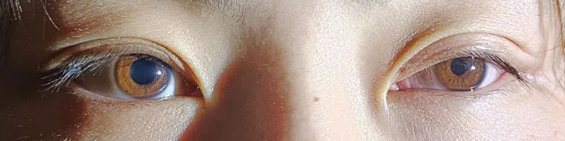
Anisocoria is a general term for a condition in which the pupil sizes of the left and right eyes differ. It is sometimes defined as a difference in pupil diameter of 0.4 mm or more 5).
Physiological anisocoria is present in about 20% of normal individuals, and the prevalence in the general population is reported to be approximately 19% 4). In physiological anisocoria, the difference in pupil size is usually 1 mm or less, the difference is nearly constant under both light and dark conditions, and the pupillary light reflex and near reflex are normal.
The causes can be broadly classified into the following two categories.
Disorders of sympathetic innervation: The pupil dilator muscle does not function adequately, resulting in insufficient dilation of the affected eye and noticeable miosis (e.g., Horner syndrome).
The causes of anisocoria range from benign physiological conditions to life-threatening conditions such as cerebral aneurysm, carotid artery dissection, and stroke, so appropriate differential diagnosis is essential.
QIs it a disease if the pupils are different sizes?
A
About 20% of normal individuals have physiological anisocoria; if the difference is 1 mm or less and the pupillary light reflex is normal, it is often not a problem. However, if it is acute onset or accompanied by headache, ptosis, diplopia, numbness, or weakness, urgent evaluation is necessary.
Xiao-Ming Li et al. Neuro-ophthalmic observation and 16-month follow-up of horner syndrome after thyroidectomy: A case report. Medicine. 2026 Jan 16; 105(3):e47236. Figure 1. PMCID: PMC12826322. License: CC BY.
Accommodation disorder: In parasympathetic disorders, contraction of the ciliary muscle is impaired, making near vision difficult.
Symptoms suggesting an emergency: If accompanied by headache, periorbital pain, diplopia, ptosis, vision loss, numbness, weakness, or ataxia, life-threatening conditions such as aneurysm, dissection, or stroke are possible, and prompt evaluation is necessary.
Changes in pupil diameter in light and dark conditions are essential for identifying the site of the lesion.
Anisocoria More Pronounced in Darkness
Sympathetic nerve disorder (Horner syndrome) is suggested. The affected eye’s pupil does not dilate sufficiently, and the difference from the healthy eye increases in darkness.
Dilation lag: The pupil of the affected eye dilates slowly even after entering a dark room.
Triad: Moderate miosis, mild ptosis, and apparent enophthalmos (narrowing of the palpebral fissure). The light reflex is normal, but dilation after constriction is delayed.
Decreased facial sweating: In postganglionic lesions, sweating on the affected side of the face and forehead is reduced.
Anisocoria More Pronounced in Light
Parasympathetic nerve disorder is suggested. The affected eye remains dilated, and the difference from the healthy eye increases in light.
Adie pupil: Moderate mydriasis and irregular shape. Segmental iris palsy and vermiform movements are characteristic. The light reflex is absent or weak, but the near response is slowly preserved (light-near dissociation).
Physiological anisocoria: Most common cause. Up to 20% of the population is affected, with a difference of 1 mm or less. It is thought to be due to temporary asymmetric supranuclear inhibition of the Edinger-Westphal nucleus.
Congenital aniridia: Occurs in 1 in 50,000 to 100,000 people. Caused by PAX6 gene mutations; 30% of sporadic cases develop Wilms tumor by age 5, requiring attention.
Drugs can cause mydriasis or miosis by acting directly or indirectly on the eye. A key feature is that it can occur regardless of the route of administration, including topical, oral, and transdermal.
The following table lists major drugs and their characteristics.
Drug
Route
Notes
Glycopyrrolate tablets
Oral (via CL)
Microparticles adhere to CL and transfer to one eye1). Mydriasis may persist up to 1 week.
Qbrexza (glycopyrronium) wipes
Topical
Improper hand hygiene leads to eye contact, resulting in 8 mm pupil diameter2).
Scopolamine transdermal patch
Transdermal
Contact with eye via fingers. Mydriatic effect may last up to 2 weeks4)
Azelastine 0.5% ophthalmic solution
Topical
Mydriasis despite being an H1 antihistamine. Recovery within 72 hours of discontinuation3)
Ipratropium bromide nebulizer
Inhalation
Eye exposure due to improper mask fitting. Recovery within 12 hours of discontinuation7)
Scopolamine powder (laboratory)
Inhalation/contact
Mydriasis + tachycardia, dizziness. Pilocarpine ineffective, recovery in 5 days4)
Other agents such as atropine, tropicamide, cyclopentolate, phenylephrine, and adrenaline eye drops also cause mydriasis. Angel’s trumpet (containing atropine, scopolamine, and hyoscyamine) can cause mydriasis even with inhalation exposure during mowing7). Agents that cause miosis include pilocarpine, brimonidine, prostaglandin analogs, opioids, and organophosphate insecticides.
Horner syndrome: Lesion of the sympathetic pathway (first to third-order neurons). Classified into central (stroke, Wallenberg syndrome), preganglionic (Pancoast tumor, mediastinal mass), and postganglionic (carotid artery dissection, cavernous sinus lesion). Raeder syndrome (trigeminal neuralgia + postganglionic Horner syndrome) requires evaluation for internal carotid artery aneurysm or middle cranial fossa tumor.
Adie tonic pupil: Damage to the ciliary ganglion or short ciliary nerves. 90% are women aged 20–40, 80% are unilateral, and 70% are accompanied by decreased deep tendon reflexes (Adie syndrome). It can also occur in Fisher syndrome, herpes zoster, neurosyphilis, spinocerebellar degeneration, and diabetes. In Ross syndrome, Adie pupil is combined with tendon reflex abnormalities, sweating abnormalities, and orthostatic hypotension.
Oculomotor nerve palsy: Compressive lesions (posterior communicating artery aneurysm, transtentorial herniation, tumor) usually involve pupillary abnormalities. Ischemic or diabetic types may have pupil sparing. Diabetic oculomotor nerve palsy is caused by atherosclerotic occlusion of nutrient vessels within the cavernous sinus and may be painful.
Dural arteriovenous fistula (CCF): Posterior drainage-type dural carotid-cavernous sinus fistula may present as isolated painful oculomotor nerve palsy as a “white-eyed carotid-cavernous sinus fistula” lacking typical anterior signs such as conjunctival injection and pulsatile proptosis6).
Pontine miosis: Bilateral severe miosis (pinpoint pupil, about 1 mm) due to pontine hemorrhage is a sign of poor prognosis.
Trigeminal autonomic cephalalgias and autoimmune autonomic ganglionopathy can also cause pupillary abnormalities.
Traumatic mydriasis (iridodialysis), paralytic mydriasis after acute angle-closure glaucoma attack, posterior synechiae due to uveitis, etc.
QCan over-the-counter eye drops or patches change pupil size?
A
Drug-induced mydriasis has been reported with antihistamine eye drops (e.g., azelastine)3), scopolamine transdermal patches for motion sickness4), and glycopyrronium wipes for hyperhidrosis2). All resolve spontaneously after discontinuing the causative agent, but it is important to inform the doctor.
Measurement of pupil diameter under both light and dark conditions is the most important examination. The normal pupil diameter is about 4 mm on average indoors (range 2–6 mm, with individual variation). In infants, it is smaller at 2–2.5 mm, and in the elderly, the pupils tend to be slightly constricted.
Pupil diameter measurement in light and dark: More pronounced in dark → sympathetic disorder; more pronounced in light → directly linked to differentiation of parasympathetic disorder.
Light reflex: Check the speed and degree of direct and consensual responses. The light source angle should be the same for both eyes.
Swinging flashlight test: Useful for detecting RAPD. Used for objective assessment of optic nerve damage. Note that if the cause of pure anisocoria is an afferent defect, anisocoria does not occur.
Performed when pharmacological testing is inconclusive or when there is clinical suspicion of aneurysm, dissection, or tumor. In acute Horner syndrome, it is appropriate to skip pharmacological testing and proceed directly to imaging. MRI/MRA is more useful than CT/CTA for detecting carotid-cavernous fistulas, and 3D time-of-flight MRA is superior for detecting flow signals within the venous sinus 6).
QIf anisocoria is found, what tests should be performed?
A
First, measurement of pupil diameter under both light and dark conditions is most important. Next, check the light reflex and near response, and perform pharmacological eye drop tests (e.g., apraclonidine, pilocarpine) as indicated to narrow down the cause. If an emergency condition such as aneurysm or dissection is suspected, proceed to head MRI/MRA without waiting for pharmacological testing.
The top priority in treatment is to rule out and appropriately manage underlying high-urgency conditions such as posterior communicating artery aneurysm, carotid artery dissection, and acute angle-closure glaucoma.
Benign/Physiological
Physiological anisocoria: No treatment needed. Only follow-up observation.
Adie pupil: Benign with a tendency for spontaneous recovery (pupil tends to become smaller over time). If photophobia is severe, use low-concentration pilocarpine (0.125–0.25%) eye drops, tinted glasses, or iris-colored contact lenses. For accommodative difficulties, reading glasses are useful.
Pharmacologic anisocoria: Resolves spontaneously upon discontinuation of the causative agent. When prescribing anticholinergic drugs to contact lens users, instruct them on thorough hand hygiene 1, 2).
Neurological Disorders
Horner syndrome: Treatment of the underlying disease (e.g., carotid artery dissection, Pancoast tumor) is prioritized. For benign causes, observation is sufficient.
Oculomotor nerve palsy: Compressive lesions due to posterior communicating artery aneurysm are indications for emergency surgery. In diabetic cases, treatment of diabetes is prioritized. Benign causes are observed.
Congenital aniridia: Photophobia is reduced with iris-colored contact lenses or tinted glasses. Artificial iris implantation is rarely indicated.
Mechanical anisocoria: Includes iridodialysis and posterior synechiae due to uveitis. Surgical correction of structural defects may be necessary.
QDoes Adie pupil require treatment?
A
Adie pupil is a benign condition with a tendency for spontaneous recovery, so observation alone is sufficient if asymptomatic. If photophobia is severe, low-concentration pilocarpine eye drops or sunglasses/tinted glasses can alleviate symptoms. However, it is necessary to rule out underlying systemic diseases such as Fisher syndrome or neurosyphilis.
The sympathetic pathway consists of three neurons.
First-order neuron: From the posterolateral hypothalamus descends through the brainstem to the ciliospinal center of Budge (C8–T2).
Second-order neuron: Exits the spinal cord, passes over the lung apex, and synapses at the superior cervical ganglion at the carotid bifurcation.
Third-order neuron: Ascends within the adventitia of the internal carotid artery, passes through the cavernous sinus, and innervates the dilator pupillae, Müller’s muscle of the upper eyelid, and the inferior tarsal muscle of the lower eyelid.
Parasympathetic Pathway (Controls Pupil Constriction and Accommodation)
Pretectal nucleus → bilateral Edinger-Westphal (EW) nuclei (anatomical basis for the consensual light reflex).
EW nucleus → parasympathetic fibers in oculomotor nerve → cavernous sinus → superior orbital fissure → synapse in ciliary ganglion.
Short ciliary nerves → innervate sphincter pupillae (5%) and ciliary muscle (95%).
The structural asymmetry, with accommodative fibers being 95 times more numerous than pupillary sphincter fibers, forms the basis for aberrant reinnervation after ciliary ganglion damage and the dissociation between light reflex and near response.
Supranuclear fibers for the near response to the EW nucleus run more ventrally than the midbrain pretectal area through which afferent fibers of the light reflex pass, so damage to the pretectal area causes dissociation between light reflex and near response (Argyll Robertson pupil).
Anticholinergics: inhibit muscarinic receptors (M3) → relaxation of sphincter pupillae → mydriasis and cycloplegia. Glycopyrrolate readily penetrates the cornea and exerts a more potent mydriatic effect than atropine even at low concentrations1).
Damage to the ciliary ganglion or short ciliary nerves → aberrant regeneration → upregulation of cholinergic receptors (denervation supersensitivity). Because accommodative fibers (95%) aberrantly reinnervate the sphincter pupillae, response to near stimuli is preserved but the light reflex is weak (light-near dissociation). Also, due to denervation supersensitivity, a normal pupil does not respond to low-concentration pilocarpine (0.1–0.125%), but an Adie pupil constricts.
Mechanism of oculomotor nerve palsy due to posterior drainage type carotid-cavernous fistula
Compression of the oculomotor nerve against the lateral wall of the cavernous sinus due to increased venous plexus pressure, venous stasis, and vascular steal are involved in a complex manner6). The oculomotor nerve runs along the lateral wall of the cavernous sinus, making it susceptible to compression by venous dilation, and the first branch of the trigeminal nerve runs along the epineurium of the oculomotor nerve, causing pain6).
7. Latest Research and Future Prospects (Investigational Reports)
For hyperhidrosis treatment, glycopyrrolate is available in conventional tablets as well as an orally disintegrating tablet (Dartisla ODT).
Adamkiewicz et al. (2024) pointed out that orally disintegrating tablets disintegrate faster than conventional tablets, potentially increasing the risk of direct eye contact with drug microparticles 1). They recommend thorough hand hygiene guidance when prescribing oral anticholinergics to contact lens wearers to prevent drug-induced anisocoria.
Awareness of Hyperhidrosis Medications and Avoidance of Unnecessary Tests
Qbrexza (glycopyrronium tosylate wipes) is a relatively new drug approved by the FDA in 2018, and lack of awareness among physicians may lead to unnecessary neuroimaging studies.
Sasher et al. (2024) highlighted through two cases the patient burden, cost, and unnecessary consumption of medical resources caused by performing neuroimaging in patients with anisocoria without a detailed medication history 2). They suggest that in patients using hyperhidrosis medications who present with sudden-onset anisocoria, drug-induced causes should be suspected first.
Limitations of the Pilocarpine Test and Diagnosis of Drug-Induced Mydriasis
In the standard diagnostic flow for drug-induced mydriasis, lack of response to high-concentration pilocarpine (1–2%) suggests drug-induced mydriasis. However, atypical responses have been reported with scopolamine-induced mydriasis.
Li et al. (2025) reported a case of drug-induced mydriasis from a scopolamine transdermal patch where temporary miosis occurred after pilocarpine (1–2%) instillation, followed by redilation 4). This finding suggests that the pilocarpine test is not always reliable for diagnosing drug-induced mydriasis, and a detailed medication history is essential.
Standardization of Preoperative Pupil Examination Protocols
Under general anesthesia, drug effects such as narcotics and muscle relaxants make interpretation of pupillary findings difficult.
Harada et al. (2023) reported a case where an undiagnosed Adie pupil was incidentally discovered after induction of general anesthesia, leading to cancellation of surgery due to suspected cerebrovascular disorder 5). They propose that incorporating standard pupil examination into preoperative systemic evaluation may prevent unnecessary surgery cancellations under anesthesia.
Adamkiewicz D, Magazin M, Thomas D. A case of pharmacologic anisocoria in systemic glycopyrrolate use from presumed local ocular inoculation. Neuro-Ophthalmology. 2024;48(1):41–45.
Sasher T, Bomar P, Feuer D, McDonald L. Anisocoria in patients with hyperhidrosis: a case series for the primary care physician. J Family Med Prim Care. 2024;13(2):797–799.
Ribeiro M, Teixeira-Martins R, Meira J. Pharmacologic anisocoria with azelastine: the importance of a good anamnesis. Cureus. 2024;16(3):e56649.
Li L, Lian L, Zhou R. Transdermal and powdered scopolamine-induced anisocoria: a report of two cases. Case Rep Ophthalmol. 2025;16:341–345.
Harada E, Sato-Boku A, Kanazawa M, Tachi N, Okuda M. A Case of Surgery Cancellation Following the Discovery of Anisocoria After Induction of General Anesthesia. Cureus. 2023;15(1):e33803. doi:10.7759/cureus.33803. PMID:36819394; PMCID:PMC9928752.
Mosleh R, Aung A, Saindane AM, Newman NJ, Biousse V. Carotid-cavernous fistula presenting as isolated painful anisocoria. Neuro-Ophthalmology. 2023;47(2):100–105.
Ekici A, Caglar B, Kara O, Oto A, Kilic N. Rare causes of anisocoria: Ipratropium bromide and Angel’s trumpet. Northern clinics of Istanbul. 2021;8(6):623-625. doi:10.14744/nci.2020.26428. PMID:35284801; PMCID:PMC8848498.
Copy the article text and paste it into your preferred AI assistant.
Article copied to clipboard
Open an AI assistant below and paste the copied text into the chat box.