Oculomotor nerve palsy is an eye movement disorder caused by damage to the third cranial nerve (oculomotor nerve). The oculomotor nerve innervates the following muscles:
Somatic extraocular muscles: medial rectus, superior rectus, inferior rectus, inferior oblique, and levator palpebrae superioris
Autonomic nerves: pupillary sphincter and ciliary muscle (parasympathetic fibers)
These disorders result in a combination of ptosis, limited eye movement, pupillary dilation, and accommodation disturbance 13. Paralysis is classified as complete or partial (superior branch palsy, inferior branch palsy). Oculomotor nerve palsy is the second most common cranial nerve palsy.
In a population-based study in Olmsted County (Fang et al. 2017), the age- and sex-adjusted annual incidence was 4.0 per 100,000, rising to 12.5 in those aged 60 and older. The frequency of causes was reported as follows 2.
In daily clinical practice, vascular causes (ischemia due to diabetes, hypertension, or arteriosclerosis) are the most common 12. In adults, paralytic strabismus due to circulatory disorders or trauma often resolves spontaneously, but in children, except for infections, brain tumors account for most cases, so it must be treated as an emergency. In children, congenital causes are the most common, followed by trauma 1.
QIs oculomotor nerve palsy a rare disease?
A
It is the second most common cranial nerve palsy and is encountered in daily clinical practice. In adults, ischemic causes are the most common, and special attention is needed in elderly patients, diabetics, and hypertensive patients.
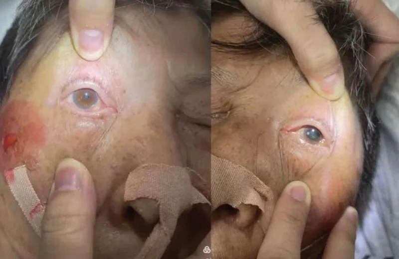
Cai Y, et al. Rare oculomotor nerve palsy after interventional treatment of anterior communicating artery aneurysm: A case report. Medicine (Baltimore). 2025. Figure 4. PMCID: PMC12599633. License: CC BY.
There is ptosis of one eye, and when the eyelid is lifted, mydriasis of the affected side can be confirmed. This photograph shows typical external ocular findings in oculomotor nerve palsy.
Diplopia: One of the most common ocular symptoms. Often presents with sudden onset of double vision.
Ptosis: Accounts for about 70% of initial symptoms. Complete paralysis of the levator palpebrae superioris results in complete closure of the palpebral fissure.
Eye pain/headache: May occur as a symptom of irritation in the first division of the trigeminal nerve. In aneurysms, particularly severe headache may be present.
Systemic neurological symptoms: Depending on the lesion site, hemiplegia, involuntary movements, or impaired consciousness may occur.
Findings differ between complete and partial palsy.
Complete Palsy
Eye position: Exotropia with mild hypotropia in primary gaze.
Eye movement: Limited adduction (does not cross midline), limited elevation, limited depression. If the trochlear nerve is intact, intorsion is observed during depression.
Pupil: Dilated, with loss of direct and consensual light reflex.
Partial Palsy
Superior branch palsy: Ptosis + superior rectus palsy (limited elevation). In cavernous sinus aneurysms, superior branch palsy is more common than inferior branch palsy.
Inferior branch palsy: Presents with a combination of pupillary fiber, inferior rectus, inferior oblique, and medial rectus involvement.
In oculomotor nerve palsy, a compensatory head posture may be adopted to avoid diplopia. If there is abduction deviation due to medial rectus palsy, the face is turned toward the affected side to maintain fusion.
QWhy is oculomotor nerve palsy with a dilated pupil (mydriasis) highly urgent?
A
The parasympathetic fibers of the oculomotor nerve (innervating the pupillary sphincter) run in the most superficial and dorsomedial layer of the nerve and are vulnerable to compression. Mydriasis suggests external compression by a posterior communicating artery aneurysm, which can rupture and cause fatal subarachnoid hemorrhage. See also the section on Causes and Risk Factors.
Vascular disorders (most common in daily practice): Ischemia due to diabetes, hypertension, and arteriosclerosis. Sudden onset, often noticed as double vision upon waking. Common in the elderly. Usually without mydriasis124.
Posterior communicating artery aneurysm (IC-PC aneurysm): The most important compressive lesion. Often presents with pupillary dilation as the first symptom. Rupture can be fatal and is a life-threatening emergency16.
Uncal herniation: Compression of the oculomotor nerve due to transtentorial herniation from increased intracranial pressure. Most common cause is intracranial hemorrhage.
Tumors: Lateral extension of pituitary tumors, meningiomas, etc. Approximately 11% in adults2.
Trauma: Approximately 12% in adults2. Often associated with severe head injury. Aberrant regeneration is common after trauma.
Inflammatory (Tolosa-Hunt syndrome): Painful ophthalmoplegia due to idiopathic granulomatous inflammation. Responds well to steroids7.
Diabetic neuropathy: Arteriosclerotic occlusion of nutrient vessels in the cavernous sinus. Microvascular ischemia is mainly due to hyalinization of arterioles and disruption of the blood-nerve barrier causing demyelination, which is reversible with remyelination4. Pupillary fibers have rich collateral circulation, so mydriasis is usually absent, but not always normal. Prognosis is good, with recovery within a few months24.
Main risk factors: diabetes, hypertension, vasculitis, infection, trauma, tumor, aneurysm.
QWhat are the characteristics of diabetic oculomotor nerve palsy?
A
Oculomotor nerve palsy due to diabetic neuropathy is caused by atherosclerotic occlusion of the nutrient vessels within the cavernous sinus. Ischemia affects the interior of the nerve, but because the pupillary fibers have abundant collateral circulation, it usually does not cause mydriasis. The prognosis is good, often recovering within a few months, and treatment of diabetes is the top priority.
History: Onset pattern (sudden or gradual), presence of eye pain or headache, history of diabetes or hypertension, diurnal variation of symptoms (important for differentiating from myasthenia gravis)
Eye movement evaluation: Limitation of movement in each direction, assessment of conjugate movements
Levator palpebrae superioris function: Assess whether ptosis is complete or partial
Pupillary light reflex and accommodation reflex: Presence or absence of direct and indirect light reflex loss
Slit-lamp examination: Check for intorsion of the eye during adduction and depression to differentiate from trochlear nerve palsy
Myasthenia gravis: Tensilon test (edrophonium 10 mg IV in 2.5 mg increments), ice test (apply ice pack to upper eyelid for 2 minutes, positive if improvement ≥2 mm, sensitivity 80–92%), assessment of diurnal variation, anti-AChR antibodies (positive rate ≤50% in ocular type)
QHow to differentiate oculomotor nerve palsy from myasthenia gravis?
A
Myasthenia gravis is characterized by diurnal variation (worsening in the evening), and the ice test (sensitivity 80–92%) and Tensilon test are useful for differentiation. Anti-AChR antibodies have a positive rate of ≤50% in ocular type, so a negative result does not rule it out. It is also important to exclude organic lesions of the oculomotor nerve and orbit on MRI/CT.
Treatment of the underlying cause is the highest priority. Treatment strategies according to the cause are shown below.
Ischemic
Natural course: Improvement usually begins within 4 weeks of onset, and complete recovery is expected within 12 weeks12. The 12-month complete recovery rate for microvascular palsy is approximately 91%, which is favorable, because demyelination due to hyalinized arterioles is reversible through remyelination4.
Pharmacotherapy: Oral vitamin B complex and circulation-improving drugs are given to promote recovery. Management of cardiovascular risk factors is also important4.
Aneurysm
Emergency management: Urgent neurosurgical treatment (clipping, coil embolization, etc.) is performed. In a report by Birchall et al., all 3 patients with posterior communicating artery aneurysms showed complete recovery of oculomotor nerve function within 1–18 days after endovascular coil embolization6.
Same-day imaging and urgent referral to a specialist are required.
Inflammatory (Tolosa-Hunt)
Steroid therapy: Prednisolone 50–60 mg/day for the first 3 days. Orbital pain improves dramatically within 24–72 hours, and cranial nerve palsy recovers over 2–8 weeks7.
Caution: Early dose reduction may cause relapse; taper gradually. Relapse occurs in up to 40% of cases7.
Traumatic
Observation: Recovery is relatively difficult. If no improvement after 6 months, consider surgical treatment5.
Diabetic: Prioritize diabetes treatment. Prognosis is good, and recovery often occurs within a few months.
Tumor / uncal herniation: Treat the underlying disease surgically or medically.
Management of residual deficits (symptomatic treatment)
Prism glasses: Consider after 6 months when symptoms have stabilized5.
Botulinum toxin: Chemical denervation by injection into the antagonist muscle temporarily reduces diplopia.
Strabismus surgery: Main goal is to correct eye alignment in primary position and reading position. In complete paralysis, combine supra-maximal recession, resection, and suturing of the lateral rectus to the lateral orbital wall periosteum, nasal transposition of the superior oblique toward the medial rectus insertion, and maximal shortening of the medial rectus5. If aberrant regeneration is present, avoid surgery on muscles with secondary reinnervation.
QHow long does it take for ischemic oculomotor nerve palsy to recover?
A
In many cases, improvement begins within 4 weeks after onset, and complete recovery is expected within 12 weeks (about 3 months). Vitamin B complex and circulation-improving drugs are administered to promote recovery. If no improvement is seen after more than six months, other causes such as trauma or management of residual deficits (strabismus surgery, prism glasses) should be considered.
6. Pathophysiology and Detailed Mechanism of Onset
The oculomotor nucleus is located in the tegmentum of the midbrain and has a complex nuclear structure. The innervation of each subnucleus is as follows.
Medial rectus, inferior rectus, and inferior oblique nuclei: ipsilateral innervation
Superior rectus nucleus: contralateral innervation (decussation site unknown)
Levator palpebrae superioris nucleus (caudal central nucleus): single nucleus innervates both sides
Autonomic nuclei (including Edinger-Westphal nucleus): located rostrally in the nuclear group, ipsilateral innervation
The fiber arrangement within the nucleus is rostral-caudal: most rostral are parasympathetic fibers, then inferior rectus and inferior oblique, and most caudal are levator palpebrae superioris and superior rectus. Mediolaterally, superior rectus and inferior oblique are lateral, while pupillary fibers and inferior rectus are medial.
Peripherally, pupillary fibers run in the most superficial layer of the oculomotor nerve, dorsomedially (superonasal), and are vulnerable to compression but relatively resistant to ischemia (due to rich collateral circulation). This anatomical characteristic forms the basis of the clinical rule: “compressive lesions → mydriasis, ischemic lesions → normal pupil” 13.
The oculomotor nerve and the posterior communicating artery run parallel in the subarachnoid space, making the nerve susceptible to compression by aneurysms. In particular, pupillary fibers are located medially among the oculomotor nerve fibers and are closest to the posterior communicating artery, so pupillary dilation often presents as an initial symptom 1.
Subarachnoid space: Compression by posterior communicating artery aneurysm is most important. Compression due to uncal herniation also occurs. Because pupillary fibers run in the most superficial layer, mydriasis appears early 1.
Cavernous sinus: Combined palsy with other cranial nerves (IV, V1, VI) is common (cavernous sinus syndrome). After the oculomotor nerve divides into superior and inferior branches, branch palsy is more likely 3.
Orbit: Accompanied by visual loss, ophthalmoplegia, and proptosis. Branch palsy is more likely 3.
Modi P, Singh J. Cranial Nerve III Palsy (Oculomotor Palsy). In: StatPearls [Internet]. Treasure Island (FL): StatPearls Publishing; 2026. PMID: 30252368. https://www.ncbi.nlm.nih.gov/books/NBK526112/
Fang C, Leavitt JA, Hodge DO, Holmes JM, Mohney BG, Chen JJ. Incidence and Etiologies of Acquired Third Nerve Palsy Using a Population-Based Method. JAMA Ophthalmol. 2017;135(1):23-28. doi:10.1001/jamaophthalmol.2016.4456. PMID: 27893002. PMCID: PMC5462106.
Shree R, Mahesh KV, Balaini N, Goel A. Oculomotor Cranial Neuropathies: Diagnosis and Management. Ann Indian Acad Neurol. 2022;25(Suppl 2):S70-S82. doi:10.4103/aian.aian_167_22. PMID: 36589037. PMCID: PMC9795710.
Galtrey CM, Schon F, Nitkunan A. Microvascular Non-Arteritic Ocular Motor Nerve Palsies—What We Know and How Should We Treat? Neuroophthalmology. 2014;39(1):1-11. doi:10.3109/01658107.2014.963252. PMID: 27928323. PMCID: PMC5123092.
Singh A, Bahuguna C, Nagpal R, Kumar B. Surgical management of third nerve palsy. Oman J Ophthalmol. 2016;9(2):80-86. doi:10.4103/0974-620X.184509. PMID: 27433033. PMCID: PMC4932800.
Birchall D, Khangure MS, McAuliffe W. Resolution of third nerve paresis after endovascular management of aneurysms of the posterior communicating artery. AJNR Am J Neuroradiol. 1999;20(3):411-413. PMID: 10219405. PMCID: PMC7056059.
Dutta P, Anand K. Tolosa-Hunt Syndrome: A Review of Diagnostic Criteria and Unresolved Issues. J Curr Ophthalmol. 2021;33(2):104-111. doi:10.4103/joco.joco_134_20. PMID: 34409218. PMCID: PMC8365592.
Copy the article text and paste it into your preferred AI assistant.
Article copied to clipboard
Open an AI assistant below and paste the copied text into the chat box.