Canaliculitis is a chronic infectious inflammation of the lacrimal canaliculi (superior and inferior) that make up the lacrimal drainage system. The most common causative agent is Actinomyces israelii, which forms concretions (dacryoliths) within the canaliculus. Rarely, it can also be caused by bacteria such as Propionibacterium, Streptococcus, or fungi (Aspergillus, Candida).
The lacrimal drainage system is a continuous structure from the punctum to the canaliculi (superior and inferior), common canaliculus, lacrimal sac, and nasolacrimal duct. Canaliculitis is an infection localized to the canaliculi within this system. It commonly occurs in the inferior canaliculus and is more frequent in middle-aged and older women.
Patients often present to the ophthalmology clinic with unilateral refractory conjunctivitis, and it may take time to reach an appropriate diagnosis and treatment. Recognizing the characteristic changes of the punctum (dilation, redness, expressible discharge) is key to early diagnosis.
Actinomycotic (most common)
Causative agent: Actinomyces israelii
Anaerobic, gram-positive filamentous bacteria. It is best adapted to the anaerobic environment within the canaliculus and forms sulfur granules. The dacryolith grows as calcium deposits on the actinomycete colony.
Nocardia is classified as an aerobic actinomycete. Fusobacterium is an anaerobic bacterium, and these can also form dacryoliths.
Fungal
Causative organisms: Aspergillus, Candida
Although rare, it tends to be refractory. Additional treatment with antifungal agents (e.g., pimaricin eye drops) may be necessary.
QWhat symptoms most commonly lead to the discovery of canaliculitis?
A
The main complaints are unilateral conjunctival injection, discharge, and epiphora. It is often suspected when a diagnosis of chronic conjunctivitis is made and does not improve with antibiotic eye drops. Characteristic findings include redness and dilation of the punctum (punch-out appearance), and purulent or sulfur granule-like discharge is expressed when the punctum is compressed. This expression finding is a clue to diagnosis.
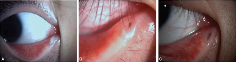
Zheng Q, Shen T, Luo H, et al. Application of lacrimal endoscopy in the diagnosis and treatment of primary canaliculitis. Medicine (Baltimore). 2019;98(33):e16789. Figure 4. PMCID: PMC6831237. DOI: 10.1097/MD.0000000000016789. License: CC BY 4.0.
Preoperative slit-lamp image showing marked swelling around the lower punctum with purulent discharge and obstruction of the punctum opening (left), and image at 3 months postoperatively showing complete resolution of swelling and discharge (right). This corresponds to the punctum dilation (punch-out appearance) and purulent discharge discussed in the section “Main Symptoms and Clinical Findings.”
The main complaints are conjunctival injection, discharge, and epiphora, similar to unilateral chronic conjunctivitis. In most cases, the lesion occurs in the lower canaliculus. Initially, antibiotic eye drops are prescribed for chronic conjunctivitis, but they do not improve, and the course is often refractory.
Discharge of sulfur granule-like material upon compression of the lacrimal punctum is a finding with high specificity for canaliculitis, and if this finding is confirmed, the clinical diagnosis is almost certain.
A fungal mass observed as a yellow to white granular material with the naked eye, formed by the deposition of calcium salts on actinomycete colonies. The term “sulfur” refers to its appearance and color, not actual sulfur. Under microscopy, a dense structure of filamentous actinomycete hyphae can be observed.
A stone-like substance formed when sulfur granules remain and grow within the lacrimal canaliculus. The presence of a concretion in the canaliculus leads to persistent chronic inflammation and is a reason why antibiotic therapy alone may not cure the condition.
It is more common in middle-aged and older women, and patients often present to ophthalmology clinics with unilateral refractory conjunctivitis. It is known to occur preferentially in the lower lacrimal canaliculus. Although precise demographic data are limited, differentiation from chronic conjunctivitis is important in patients presenting with epiphora and discharge.
In cases of lacrimal duct obstruction, stones (dacryoliths) frequently coexist within the lacrimal passages. According to the guidelines for lacrimal endoscopy, dacryocystoliths are reported to occur in 7.5% of nasolacrimal duct obstruction cases 1), indicating a predisposition to stone formation throughout the lacrimal system.
There are cases where punctal plugs used for dry eye treatment migrate into the lacrimal canaliculus, causing foreign body reaction and inflammation that leads to canaliculitis 1). In patients with a history of punctal plug placement who present with refractory conjunctivitis, canaliculitis due to plug migration should be considered.
When unilateral refractory conjunctivitis persists, it is important to actively suspect canaliculitis. The diagnostic procedure in an outpatient setting is as follows.
Step 1: Visual inspection of the punctum
Check for redness and dilation (punch-out appearance) of the punctum.
Step 2: Punctal compression test
Compress the punctum along the canaliculus to check for discharge. If purulent or sulfur granule-like discharge is expressed, a clinical diagnosis of canaliculitis is established.
Step 3: Lacrimal irrigation
Perform lacrimal irrigation. In canaliculitis, obstruction of the lacrimal passage is often absent. Since dacryocystitis is often accompanied by obstruction, patency testing is useful for differentiation.
Step 4: Microbiological examination
Collect purulent discharge expressed from the canaliculus and perform Gram staining, smear microscopy, and anaerobic culture to identify the causative organism.
Gram-positive, branching filamentous organisms (characteristic of Actinomyces)
Allows rapid presumptive diagnosis of actinomycotic infection
Anaerobic culture
Culture under anaerobic conditions
Actinomyces are anaerobic, so anaerobic culture is necessary
Aerobic culture
Standard culture
Detection of Nocardia, Candida, Aspergillus, etc.
The smear microscopy findings of Actinomyces are characteristic, and the presumptive diagnosis of actinomycotic canaliculitis can be made by identifying Gram-positive branched filamentous organisms.
Dacryoendoscopy allows direct visualization of concretions and inflammatory findings within the canaliculus. It is useful for confirming the exact location and size of concretions and for verifying complete removal 1). Removal of concretions under dacryoendoscopic guidance enables direct visualization and contributes to preventing postoperative recurrence 2).
QHow do you differentiate canaliculitis from dacryocystitis?
A
Canaliculitis is characterized by swelling around the punctum and punctal dilation, and lacrimal irrigation often shows no obstruction. In contrast, dacryocystitis primarily involves swelling of the lacrimal sac area (below the inner canthus), and pressure on the sac causes pus to reflux from the punctum, with obstruction confirmed on irrigation. The presence or absence of local punctum findings (dilation, discharge on pressure) is the key differentiating feature.
Complete physical removal of the fungal mass (dacryolith) within the canaliculus is the cornerstone of treatment. The dacryolith is a dense fungal mass into which antibiotics penetrate poorly; antibiotic eye drops alone cannot remove the mass and will not lead to cure. Surgical incision and curettage are essential.
Step 1: Canaliculotomy and Removal of Fungal Mass
Perform canaliculotomy under local anesthesia.
Thoroughly scrape the contents of the lacrimal canaliculus with a curette to completely remove fungal masses and dacryoliths. Residual fungal masses are the main cause of recurrence, so complete removal is most important.
When using a lacrimal endoscope, it is possible to remove dacryoliths and confirm complete removal under direct visualization 1).
Step 2: Postoperative antibiotic eye drops and lacrimal canalicular irrigation
Continue new quinolone eye drops postoperatively.
Administer levofloxacin ophthalmic solution 0.5% (Cravit®) 4 to 6 times a day. Irrigation of the lacrimal canaliculus with new quinolone eye drops is also useful as postoperative management.
In cases of fungal canaliculitis, consider adding antifungal agents (e.g., pimaricin eye drops).
Step 3: Systemic antibiotics and follow-up
For actinomycotic infections, administer systemic penicillin for several weeks.
Continue amoxicillin 250–500 mg (e.g., Sawacillin®) three times a day for several weeks. Perform repeated lacrimal irrigation postoperatively to prevent recurrence. Adjust antibiotics appropriately based on culture results.
Using a lacrimal endoscope allows direct visualization and removal of the dacryoliths at their exact location. It enables confirmation of complete removal and is considered useful for preventing recurrence2).
Management of migrated punctal plugs
If the cause is a migrated punctal plug, removal via punctoplasty or attempted expulsion into the nasal cavity under lacrimal endoscopy is performed1).
For recurrence due to incomplete removal of fungal mass, reoperation (re-incision and curettage) is performed. If lacrimal duct obstruction progresses, lacrimal tube intubation or DCR (dacryocystorhinostomy) may be necessary.
QCan it be cured with antibiotic eye drops alone?
A
Usually, eye drops alone are not curative. The fungal mass (dacryolith) formed in the canaliculus has a dense structure, making it difficult for antibiotics to reach the organisms inside. The mainstay of treatment is physical complete removal of the fungal mass by incising the canaliculus under local anesthesia and using a sharp spoon. Postoperatively, combining antibiotic eye drops (new quinolones) with systemic administration (penicillins for actinomycosis) increases the cure rate.
QDoes canaliculitis recur?
A
If the fungal mass is completely removed, the prognosis is good and recurrence is rare. Conversely, if removal is incomplete and dacryoliths remain in the canaliculus, recurrence is likely. Using lacrimal endoscopy to confirm complete removal of the dacryolith is useful for preventing recurrence. In case of recurrence, re-incision and curettage are performed.
The canaliculus begins at the punctum and runs horizontally along the eyelid margin. The superior and inferior canaliculi are each about 8–10 mm long, merge to form the common canaliculus, and then open into the lacrimal sac. The lumen of the canaliculus is flat, with a normal diameter of about 0.5–1 mm.
The lumen of the canaliculus has limited communication with the outside air, making it prone to an anaerobic environment. Actinomyces israelii is an obligate or facultative anaerobe that adapts and proliferates easily in this environment. This anatomical characteristic allows actinomycetes, which do not grow well in open areas (skin, conjunctiva), to colonize preferentially in the canaliculus.
Sulfur Granule Formation and Progression to Dacryolith
Colonization by Actinomyces: Actinomyces israelii colonizes the canaliculus.
Formation of sulfur granules: Actinomyces form dense colonies during metabolic activity, creating bacterial aggregates called sulfur granules. They are named for their yellow-white, granular appearance resembling sulfur.
Calcium deposition: Calcium salts deposit on the colonies, which grow into concretions (dacryoliths).
Persistent chronic inflammation: The concretion remains in the canaliculus, continuously irritating surrounding tissues and causing inflammation.
Granulation tissue formation: Chronic inflammation leads to granulation tissue formation on the canalicular wall.
Mechanism of antibiotic eye drop monotherapy ineffectiveness
The structure of a concretion is close to a solid mass with calcium deposited in dense colonies, making it difficult for antibiotic molecules to reach bacteria inside the aggregate. Additionally, the biofilm-like structure on the surface hinders drug penetration. Therefore, antibiotic eye drops alone can only partially eliminate surface bacteria but cannot remove the entire aggregate, leading to regrowth and recurrence after discontinuation. This is why surgical removal of the aggregate is the only definitive treatment.
Dacryoendoscopy is a technique that inserts a thin fiberscope into the lacrimal passage to directly visualize the lumen 1). In canaliculitis, it allows direct confirmation of the presence, location, and size of concretions, enabling verification of complete removal 2). Compared to conventional blind canalicular incision and curettage, more reliable removal of concretions is expected. According to the guidelines for dacryoendoscopy, complete removal of concretions is directly linked to cure in canaliculitis with lacrimal stones 1), and the spread of endoscopic technology contributes to improved treatment outcomes.
Cases have been reported where migration of punctal plugs, widely used for dry eye treatment, causes canaliculitis 1). After the plug falls into the canaliculus, it acts as a foreign body, becoming a nidus for inflammation and infection. In patients with punctal plugs who develop refractory conjunctivitis, this condition should be considered. Management options include removal via punctoplasty or endoscopic transnasal drainage.
Accumulation of comparative data on approaches for lacrimal canaliculotomy (one-snip vs two-snip method)
Establishment of optimal antifungal drug protocols for fungal canaliculitis
Quantitative evaluation of long-term recurrence rates and surgical outcomes
Comprehensive identification of causative bacteria by next-generation sequencing (16S rRNA analysis) and investigation of optimal treatment by bacterial species
Ali MJ, Alam MS, Naik MN. Dacryoendoscopic features in a case of canaliculitis with concretions. Ophthalmic Plast Reconstr Surg. 2017;33:228-229. doi:10.1097/iop.0000000000000864.
Copy the article text and paste it into your preferred AI assistant.
Article copied to clipboard
Open an AI assistant below and paste the copied text into the chat box.