Lagophthalmos is a condition in which blinking and eyelid closure are incomplete, leaving the eyeball exposed. The term originates from the Greek word “lagos” (hare), named after the habit of hares sleeping with their eyes open.
Exposure of the eyeball disrupts the tear film, leading to dry eye and corneal infection. In advanced cases, it can lead to corneal perforation and blindness. Conjunctival hyperemia and eye pain also occur 2).
Lagophthalmos is broadly classified into the following four types.
The most common cause is paralytic, among which facial nerve palsy (especially Bell’s palsy) accounts for the majority. In paralytic lagophthalmos, paralysis of the orbicularis oculi muscle causes the upper eyelid margin to be elevated with skin laxity (pseudoptosis), and the lower eyelid to become droopy or everted, resulting in incomplete eyelid closure.
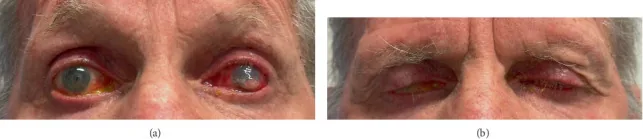
Richardson P, et al. Severe Microbial Keratitis Secondary to Prostaglandin-Associated Periorbitopathy. Case Rep Ophthalmol Med. 2025;2025:5635118. DOI: 10.1155/crop/5635118. Figure 1. License: CC BY. PMCID: PMC12527608.
At maximum opening (left), conjunctival injection in both eyes and a corneal ulcer in the lower left eye are observed. At maximum effort to close (right), incomplete eyelid closure with exposure of the lower cornea and conjunctiva is clearly shown. This corresponds to the incomplete eyelid closure and exposure keratopathy discussed in the section “Main Symptoms and Clinical Findings.”
As for corneal findings, corneal epithelial damage due to incomplete eyelid closure is characterized by being compartmental in the lower part, with almost no epithelial damage above a certain height. When compartmental corneal epithelial damage limited to the lower part is observed, incomplete eyelid closure or nocturnal lagophthalmos should be strongly suspected.
Bell’s phenomenon (upward rotation of the eyes during sleep) normally protects the cornea, but when eyelid closure is severely incomplete, the lower cornea becomes exposed, leading to inferior corneal damage due to nocturnal lagophthalmos. Nocturnal lagophthalmos often presents with few daytime symptoms, with ocular symptoms appearing in the morning 2).
Thyroid eye disease (Graves’ disease): Severe proptosis can lead to inability to close the eyelids.
Orbital tumor/Orbital hematoma: Mechanical lagophthalmos due to forward displacement of the globe.
High myopia: Axial elongation may cause proptosis leading to lagophthalmos.
Physiological (nocturnal lagophthalmos)
Also present in healthy individuals: During sleep, voluntary eyelid closure is absent, and Bell’s phenomenon exposes the lower cornea.
Prevalence: Observed in a certain proportion of healthy individuals. Symptoms are more likely to become apparent in dry environments or with use of sleeping pills.
If the patient complains of morning eye pain or photophobia, suspect nocturnal lagophthalmos.
Risk factors for Bell’s palsy include diabetes, pregnancy, and immunodeficiency. Lagophthalmos frequently occurs after facial nerve palsy, and the risk of corneal complications increases with the severity of palsy10). In a study of 2,500 cases of Bell’s palsy, complete recovery within 6 months was reported in 71% of patients1).
The following assessments are performed for eyelid closure evaluation:
Measure and record the residual palpebral fissure (mm) during strong and gentle eyelid closure
Observation not only in the sitting position but also in the supine position: Incomplete closure may become more apparent in the supine position
Evaluation of unconscious blinking: The degree of closure differs between voluntary closure and blinking
Confirmation of lagophthalmos during sleep: Ask family members or partners to take a photo of the eyes during sleep with a mobile device for comparison
Detects sectoral epithelial damage in the lower cornea. Also used to assess the area of exposed cornea. Schirmer test is performed to evaluate concomitant dry eye.
Evaluation of Facial Nerve Palsy Using the House-Brackmann Classification
Eyelid retraction: In thyroid eye disease, lagophthalmos is accompanied by eyelid retraction. Eyelid retraction is a condition where the upper eyelid margin is above the corneal limbus.
Blepharospasm: A disease characterized by increased eyelid closure due to involuntary contraction of the orbicularis oculi muscle, opposite to lagophthalmos.
Pseudoptosis in Bell’s palsy: The skin becomes lax due to orbicularis oculi muscle paralysis, appearing ptotic, but levator function is normal.
QIs treatment necessary if the eyes do not close only at night?
A
Even nocturnal lagophthalmos alone can cause corneal epithelial damage. Corneal protection is achieved by applying ophthalmic ointment or taping before sleep. If symptoms persist, an ophthalmology consultation is recommended. If eye pain or photophobia occurs in the morning, strongly suspect nocturnal lagophthalmos and confirm inferior corneal epithelial damage with fluorescein staining.
Artificial tears: Sodium hyaluronate 0.1% eye drops, diquafosol sodium 3% eye drops, etc., are instilled frequently (4 to 6 or more times a day).
Ophthalmic ointments: Antibiotic ophthalmic ointments (e.g., ofloxacin ophthalmic ointment) are applied before bedtime. They are also expected to provide physical protection by keeping the eyelids closed.
Example daytime prescription: Artificial tears are instilled frequently, and ophthalmic ointment is applied as needed.
At bedtime: Apply ophthalmic ointment and perform taping as needed.
Taping:
Condition
Taping method
Mild (when eyelid opening is needed during the day)
Tape in two directions: lifting the eyebrow upward and pulling the lower eyelid upward and outward toward the ear.
Severe (nighttime or strong corneal epithelial damage)
After applying ophthalmic ointment, tape vertically from the upper eyelid to the lower eyelid while looking downward to achieve complete eyelid closure.
Moisture chamber glasses: Attach a moisture chamber to the eyeglass frame to maintain humidity around the eyes and reduce corneal dryness.
Gold plate insertion (lid loading): Insert a 0.8–1.6 g gold or platinum plate onto the anterior surface of the upper eyelid tarsal plate. This assists eyelid closure by gravity and is most effective in upright and sitting positions. Complications include foreign body sensation, migration, infection, and plate visibility 3, 4, 5).
Upper eyelid levator muscle lengthening: An auxiliary surgery for orbicularis oculi muscle paralysis.
Fascial graft: Reinforcement of orbicularis oculi function using temporal fascia, etc.
Lateral tarsal strip: Corrects lower eyelid laxity and ectropion. Shortens the lateral lower eyelid to restore tension.
Wedge resection: Corrects laxity by shortening the lower eyelid.
Conchal cartilage graft: Reinforces the supporting tissue of the posterior lamella of the lower eyelid.
Tarsorrhaphy:
Partial suturing of the eyelids laterally (lateral tarsorrhaphy) or medially to reduce the area of exposed ocular surface. Useful for corneal protection in the acute phase, performed temporarily or permanently. If recovery of facial nerve palsy is expected, reversible temporary tarsorrhaphy is chosen6).
In thyroid eye disease, symptomatic treatment with artificial tears and eye ointment stabilizes the ocular surface, and orbital decompression after the inflammatory phase resolves the root cause. For orbital tumors, tumor excision is curative.
Acute phase (within 72 hours of onset) pharmacotherapy:
Steroids: Start with prednisolone 60 mg/day and taper. Early administration has been reported to improve recovery rates in RCTs 8)
Antiviral drugs: Valacyclovir (1,000 mg/day for 7 days) is used in combination with steroids 9)
Ophthalmologic management:
Protect the cornea with artificial tears, eye ointment, and taping from the acute phase
Initiate aggressive protection for HB grade IV or higher
If residual paralysis is fixed after more than 6 months of follow-up, consider surgical intervention 6, 10)
QWhen lagophthalmos occurs due to facial nerve palsy, what should be done first?
A
First, protect the cornea with frequent instillation of artificial tears and application of eye ointment before bedtime. Taping to assist eyelid closure is also effective. If corneal epithelial damage progresses, promptly consult an ophthalmologist. In Bell’s palsy, early administration of steroids and antiviral drugs is important for nerve recovery, and priority should be given to visiting an otolaryngologist or neurologist within 72 hours of onset.
QHow long does the effect of gold plate surgery last?
A
It functions permanently, but rarely, plate migration or infection may occur, requiring reoperation. Platinum chains are said to have better flexibility and lower risk of migration compared to gold plates 12). Note that since it assists eyelid closure using gravity, its effect is reduced in the supine position (during sleep).
QHow long does it take for lagophthalmos to heal?
A
It varies greatly depending on the cause. In Bell’s palsy, 70–85% recover completely with appropriate treatment, but it usually takes 3–6 months 1). If there is no improvement after 6 months, surgical intervention is considered. For cicatricial or mechanical lagophthalmos, treatment of the underlying disease is fundamental, and spontaneous recovery is often not expected.
6. Pathophysiology and Detailed Mechanism of Onset
The facial nerve (cranial nerve VII) runs through the temporal bone, exits the stylomastoid foramen, and branches into multiple peripheral branches within the parotid gland. The orbicularis oculi muscle is innervated by the temporal and zygomatic branches, and its contraction closes the eyelid. In Bell’s palsy, edema and ischemia within the facial nerve canal are the main causes of nerve damage 7).
Paralytic: Facial nerve (VII) palsy → orbicularis oculi muscle paralysis → incomplete eyelid closure. In the upper eyelid, skin laxity occurs, giving the appearance of pseudoptosis. In the lower eyelid, drooping and ectropion occur, worsening the incomplete closure.
Cicatricial: Scar contracture of the anterior lamella (skin and orbicularis muscle) physically prevents eyelid closure. It may progress rapidly after thermal or chemical burns.
Mechanical: Proptosis prevents the eyelids from covering the eye. In thyroid eye disease, edema and fibrosis of orbital fat and extraocular muscles push the eyeball forward.
Nocturnal Lagophthalmos (Physiologic): Voluntary eyelid closure disappears during sleep, and physiologic upward eye rotation (Bell’s phenomenon) exposes the lower cornea. It becomes apparent when complete closure is not possible without voluntary orbicularis contraction.
In Bell’s palsy, 70–85% recover completely with appropriate treatment (steroids + antivirals). If residual palsy persists for more than 6 months, surgical treatment for fixed paralytic lagophthalmos is considered. In thyroid eye disease, after symptomatic treatment during the inflammatory (active) phase, orbital decompression and eyelid surgery are performed during the non-inflammatory (stable) phase. Severe corneal exposure carries a risk of corneal perforation and may require emergency procedures such as tarsorrhaphy 6).
Nerve Reconstruction:
For irreversible facial nerve palsy, nerve transfer such as masseter-to-facial nerve transfer has been reported. A comparison between the masseter nerve and hypoglossal nerve groups showed better recovery of spontaneous facial movement in the former 13). Crossface nerve graft (facial nerve to facial nerve anastomosis) is excellent for restoring symmetry of facial expression but requires time for nerve regeneration.
Platinum Chain Lid Loading:
Developed as an improved version of the gold plate. Comparative studies show superior flexibility, less cosmetic discomfort during eyelid opening, and lower risk of migration 12).
Botulinum toxin injection:
This method induces temporary ptosis by injecting botulinum toxin into the levator palpebrae superioris muscle, thereby assisting eyelid closure. It may be positioned as a temporary measure during the acute phase of Bell’s palsy.
Artificial nerve grafting / stem cell therapy:
Basic research aimed at facial nerve regeneration is ongoing. Although not yet clinically applied, it is attracting attention as a future therapeutic strategy.
Peitersen E. Bell’s palsy: the spontaneous course of 2,500 peripheral facial nerve palsies of different etiologies. Acta Otolaryngol Suppl. 2002;(549):4-30. doi:10.1080/000164802760370736.
Seiff SR, Sullivan JH, Freeman LN, Ahn J. Pretarsal fixation of gold weights in facial nerve palsy. Ophthalmic plastic and reconstructive surgery. 1989;5(2):104-9. doi:10.1097/00002341-198906000-00005. PMID:2487203.
Kartush JM, Linstrom CJ, McCann PM, Graham MD. Early gold weight eyelid implantation for facial paralysis. Otolaryngology—head and neck surgery : official journal of American Academy of Otolaryngology-Head and Neck Surgery. 1990;103(6):1016-23. doi:10.1177/019459989010300622. PMID:2126116.
Terzis JK, Kyere SA. Experience with the gold weight and palpebral spring in the management of paralytic lagophthalmos. Plastic and reconstructive surgery. 2008;121(3):806-815. doi:10.1097/01.prs.0000299919.18076.b4. PMID:18317130.
Rahman I, Sadiq SA. Ophthalmic management of facial nerve palsy: a review. Surv Ophthalmol. 2007;52(2):121-144. doi:10.1016/j.survophthal.2006.12.009.
Ioannis Mavrikakis. Facial Nerve Palsy: Anatomy, Etiology, Evaluation, and Management. Orbit. 2008;27(6):466-474. doi:10.1080/01676830802352543.
Sullivan FM, Swan IR, Donnan PT, et al. Early treatment with prednisolone or acyclovir in Bell’s palsy. N Engl J Med. 2007;357(16):1598-1607.
Engström M, Berg T, Stjernquist-Desatnik A, Axelsson S, Pitkäranta A, Hultcrantz M, et al. Prednisolone and valaciclovir in Bell’s palsy: a randomised, double-blind, placebo-controlled, multicentre trial. The Lancet. Neurology. 2008;7(11):993-1000. doi:10.1016/S1474-4422(08)70221-7. PMID:18849193.
Hohman MH, Hadlock TA. Etiology, diagnosis, and management of facial palsy: 2000 patients at a facial nerve center. Laryngoscope. 2014;124(7):E283-E293.
House JW, Brackmann DE. Facial nerve grading system. Otolaryngol Head Neck Surg. 1985;93(2):146-147. doi:10.1177/019459988509300202.
Bladen JC, Norris JH, Malhotra R. Cosmetic comparison of gold weight and platinum chain insertion in primary upper eyelid loading for lagophthalmos. Ophthalmic plastic and reconstructive surgery. 2012;28(3):171-5. doi:10.1097/IOP.0b013e3182467bf7. PMID:22460670.
Hontanilla B, Marré D. Comparison of hemihypoglossal nerve versus masseteric nerve transpositions in the rehabilitation of short-term facial paralysis using the Facial Clima evaluating system. Plastic and reconstructive surgery. 2012;130(5):662e-672e. doi:10.1097/PRS.0b013e318267d5e8. PMID:23096620.
Copy the article text and paste it into your preferred AI assistant.
Article copied to clipboard
Open an AI assistant below and paste the copied text into the chat box.