When blunt external force is applied to the eye, the orbital bones may fracture. The orbital floor and medial wall are very thin and are common sites for this condition. It is called an orbital blowout fracture because the bone is displaced significantly toward the paranasal sinuses.
Approximately 10% of all facial fractures are isolated orbital wall fractures, and the orbit is involved in 30–40% of all facial fractures. 2) Isolated orbital floor fractures account for 22–47% of all orbital fractures. 2)
In terms of sex and age distribution of injured patients (analysis of 268 cases), 72% were male and 28% female, with an average age of 36 years. 2)
The orbital floor and medial wall are particularly thin and prone to fracture.
Site
Bone thickness characteristics
Orbital floor (infraorbital nerve canal area)
Approximately 0.23 mm (extremely thin)
Orbital floor (posteromedial area)
Average 0.37 mm
Orbital floor (lateral area)
Average 1.25 mm
Medial orbital wall (lamina papyracea)
Thinnest among orbital walls
In medial orbital wall fractures, the lamina papyracea is damaged. Isolated occurrence is relatively rare, and it often occurs together with orbital floor fractures or as part of a complex fracture.
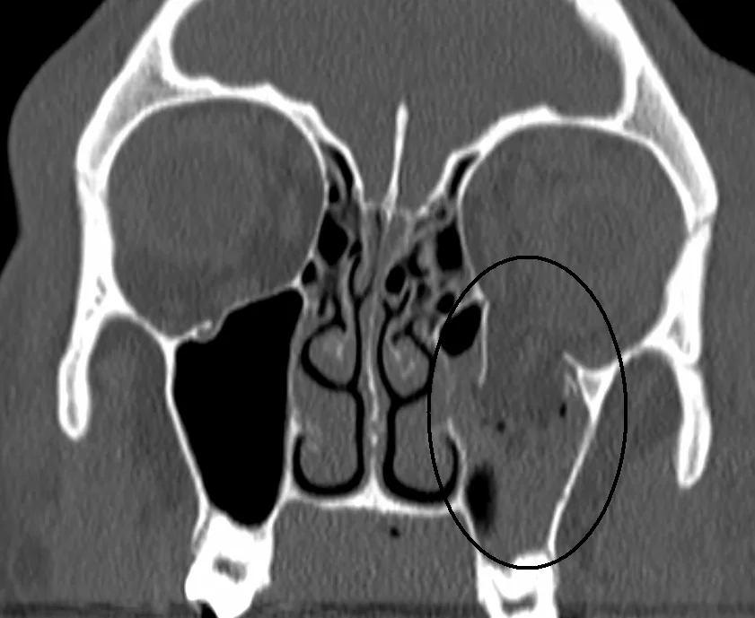
Coronal CT (bone window) shows a black circle indicating the fracture site of the left orbital floor. Orbital fat herniates into the maxillary sinus through the fracture opening. This corresponds to the herniation of orbital contents into the paranasal sinuses in open-type fractures discussed in section “1. What is orbital blowout fracture?”
Fractures are classified into open and closed types based on imaging. In open-type fractures, bone fragments and soft tissues are significantly displaced into the paranasal sinuses. In contrast, closed-type fractures show subtle imaging changes despite prominent clinical symptoms, and the missing rectus sign, where the entrapped extraocular muscle appears to disappear at the fracture site, is characteristic. Closed-type (trapdoor) fractures are more common in young individuals due to higher bone elasticity.
QAre orbital floor fracture and medial orbital wall fracture different diseases?
A
Both are types of orbital blowout fracture and are not separate diseases. Orbital floor fractures mainly cause vertical eye movement disorders and hypoesthesia of the cheek and upper lip. Medial orbital wall fractures primarily cause horizontal eye movement disorders, epistaxis, and subcutaneous emphysema. Combined fractures are also common.
Diplopia: Associated with eye movement disorders. It worsens with vertical movement in orbital floor fractures and with horizontal movement in medial wall fractures.
Enophthalmos: Progresses over days to weeks after injury as swelling subsides.
Eye pain: Occurs with eye movement; the direction (vertical or horizontal) depends on the fracture site.
Hypoesthesia or paresthesia: In inferior wall fractures involving the infraorbital groove, paralysis of the second branch of the trigeminal nerve causes sensory disturbance on the affected cheek and upper lip.
Epistaxis: Blood accumulated in the paranasal sinuses due to fracture flows into the nasal cavity. Characteristic of medial wall fractures.
Subcutaneous emphysema: Periorbital swelling worsens after nose blowing, suggesting communication between the orbit and nasal cavity.
Eyelid symptoms: Eyelid contusion, edema, and subcutaneous hemorrhage.
In closed fractures, which are more common in young individuals, systemic symptoms such as severe eye pain, nausea, vomiting, syncope, and bradycardia due to the vagal reflex are frequently associated. Caution is required as these may be misdiagnosed as symptoms of increased intracranial pressure.
Preoperative clinical findings (among 262 cases): enophthalmos 33.6%, diplopia 65.8%, limitation of eye movement 55.1%, and infraorbital nerve hypoesthesia 46.2%. 2)
The characteristics of findings by fracture type are shown below.
Open Fracture
Displacement of bone fragments: Bone fragments and soft tissue are significantly displaced into the paranasal sinuses.
Enophthalmos: The eyeball moves posteriorly due to increased orbital volume. It becomes more prominent as swelling subsides.
Prognosis: If there is no incarceration, the prognosis for eye movement is relatively good.
Closed Fracture (Trapdoor)
Subtle imaging changes: Slight displacement at the fracture site or the missing rectus sign are characteristic findings.
Associated systemic symptoms: Vagal reflex due to tissue incarceration causes severe eye pain, nausea, vomiting, syncope, and bradycardia.
High urgency: If accompanied by extraocular muscle entrapment, there is a risk of muscle necrosis, and emergency surgery is indicated.
Orbital compartment syndrome (retrobulbar hemorrhage): Causes painful proptosis, increased intraocular pressure, and decreased vision due to optic nerve compression. It is a rare but possible emergency in severe cases.
QWhy does a child vomit after trauma?
A
When an extraocular muscle is incarcerated in the fracture site, the oculocardiac reflex (vagal reflex) occurs, causing nausea, vomiting, and bradycardia. It is easily mistaken for symptoms of increased intracranial pressure, and transfer to neurosurgery or pediatrics may delay diagnosis. In children presenting with vomiting after trauma, orbital fracture should be actively suspected.
Blunt trauma to the eye or periorbital area is the cause. Causes of injury (analysis of 268 cases): assault 35.1% (most common), falls 21.6%, sports 19.0%, traffic accidents 13.8%, workplace accidents 1.1%. 2)
There are two theories regarding the mechanism of fracture.
Hydraulic theory
Increased intraorbital pressure due to impact: A fist or ball directly strikes the eyeball, displacing it posteriorly.
Rupture of the weakest point: The sudden rise in intraorbital pressure causes the thinnest area just above the infraorbital neurovascular bundle to blow out.
Soft tissue herniation: Orbital contents herniate into the paranasal sinuses through the fracture site.
Buckling theory
Propagation of pressure waves: Pressure waves generated by blunt trauma to the cheek propagate posteriorly through the bone.
Bone compression and buckling: Anteroposterior bone compression causes the weakest part of the orbital wall to buckle, pushing out bone fragments.
Direct bone deformation: Unlike the hydraulic theory, direct impact to the eyeball is not necessarily required.
Both theories are considered valid based on cadaver studies. The mechanisms are thought to involve increased intraorbital pressure from external force causing fracture, and a contrecoup fracture where a distant point from the impact site fractures.
QWhy did the area around my eye swell after blowing my nose?
A
When the orbital wall is fractured and the orbit communicates with the paranasal sinuses, blowing the nose can force air into the orbit, causing orbital emphysema. This rapidly worsens periorbital swelling. Patients are instructed not to blow their nose for at least two weeks after injury.
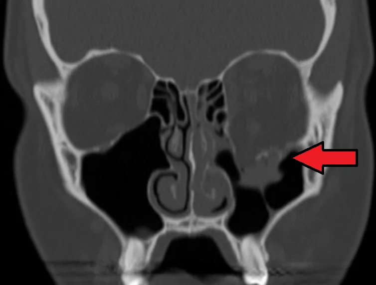
Coronal CT (bone window) shows the left orbital floor fracture site indicated by the red arrow. The continuity of the orbital floor is disrupted, and orbital contents are herniating into the maxillary sinus. This corresponds to the definitive diagnosis of orbital floor fracture using orbital CT (coronal, bone window) discussed in section “4. Diagnosis and Examination Methods.”
A complete ophthalmic examination is essential. The first step is to rule out complications that threaten visual function, such as globe rupture or retinal detachment.
Orbital CT is mandatory for definitive diagnosis. Soft tissue window is useful for observing the positional relationship, displacement, herniation, incarceration, and strangulation of bone and soft tissues, while bone window is useful for detecting subtle fractures. Both windows should be ordered.
CT bone window: Useful for detecting subtle fractures. Allows accurate assessment of fracture morphology.
CT soft tissue window: Useful for observing incarceration, herniation, and strangulation. Adjusting the window width makes it easier to identify orbital fat and orbital emphysema.
Coronal and sagittal views: Essential for evaluating the inferior wall. Axial views alone are insufficient.
MRI: Not first-line due to the possibility of intraocular metallic foreign bodies after trauma. Used adjunctively when additional evaluation of paranasal sinus mucoceles, etc., is needed.
When orbital volume increases by 13% or more, the risk of enophthalmos is high. CT utilization rate was 88%, MRI 0.5% (UK survey). 4)
Extraocular muscle palsy, hemorrhage, or contusion without fracture
QWhy is CT preferred over MRI?
A
After trauma, there may be intraocular metallic foreign bodies, and MRI is not the first choice due to safety concerns. CT is excellent for bone visualization and is most suitable for evaluating fractures, entrapment, and retrobulbar hemorrhage. MRI is used adjunctively when additional evaluation such as sinus mucocele is needed.
In closed fractures with extraocular muscle entrapment, there is a risk of muscle necrosis, and emergency reduction surgery should be performed within 24 hours of injury. If orbital soft tissue other than muscle is entrapped, surgery should be performed as early as possible (usually within 2 weeks).
A UK survey found that 54% chose surgery 6–10 days after injury. 4) Surgical delay worsens prognosis. The residual enophthalmos rate is about 20% when repair is performed within 14 days, compared to about 72% when repair is performed after 6 months or more. 4)
Performed under general anesthesia. The orbital rim periosteum is approached percutaneously or transconjunctivally. After periosteal incision, the surgical field is developed posteriorly into the orbit. All prolapsed soft tissues are reduced into the orbit, displaced bone fragments are repositioned, and the defect is repaired with bone reconstruction materials. Damaged periosteum is sutured together or reconstructed with silicone or absorbable plates.
In a UK survey, approach selection was: subciliary 41%, infraorbital 37%, transconjunctival 7%. 4)
Open Approach
Indications: Combined medial wall and orbital floor fractures.
Advantages: Slight benefits in operative time, hospital stay, and cost.
Complications: Eyelid malposition, infraorbital nerve hypoesthesia. Subciliary incision carries a risk of cicatricial ectropion.
Endoscopic Approach
Procedure: Uncinate process resection → ethmoidectomy → identification of fracture site → reduction of herniated tissue → implant placement.
Advantages: Can be used for early repair, less globe traction. Suitable for medial wall and trapdoor fractures. Less soft tissue injury and hypoesthesia. 2)
Confirmation: Forced duction test and pulse test to verify eye movement and implant position.
The characteristics and complication rates of major reconstruction materials are shown below.
Material
Characteristics
Material-related complication rate
Titanium mesh
Good rigidity and formability, suitable for large defects
2.4% (741 cases)3)
Porous polyethylene (Medpor)
Fixed by tissue ingrowth, low infection rate
Not reported (326 cases)3)
Absorbable materials (Poly-L/D-lactic acid, PLLA)
Suitable for small to medium defects, low complications
3.4% (176 cases)3)
Autologous bone (skull, iliac bone, etc.)
High biocompatibility but resorption is a concern
Donor site complications present3)
Silicone
Inexpensive, easy to handle
17.5% (530 cases, highest value) 3)
In a UK survey, 66% preferred silicone, but 66% of surgeons considered it inferior. 4) Reasons for implant removal were extrusion 70% and infection 46%. 4)
Steroids: Short-term administration. Prednisone 0.75–1.0 mg/kg/day for 5–7 days. Alternatively, intraoperative dexamethasone 20 mg IV, followed by prednisone 0.75–1.0 mg/kg/day for 3–5 days. In a UK survey, 53% prescribed them. 4)
Antibiotics: Perioperative antibiotic prophylaxis is common. Amoxicillin-clavulanate is most frequently used. 2) Prophylaxis is debated; 47% prescribed at diagnosis. 4)
A systematic review of 444 cases reported that surgery improved enophthalmos in 85.2%, diplopia in 74.8%, eye movement restriction in 61.6%, and sensory disturbance in 61.1%. 2)
QIs surgery always necessary for orbital fractures?
A
Surgery is not always necessary. Mild diplopia and movement disorders often improve spontaneously, and observation may be chosen. Closed fractures with extraocular muscle entrapment, persistent diplopia, or enophthalmos are main indications for surgery.
Two mechanisms have been proposed for orbital blowout fractures, both confirmed by cadaver studies: increased intraorbital pressure due to external force, and a contrecoup fracture at a site distant from the point of impact.
Mechanism of Diplopia and Ocular Movement Disorders
Entrapment of orbital septa: Orbital septa run vertically and horizontally within the orbital fat, and even entrapment of septa near the extraocular muscles at the fracture site can cause restriction of eye movement. 1)
Multifactorial causes: Due to a combination of trauma, soft tissue damage, fibrosis, nerve contusion, etc. The direct relationship between orbital floor fracture and ocular movement disorders is considered to have insufficient definitive evidence. 1)
Orbital floor and medial wall fractures cause herniation of orbital contents into the paranasal sinuses. Expansion of the orbital volume leads to posterior displacement of the globe, resulting in enophthalmos. It is said that enophthalmos occurs when the orbital volume increases by 13% or more.
Open type (comminuted fracture): Fracture fragments, bone, and orbital contents (extraocular muscles, fat, soft tissue) are significantly displaced or herniated into the paranasal sinuses. This is the most common type.
Closed type (trapdoor): Due to bone elasticity, when the bone returns to its original shape, the extraocular muscles and surrounding soft tissues become entrapped in the crack. Common in young patients.
Hinge fracture: The bone fragment is displaced like a hinge.
Blow-in fracture: The bone fragment protrudes into the orbit, conversely reducing the orbital volume.
The medial orbital wall (lamina papyracea) is the thinnest of the orbital walls, and orbital fractures are classified as pure internal fractures (blow-out fractures) that by definition do not involve the orbital rim.
The use of custom-made implants based on CT images tailored to individual fracture patterns is advancing. Reduction in surgery time has been reported, with a significant reduction in the pre-contoured method (57.3±23.4 minutes) compared to the freehand contouring method (99.8±28.9 minutes). 2)
Endoscopic transnasal and transmaxillary approaches are associated with less soft tissue damage and less infraorbital nerve hypoesthesia compared to conventional percutaneous approaches. 2) They are considered particularly effective for medial wall fractures and trapdoor fractures, and wider adoption is expected.
In a narrative review of 66 studies and 3870 cases by Sivam & Enninghorst (2022), neobone formation was confirmed after complete absorption of Poly-L/D-lactic acid, and significant improvements in eye movement, diplopia, and enophthalmos were reported. 3)
A systematic review by de Santana et al. (2024) concluded that current evidence for a direct association between orbital floor fractures and ocular motility disorders is insufficient. 1) Further multicenter studies are needed to elucidate the pathophysiology.
de Santana IHG, Viana MRM, Palhano-Dias JC, Ferreira-Júnior O, Sant’Ana E, Shinohara ÉH, et al. Orbital floor fracture (blow out) and its repercussions on eye movement: a systematic review. European journal of medical research. 2024;29(1):427. doi:10.1186/s40001-024-02023-y. PMID:39164786; PMCID:PMC11334373.
Miran B, Toneatti DJ, Schaller B, Kalaitsidou I. Management Strategies for Isolated Orbital Floor Fractures: A Systematic Review of Clinical Outcomes and Surgical Approaches. Diagnostics (Basel, Switzerland). 2025;15(23). doi:10.3390/diagnostics15233024. PMID:41374405; PMCID:PMC12690994.
Sivam A, Enninghorst N. The Dilemma of Reconstructive Material Choice for Orbital Floor Fracture: A Narrative Review. Medicines (Basel, Switzerland). 2022;9(1). doi:10.3390/medicines9010006. PMID:35049939; PMCID:PMC8778999.
Courtney DJ, Thomas S, Whitfield PH. Isolated orbital blowout fractures: survey and review. The British journal of oral & maxillofacial surgery. 2000;38(5):496-504. doi:10.1054/bjom.2000.0500. PMID:11010781.
Copy the article text and paste it into your preferred AI assistant.
Article copied to clipboard
Open an AI assistant below and paste the copied text into the chat box.
