Orbital fracture (blowout fracture) is a traumatic condition in which the bones of the orbit are fractured by blunt force to the eye. Two mechanisms are proposed: increased intraorbital pressure causing fracture, and contrecoup fracture at a site distant from the impact.
The orbital floor and medial wall are very thin and are common sites of fracture. In these areas, the bone often displaces significantly toward the paranasal sinuses, leading to the term blowout fracture.
Approximately 10% of all facial fractures are isolated orbital wall fractures, most of which occur in the orbital floor. The orbit is involved in 30–40% of all facial fractures, and isolated orbital floor fractures account for 22–47% of all orbital fractures. 2) Among 268 cases analyzed, 72% were male and 28% female, with a mean age of 36 years. 2)
As a structural feature of the orbital floor, the bone thickness at the site of the infraorbital neurovascular bundle is only 0.23 mm, and the posteromedial area averages 0.37 mm, making it extremely thin. The lateral area averages 1.25 mm, more than five times thicker. The medial orbital wall and the infraorbital groove are particularly thin and are common sites of fracture.
QAre orbital floor fracture and blowout fracture the same thing?
A
They are essentially synonymous. “Blowout fracture” refers to the phenomenon where a blow to the eye increases intraorbital pressure, causing the thinnest part of the orbital floor to blow out. Orbital floor fracture indicates the typical fracture site.
Fractures are classified into open and closed types based on CT imaging findings.
Open Fracture
Displacement of fracture fragments: Imaging shows significant displacement of bone fragments and soft tissue into the paranasal sinuses.
Enophthalmos: The eyeball moves posteriorly due to expansion of the orbital volume. This becomes more noticeable as swelling subsides after injury.
Irreversible changes: Tissue position does not change with observation. Early reduction surgery before scarring is desirable.
Closed Fracture (Trapdoor Type)
Minimal imaging changes: Slight bone displacement or the missing rectus sign (where the extraocular muscle appears to disappear) are characteristic findings.
Associated systemic symptoms: The extraocular muscle is entrapped in the fracture site, causing severe eye pain, nausea, vomiting, syncope, and bradycardia due to the vagal reflex. This may be misdiagnosed as increased intracranial pressure.
High urgency: When accompanied by extraocular muscle entrapment, there is a risk of muscle necrosis, and emergency surgery within 24 hours of injury is indicated. This is more common in young people and children.
Diplopia: Occurs with eye movement disorders. Worsens with vertical eye movements.
Enophthalmos: Progresses over days to weeks after injury as swelling subsides.
Eye pain: Characteristically occurs with vertical eye movements.
Hypesthesia and paresthesia: In inferior wall fractures, paralysis of the second branch of the trigeminal nerve (infraorbital nerve) causes hypesthesia and paresthesia from the cheek to the upper lip on the affected side.
Eyelid symptoms: Eyelid contusion, eyelid edema, and subcutaneous hemorrhage of the eyelid.
Orbital emphysema: Air from the paranasal sinuses enters the orbit during nose blowing, worsening eyelid swelling and eye movement disorders.
If there is tissue incarceration at the fracture site, vertical eye movements may cause nausea and bradycardia (oculocardiac reflex).
QWhy does diplopia occur in orbital fractures?
A
The main cause is restriction of movement due to incarceration of extraocular muscles and orbital tissues at the fracture site. Even if only the orbital septa within the orbital fat are trapped near the extraocular muscles, eye movement restriction can occur. The cause of diplopia is not the fracture alone but a combination of factors including trauma, soft tissue injury, fibrosis, and nerve contusion. 1)
Blunt trauma to the eye and periorbital area is the cause. The causes of injury (analysis of 268 cases) were assault 35.1% (most common), falls 21.6%, sports 19.0%, traffic accidents 13.8%, and workplace accidents 1.1%. 2)
In children, the main causes are falls, knee strikes, traffic accidents, and fist blows. Because the bone walls are thin, closed fractures are more likely to occur, and the common site is the maxilla and ethmoid bone (orbital floor).
Two theories have been proposed for the mechanism of fracture, both confirmed as valid in cadaver studies.
Hydraulic theory: A fist or ball directly strikes the eye, causing a sudden increase in intraorbital pressure that blows out the weakest area (directly above the infraorbital neurovascular bundle).
Buckling theory: A blow to the cheek transmits a pressure wave posteriorly, compressing the bone in the anteroposterior direction, causing the weakest area to buckle and the bone fragment to be pushed downward.
A complete ophthalmic examination is essential. The first step is to rule out complications that threaten visual function, such as globe rupture and retinal detachment.
Orbital CT is mandatory for definitive diagnosis. Thin-slice (1.0–1.5 mm) axial CT with coronal reconstruction is standard. Instruct the radiology department to acquire both bone and soft tissue windows.
CT bone window: Useful for observing fine fractures. Allows accurate assessment of fracture morphology.
CT soft tissue window: Useful for observing the positional relationship between bone and soft tissue, as well as findings of displacement, herniation, incarceration, and strangulation.
Coronal and sagittal views: Essential for detailed assessment of inferior wall fractures. Axial views alone are insufficient.
MRI: Used complementarily when detailed observation of soft tissues is needed.
When orbital volume increases by 13% or more, the risk of enophthalmos is high. Note that radiological herniation of the inferior rectus muscle does not necessarily predict clinical motility disturbance.
A cross-sectional survey of 187 oral surgeons in the UK reported the following frequencies of test use: CT 88%, X-ray 83%, Hess chart 75%, visual acuity test 65%, and ophthalmology consultation 60%. 4)
Hess chart and binocular single vision field test: Objectively evaluate eye movement and diplopia. Also used for pre- and post-treatment comparison.
Forced duction test: Imaging and clinical findings are usually sufficient to determine surgical indication; this test is not recommended to be performed actively because it is painful when performed awake.
Differential diagnoses include orbital congestion, extraocular muscle palsy, nerve palsy, diplopia due to loss of fusion, and medial orbital wall fracture. Note that incarceration is a purely clinical diagnosis, not a radiological one.
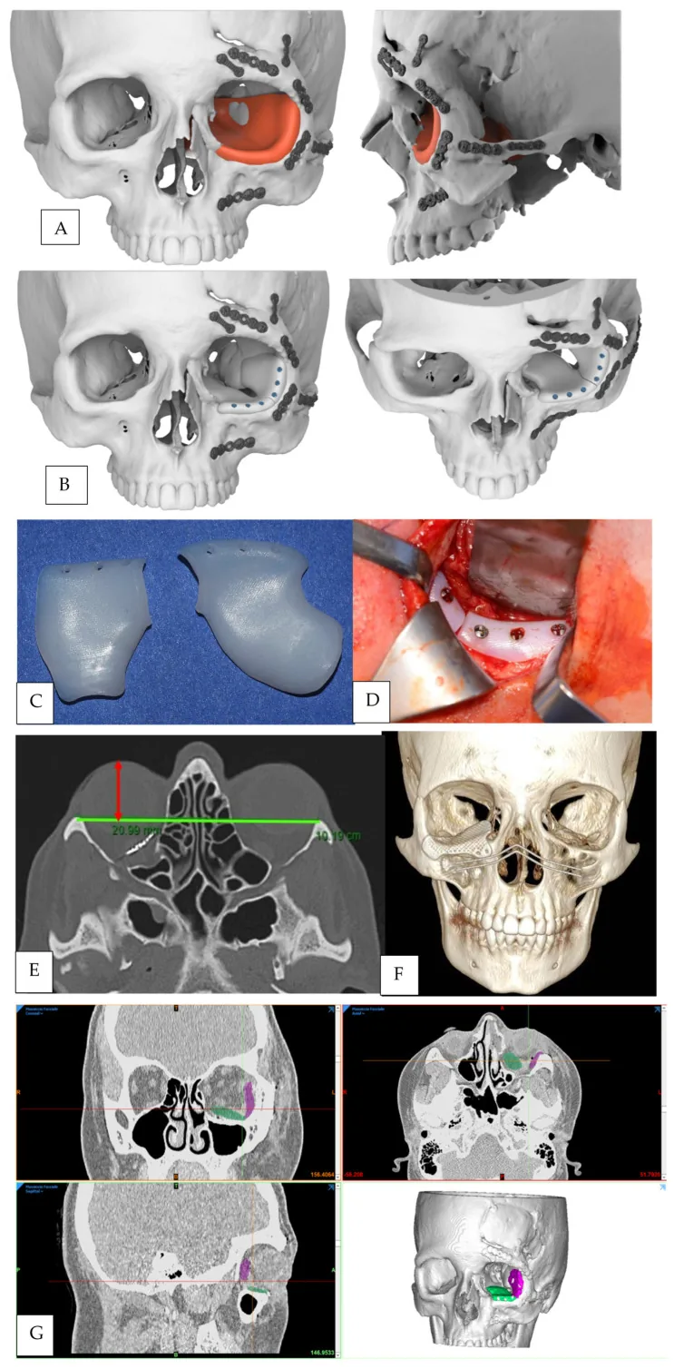
D’Alpaos D, Badiali G, Ceccariglia F, Tarsitano A. Delayed Orbital Floor Reconstruction Using Mirroring Technique and Patient-Specific Implants: Proof of Concept. J Pers Med. 2024;14(5):459. Figure 1. PMCID: PMC11122088. DOI: 10.3390/jpm14050459. License: CC BY 4.0.
Multi-panel diagram showing the sequence of orbital fracture repair. (A) 3D CT reconstruction of the affected side using mirroring of the healthy side for skeletal planning, (B) CAD design of a left orbital floor implant, (C) High-density polyethylene (HDPE) implant (in two parts), (D) Intraoperative implant insertion via transconjunctival approach, (F) Postoperative 3D CT with custom titanium mesh. Corresponds to orbital wall reconstruction (titanium mesh, porous polyethylene) and pre-/postoperative CT evaluation discussed in section “5. Standard Treatment”.
For cases with mild ocular motility disorders or diplopia and predominantly reversible imaging changes, observation is recommended. In large open fractures, edema subsides within 2 weeks after injury, making enophthalmos prominent; therefore, prior explanation to the patient is important.
It is important not to casually observe cases with clear surgical indications, but to transfer them to a facility capable of surgery.
Surgery is performed under general anesthesia. The infraorbital rim periosteum is approached via a subciliary skin incision or a lower fornix conjunctival incision. After periosteal incision, the surgical field is extended posteriorly into the orbit, and the incarcerated orbital tissue is gently reduced into the orbit using micro forceps. Displaced bone fragments are returned to their original position and reconstructed with bone reconstruction materials. If the bone is too fragmented to use, artificial bone is used for augmentation. Damaged periosteum is sutured or reconstructed with silicone or absorbable plates.
A UK survey reported that the subciliary approach was chosen in 41% of cases, infraorbital rim incision in 37%, and transconjunctival incision in 7%. 4) In children, a transconjunctival fornix approach is also used. The transmaxillary sinus (paranasal sinus) approach is another option, including balloon placement in the maxillary sinus.
Regarding surgical timing, a UK survey of 187 patients found that surgery 6–10 days after injury was most common (54%), with 1–5 days in 19% and 11–14 days in 16%. 4)
The characteristics and complication rates of major reconstruction materials are shown below.
Material
Characteristics
Material-related Complication Rate
Titanium mesh
Good rigidity and formability, suitable for large defects
2.4% (741 cases) 3)
Porous polyethylene (Medpor)
Fixed by tissue ingrowth, low infection rate
Not reported (326 cases) 3)
Absorbable material (Poly-L/D-lactic acid)
Suitable for small to medium defects, low complication rate
3.4% (176 cases) 3)
Autologous bone (skull, iliac bone, etc.)
High biocompatibility but absorption is a challenge
Donor site complications possible 3)
Silicone
Inexpensive, easy to handle
17.5% (530 cases, highest) 3)
In a survey of 187 UK physicians, silicone was the most frequently chosen material (66%), but 66% of physicians who expressed concerns about silicone rated it as inferior. 4)
Steroids: Short-term administration. Prednisone 0.75–1.0 mg/kg/day for 5–7 days. Alternatively, intraoperative Decadron 20 mg IV, followed by prednisone 0.75–1.0 mg/kg/day for 3–5 days. In a UK survey, steroids were used in 53% of cases. 4)
Antibiotics: Prophylactic antibiotics were used in 47%, perioperative in 53%, and postoperative for 5 days in 63% of cases. 4) Amoxicillin-clavulanate is the most commonly used. 2)
A systematic review of 444 cases reported that surgery improved enophthalmos in 85.2%, diplopia in 74.8%, restricted eye movement in 61.6%, and sensory disturbance in 61.1% of cases. 2)
QIs surgery always necessary for orbital fractures?
A
Surgery is not always necessary. Mild diplopia and motility disturbances often improve spontaneously, and observation may be chosen. Closed fractures with extraocular muscle entrapment, persistent diplopia, or enophthalmos are the main indications for surgery.
QAre orbital fractures in children different from those in adults?
A
In children, closed (trapdoor) fractures are more common. Because the bone wall is thin, extraocular muscles are easily entrapped in the fracture site, and characteristic symptoms such as nausea, vomiting, syncope, and bradycardia due to the vagal reflex may appear. These systemic symptoms can be misdiagnosed as signs of increased intracranial pressure, so caution is needed. To avoid muscle necrosis, emergency surgery within 24 hours of injury is required when extraocular muscle entrapment is confirmed.
The orbital floor and medial wall are adjacent to the maxillary and ethmoid sinuses, and the bone is very thin. This anatomical vulnerability makes them prone to fracture under external force.
Hydraulic theory
Increased intraorbital pressure due to impact: A fist or ball directly strikes the eye, displacing the globe posteriorly.
Rupture of the weakest point: The sudden rise in intraorbital pressure causes the thinnest area just above the infraorbital neurovascular bundle to blow out.
Herniation of soft tissue: Orbital contents (fat and muscle) herniate through the fracture into the paranasal sinuses.
Buckling theory
Propagation of pressure waves: A blunt trauma to the cheek generates pressure waves that travel posteriorly through the bone.
Compression and buckling of bone: Anteroposterior bone compression causes the weakest part of the orbital floor to buckle, pushing bone fragments downward.
Direct bone deformation: Unlike the hydraulic theory, direct impact to the eyeball is not necessarily required.
The mechanisms of diplopia and ocular movement disorders are as follows.
Restriction of extraocular muscle movement: Entrapment of the extraocular muscle at the fracture site is the main cause. Damage or strangulation of the extraocular muscle itself can also cause severe contractile dysfunction.
Entrapment of orbital septa: Orbital septa run vertically and horizontally within the orbital fat, and even entrapment of the septa near the extraocular muscles at the fracture site can restrict eye movement.
Multifactorial causes: The cause of diplopia is not solely the fracture but a combination of factors including trauma, soft tissue injury, fibrosis, and nerve contusion. 1)
The mechanism of enophthalmos is as follows. Herniation of orbital tissue into the paranasal sinuses through the fracture expands the orbital volume, causing the eyeball to move posteriorly, resulting in enophthalmos. Injury to the second branch of the trigeminal nerve (infraorbital nerve) in a floor fracture can cause sensory disturbance from the cheek to the upper lip.
Prognosis is usually good if reduction surgery is performed early with an appropriate technique.
7. Latest Research and Future Perspectives (Investigational Reports)
The use of custom-made implants based on CT images tailored to individual fracture patterns is advancing. Reduction in surgical time has been reported, with a significant reduction in the preformed group (57.3±23.4 minutes) compared to the freehand molding group (99.8±28.9 minutes). 2)
Endoscopic transnasal and transmaxillary approaches are associated with less soft tissue damage and less infraorbital nerve hypoesthesia compared to conventional percutaneous approaches. 2)
After complete absorption of Poly-L/D-lactic acid, neobone (new bone) formation was confirmed, and a study of 94 cases reported significant improvement in eye movement, diplopia, and enophthalmos. 3)
At present, definitive evidence for a direct association between orbital floor fractures and ocular motility disorders is considered insufficient. 1) Future multicenter collaborative studies are needed to elucidate the pathophysiology.
de Santana IHG, Viana MRM, Palhano-Dias JC, Ferreira-Júnior O, Sant’Ana E, Shinohara ÉH, et al. Orbital floor fracture (blow out) and its repercussions on eye movement: a systematic review. European journal of medical research. 2024;29(1):427. doi:10.1186/s40001-024-02023-y. PMID:39164786; PMCID:PMC11334373.
Miran B, Toneatti DJ, Schaller B, Kalaitsidou I. Management strategies for isolated orbital floor fractures: a systematic review of clinical outcomes and surgical approaches. Diagnostics. 2025;15:3024.
Sivam A, Enninghorst N. The Dilemma of Reconstructive Material Choice for Orbital Floor Fracture: A Narrative Review. Medicines (Basel). 2022;9(1):6. doi:10.3390/medicines9010006. PMID:35049939; PMCID:PMC8778999.
Courtney DJ, Thomas S, Whitfield PH. Isolated orbital blowout fractures: survey and review. Br J Oral Maxillofac Surg. 2000;38:496-504.
Copy the article text and paste it into your preferred AI assistant.
Article copied to clipboard
Open an AI assistant below and paste the copied text into the chat box.