Lyme disease is a multi-organ infection caused by spirochetes of the genus Borrelia (mainly B. burgdorferi). It is transmitted through the bite of ticks of the genus Ixodes and causes symptoms in the skin, eyes, nervous system, joints, and heart. It is a zoonotic disease classified as a Category IV infectious disease under the Infectious Diseases Control Law.
Pathogen and Vector
B. burgdorferi is a spiral-shaped microorganism 20–30 μm long and 0.2–0.3 μm wide, with 7–11 flagella providing high motility. It requires complex media such as Barbour-Stoenner-Kelly (BSK II) medium.
Ticks have a two-year lifespan with three stages: larva, nymph, and adult. Nymphs are the main source of infection for humans. Animal reservoirs include rodents, deer, and birds.
Epidemiology and Vector Species in Japan
In Japan, Ixodes persulcatus and Ixodes ovatus are the vectors of Lyme disease. Asymptomatic infections are common. The main endemic area is northern Japan, centered on Hokkaido, which corresponds to the geographic distribution of Ixodes persulcatus. Cases have also been reported in Honshu, but the majority of symptomatic cases are reported from northern Japan.
Global epidemiology
Globally, Lyme disease is widespread in Europe and North America, with approximately 128,888 cases reported annually in Europe. Incidence rates are particularly high in Estonia, Lithuania, Slovenia, and Switzerland, exceeding 100 cases per 100,000 population per year 1). Recent European surveillance data show a significant increase in disease burden over the past 15 years, and expansion into new areas has been documented 1).
QWhy is Lyme disease more common in Hokkaido, Japan?
A
In Japan, the vectors are Ixodes persulcatus and Ixodes ovatus, which carry Borrelia species. The main reason is that the geographic distribution of these ticks is concentrated in northern Japan, centered on Hokkaido. Although cases have been reported in Honshu, most symptomatic cases are reported from northern Japan.
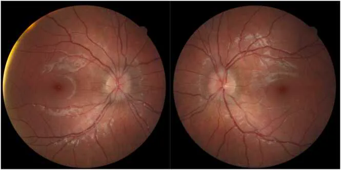
Barten M, Strand E, Knight T. Optic disc edema associated with neuroborreliosis. IDCases. 2025;42:e02438. Figure 1. PMID: 41473482; PMCID: PMC12747239; DOI: 10.1016/j.idcr.2025.e02438. License: CC BY.
Color fundus photographs showing mild bilateral optic disc edema, with the right eye (OD) on the left and the left eye (OS) on the right. This corresponds to “optic disc edema” discussed in section “2. Main symptoms and clinical findings.”
Stage 3: Chronic arthritis (especially of the knee), encephalomyelitis, demyelination, and other neurological symptoms. Ocular symptoms include corneal parenchymatitis and episcleritis.
Chronic arthritis, encephalomyelitis, progression of demyelinating lesions
Erythema migrans begins as a red papule and, as it expands, forms a “target-like” lesion with a bright red outer border and a clearing center. About 20% of patients do not show the typical target-like lesion. Chronic erythema after a tick bite is an important diagnostic clue.
Cranial nerve palsy: Palsies of cranial nerves II, III, IV, VI, and VII (facial). Bilateral facial nerve palsy is characteristic5). May be unilateral or bilateral, appearing sequentially or simultaneously.
Keratitis: The most common ocular symptom in the third stage.
Corneal findings: Bilateral, patchy, focal, stromal or subepithelial infiltrates. Ill-defined borders, appearing months to years after initial infection. Good response to topical steroids (suggesting immune-mediated reaction).
Others: Episcleritis, pupillary abnormalities, extraocular muscle palsy, and ptosis (rare)5).
QWhat is the most common ocular manifestation of Lyme disease?
A
Intermediate uveitis in the second stage is the most common ocular manifestation. It may also present as anterior, posterior, or panuveitis, but intermediate uveitis with predominant vitritis is typical. In the third stage, corneal stromal keratitis is the most common ocular symptom.
Infected ticks attach to the host (human) and transmit spirochetes via salivary secretions. After attaching to host cell proteoglycans, the microorganism disseminates lymphohematogenously to multiple organs including skin, musculoskeletal system, nervous system, and eyes4). Removal of the tick within 24–48 hours of attachment significantly reduces the risk of infection.
Risk factors
Exposure to outdoor areas where ticks (genus Ixodes) live (grasslands, forests, mountains)
Stay or travel history in endemic areas (Hokkaido, northern Japan)
Outdoor activities from summer to autumn (May to October) such as camping, farming, and forest work
Contact with wild animals such as rodents and deer
Endemic areas and infection risk in Japan
In Hokkaido, the density of Ixodes persulcatus is high, and the Borrelia carriage rate is relatively high. Infections have also been reported in the Tohoku and Hokuriku regions, such as Nagano, Niigata, and Iwate. The risk of infection is low in urban areas, but cases outside endemic areas have been reported due to the spread of outdoor recreation.
Erythema migrans has high diagnostic value, and it is important to obtain a history of exposure to endemic areas and tick bites. A history of tick bite can be a clue for diagnosis, but diagnosis based on ocular findings alone is difficult, and serological testing is essential. The combination of systemic symptoms (chronic erythema, arthritis, neurological symptoms) is key to diagnosis.
Enzyme immunoassay (first step): Detection of IgM and IgG antibodies. IgM peaks within the first month of infection. Sensitivity is low in the first few weeks of infection.
Western blot (second step): Performed when enzyme immunoassay is equivocal or positive.
Western blot interpretation criteria (CDC criteria) 2):
IgM positive: presence of 2 of 3 bands at 23, 30, 41 kDa (caution: combination of 23 and 41 kDa may be false positive)
IgG positive: presence of 5 of 10 bands at 18, 23, 28, 30, 39, 41, 45, 58, 66, 93 kDa
ELISA detects antibodies (IgM, IgG) against Borrelia species, but sensitivity is low in early infection. Cerebrospinal fluid testing should be considered in cases with neurological symptoms.
PCR: Amplifies genomic and plasmid DNA from tissues including intraocular fluid 4). More sensitive than culture and used for definitive diagnosis of ocular Lyme disease.
Culture: Isolation is easy mainly from erythema lesions. Use BSK II medium.
Differentiation from syphilis: Lyme disease may cause false-positive syphilis serology (FTA-ABS).
Persistence of antibodies after treatment: IgG and IgM responses may persist for years after antibiotic treatment. A positive IgM response alone cannot be interpreted as evidence of recent infection or reinfection.
Other differential diagnoses: Colorado tick fever, ehrlichiosis, Rocky Mountain spotted fever, tick-borne relapsing fever, tularemia, and other tick-borne diseases with ocular symptoms need to be differentiated. JIA-associated uveitis and HLA-B27-associated anterior uveitis should also be excluded.
QWhy does the syphilis test become false positive in the diagnosis of Lyme disease?
A
Borrelia burgdorferi belongs to the family Spirochaetaceae and is a microorganism of the same order Spirochaetales as Treponema pallidum. Because their antigens cross-react, patients with Lyme disease may show false-positive results in syphilis lipid antigen tests (RPR, VDRL) and fluorescent treponemal antibody absorption test (FTA-ABS). For differentiation, it is necessary to combine tests specific to T. pallidum.
Early disease, facial nerve palsy (oral administration)3)
Choose one of the following:
Doxycycline: 100 mg orally twice daily for 10–21 days (first choice)
Amoxicillin: 500 mg orally three times daily for 14–21 days (alternative for children and pregnant women)
Cefuroxime: 500 mg orally twice daily for 14–21 days
Pediatric doses: amoxicillin 25–50 mg/kg/day (max 500 mg three times daily), cefuroxime 30 mg/kg/day (max 500 mg twice daily)3).
Meningitis, recurrent arthritis, central or peripheral nervous system disease (intravenous administration)3)
Ceftriaxone: 2 g/day intravenously once daily for 14–28 days
For neurological symptoms (neuroborreliosis), high-grade atrioventricular block, or severe arthritis, intravenous therapy is chosen. Ceftriaxone 2 g single daily dose is standard, and the duration is determined while evaluating treatment response.
Performed after systemic antibiotic treatment has been initiated.
Steroid eye drops: Used for inflammation of the anterior segment of the eye (iridocyclitis, anterior uveitis).
Mydriatic agents (cycloplegics): Added to prevent posterior synechiae, relieve pain, and reduce photophobia.
Posterior uveitis and vitreitis: Monitor while continuing systemic antibiotic treatment. If inflammation is severe, consider systemic steroid administration.
With early antibiotic treatment (stage 1), the prognosis is good, and complete recovery can be expected in most cases. If the disease progresses to stages 2 or 3, neurological and joint sequelae are more likely. Most ocular symptoms improve with antibiotic treatment, but in optic neuritis, visual recovery may be incomplete. In cases of ocular Lyme disease with choroidal neovascular membrane (CNVM), visual prognosis has been reported to be poor 5).
Spirochetes proliferate in the salivary glands of infected ticks and are injected into the host skin during blood feeding. Infection begins 12–24 hours after tick attachment, and the risk of infection increases significantly after 36–48 hours of attachment.
Borrelia attaches to host proteoglycans via surface glycosaminoglycans (such as decorin-binding proteins) 4). After attachment, the bacteria invade tissues using host extracellular matrix components. Subsequently, the bacteria disseminate lymphogenously or hematogenously throughout the body, with particular affinity for the following tissues:
Skin (erythema migrans: local inflammatory reaction)
Joint synovium (arthritis: immune complex deposition and CD4-positive T cell infiltration)
Peripheral and central nervous system (neuroborreliosis: inflammation of brain parenchyma, meninges, and nerve roots)
Cardiac conduction system (atrioventricular block: myocarditis, conduction pathway disorder)
Ocular tissues (uvea, cornea, optic nerve)
Mechanism of ocular involvement
Uveitis in the eye involves both direct bacterial invasion and the host immune response 5). Two mechanisms coexist: a “direct infection type” in which spirochetes directly invade the uvea, and an “immune-mediated type” due to the immune response to infection.
In the third stage, keratitis responds well to topical steroids alone, suggesting that the immune response (inflammation) is the main factor rather than the infection itself. Bilateral facial nerve palsy results from cranial neuritis caused by Borrelia and is a characteristic finding of disseminated infection in the second stage. Rare cases of ocular Lyme disease with choroidal neovascular membrane (CNVM) have also been reported 5).
Ocular complications in neuroborreliosis
In Lyme neuroborreliosis, Borrelia crosses the blood-brain barrier and invades the central nervous system. This leads to optic neuritis, oculomotor nerve palsy, and supranuclear gaze disorders. Detection of antibodies in cerebrospinal fluid and increased cell count aid in diagnosis.
7. Latest research and future perspectives (reports under investigation)
Phase 3 clinical trials of the Lyme disease vaccine (VLA15) jointly developed by Valneva and Pfizer are ongoing in Europe and North America. The European Lyme borreliosis disease burden study also serves as its foundation 1), and from the perspective of preventive medicine, it is entering a new phase. The target age (5 years and older), vaccination schedule, and duration of effectiveness are currently under evaluation.
Increasing trend and epidemiological dynamics in Europe
According to European surveillance data, the disease burden of Lyme disease has increased significantly in endemic areas over the past 15 years, and expansion to new regions has also been reported 1). This epidemiological change is linked to the expansion of tick habitats due to climate change and an increase in the population engaging in outdoor recreation.
Improvement of PCR and Multiplex Diagnostic Techniques
PCR methods for detecting Borrelia DNA in various specimens, including intraocular fluid samples, have been improved, and their application in cases where serological diagnosis is difficult is being studied 4). Specimen analysis using next-generation sequencing (NGS) is also expected to improve diagnostic accuracy.
In pediatric Lyme disease, the pattern of ocular complications may differ from that in adults, and studies are ongoing to evaluate the long-term impact on visual function 5). In children, facial nerve palsy tends to become apparent early and may prompt an ophthalmology consultation.
Marques AR, Strle F, Wormser GP.. Comparison of Lyme Disease in the United States and Europe. Emerg Infect Dis. 2021;27(8):2017-2024. doi:10.3201/eid2708.204763. PMID:34286689; PMCID:PMC8314816.
Mead P, Petersen J, Hinckley A. Updated CDC Recommendation for Serologic Diagnosis of Lyme Disease. MMWR Morb Mortal Wkly Rep. 2019;68(32):703. doi:10.15585/mmwr.mm6832a4. PMID:31415492; PMCID:PMC6818702.
Lantos PM, Rumbaugh J, Bockenstedt LK, et al. Clinical Practice Guidelines by the IDSA/AAN/ACR: 2020 Guidelines for the Prevention, Diagnosis, and Treatment of Lyme Disease. Clin Infect Dis. 2021;72(1):e1-e48.
Radolf JD, Caimano MJ, Stevenson B, Hu LT.. Of ticks, mice and men: understanding the dual-host lifestyle of Lyme disease spirochaetes. Nat Rev Microbiol. 2012;10(2):87-99. doi:10.1038/nrmicro2714. PMID:22230951; PMCID:PMC3313462.
Krause PJ, Bockenstedt LK. Cardiology patient pages. Lyme Disease and the Heart. Circulation. 2013;127(7):e451-e454. doi:10.1161/circulationaha.112.101485.
Copy the article text and paste it into your preferred AI assistant.
Article copied to clipboard
Open an AI assistant below and paste the copied text into the chat box.