Goldmann kinetic perimetry (GP) is a visual field test in which a target of fixed luminance and size is moved from the periphery toward the center, and the boundary points where the patient first perceives it are connected to draw isopters (lines of equal sensitivity). By superimposing multiple isopters, the sensitivity distribution of the entire visual field can be understood.
While static perimetry (e.g., Humphrey Field Analyzer: HFA) is sensitive for detecting localized abnormalities within the central 30°, GP excels in evaluating the entire visual field extending to nasal 60° and temporal 100° or more 1). Although kinetic perimetry is less suitable for quantitative progression assessment, it is appropriate for patients in whom static perimetry is difficult to perform 1).
The patient profiles for which GP is particularly recommended are as follows.
Elderly individuals, children, and other patients who have difficulty maintaining sustained attention
Patients with low vision, cataracts, or other media opacities that make it difficult to identify small targets
Patients with moderate to advanced glaucoma suspected of having extensive abnormalities beyond the measurement range of static perimetry
Patients with retinal diseases, optic nerve diseases, intracranial lesions, or other conditions expected to cause extensive visual field defects
Patients undergoing visual field testing for the first time (who are not yet familiar with static perimetry)
Advanced glaucoma, retinal diseases, neuro-ophthalmology, children
Early glaucoma, quantitative assessment of visual field defects
Psychogenic visual field disorders
Useful for evaluation
Difficult to evaluate
QHow to choose between GP and HFA (static perimetry)?
A
HFA is superior for detecting localized visual field defects within the central 30° (e.g., arcuate scotoma, nasal step in early glaucoma). In contrast, GP is suitable when evaluation of the entire visual field including the periphery is needed (e.g., advanced glaucoma, retinitis pigmentosa, pituitary adenoma). Additionally, static perimetry is often difficult to perform in elderly patients, children, those with low vision, or cataract patients, and GP is useful in such cases as well.
2. Types of test targets and examination parameters
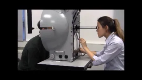
Clinical photograph of an examiner performing visual field testing with a Goldmann kinetic perimeter while presenting targets to the patient. Corresponds to the examination procedures and precautions discussed in section “3. Examination technique and precautions.”
To ensure examination accuracy, perform the following luminance checks without fail. If the prerequisites are not met, follow-up of the visual field becomes impossible.
Target luminance: Illuminate the V/4e target with a luminance meter and adjust to 1000 asb
Background luminance: Project the V/1e target onto the measuring plate and adjust with the background luminance control to match the target luminance
Drooping of the upper eyelid can obstruct the visual field; manage with taping
Occlusion of the non-examined eye
Use an eye patch and be careful not to press on the eyeball
Posture adjustment
Uncomfortable posture leads to decreased concentration. Perform in a comfortable position
Verbal encouragement
Continuously check fixation on the fixation point and provide repeated verbal cues
Bjerrum area
Pay attention to scotomas within the central 30° and arcuate areas extending from the blind spot.
QWhat are the key points to improve the accuracy of Goldmann perimetry?
A
First, it is essential to perform a luminance check (V/4e → 1000 asb) before the examination. During the test, do not neglect fixation monitoring; confirming the Mariotte blind spot early can detect poor fixation. Ptosis and uncorrected refractive errors also significantly affect results, so prior confirmation is important. Additionally, since accuracy varies with the examiner’s experience, standardization of stimulus movement speed and procedures is required.
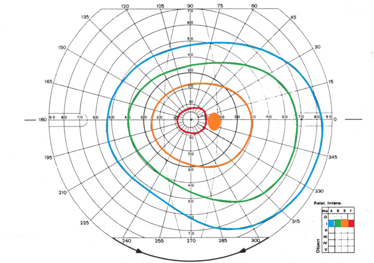
Photograph of a Goldmann visual field chart for the right eye with multiple isopters. Corresponds to the reading of isopters and visual field abnormality patterns discussed in section “4. Interpretation of Results and Typical Patterns.”
A normal visual field shows a “hill of vision” with highest sensitivity at the center. By evaluating the spacing, shape, and symmetry of each isopter, the location and nature of visual field abnormalities can be identified.
During screening, it is important not to overlook the following three patterns: hemianopsia, concentric constriction, and ring scotoma.
Bitemporal hemianopsia: Typically caused by compression of the optic chiasm midline, such as from a pituitary adenoma. Because nasal nerve fibers cross, the temporal visual field is lost.
Homonymous hemianopsia: Visual field defect on the same side as the lesion due to pathology posterior to the optic chiasm (optic radiations or occipital lobe). Occurs in cerebrovascular disease and brain tumors.
Concentric Constriction and Ring Scotoma
Ring scotoma: A characteristic pattern seen in early stages of retinitis pigmentosa (RP). Central and peripheral visual fields are preserved, but the intermediate annular area is defective.
Concentric visual field constriction: As RP progresses, the ring scotoma expands and merges, causing the visual field to shrink from the periphery. End-stage glaucoma also shows a similar pattern.
Glaucomatous visual field defects
Paracentral scotoma: A scotoma occurring in the Bjerrum area (arcuate area within 30° of the center). It starts from the blind spot and expands in an arcuate shape.
Nasal step: A horizontal step-like defect in the nasal visual field. A characteristic pattern in early to moderate glaucoma.
Brain tumors and cerebrovascular disorders: Various visual field patterns depending on the lesion site. Bitemporal hemianopia at the optic chiasm, homonymous hemianopia posteriorly.
Retinitis pigmentosa (RP): Progression from ring scotoma to concentric visual field constriction can be evaluated in the full visual field. Also used for assessing eligibility for visual assistive devices.
Advanced glaucoma (MD < −20 dB approximately): Switch to GP when extensive abnormalities occur that cannot be measured by static perimetry.
Optic nerve diseases: Useful when central scotoma is extensive and fixation is difficult.
Intracranial diseases (pituitary adenoma, brain tumor, cerebral infarction): Suitable for evaluating the entire visual pathway from the optic chiasm to the occipital lobe.
QWhat is the difference between concentric visual field constriction and hemianopia?
A
Concentric visual field constriction is a pattern in which the entire visual field shrinks concentrically, characteristic of retinitis pigmentosa and end-stage glaucoma. Vision is lost in all directions from the center to the periphery. In contrast, hemianopia is a pattern in which the visual field is lost to the left/right or up/down with a vertical or horizontal border, caused by lesions in the optic chiasm, optic radiations, or occipital lobe. In GP, the shapes of the isopters are clearly different between the two, making differentiation relatively easy.
GP shows non-organic patterns such as spiral visual field, tubular visual field (visual field angle does not change with distance), or concentric contraction
Suspect when visual field defect cannot be explained anatomically
6. Measurement Principles and Technical Background
In kinetic perimetry, a stimulus of fixed luminance and size is moved at a constant speed from the periphery toward the center of the visual field. The point where the patient first detects the stimulus becomes a point on the isopter. By repeating this process from multiple directions, a single isopter (line of equal sensitivity) is completed. By changing the luminance or size of the stimulus, multiple isopters are plotted, constructing a sensitivity map of the entire visual field.
While static perimeters measure thresholds at fixed test points, Goldmann perimetry directly plots isopters themselves. Static perimetry is sensitive for detecting localized threshold reductions but has limitations in assessing the peripheral visual field extensively. Goldmann perimetry plays a complementary role, and in moderate to advanced glaucoma, it may be the only means of visual field assessment.
The normal visual field shows a hill-shaped sensitivity distribution (island of vision) with highest sensitivity at the center. Larger and brighter stimuli produce isopters that extend further outward, allowing measurement of the visual field’s “height” at different levels. The Mariotte blind spot (physiologic blind spot) corresponds to the optic disc and is identified as an absolute scotoma located approximately 15° temporal and slightly inferior to the fixation point.
Automated perimeters such as the Octopus 900 are equipped with a Kinetic mode that enables computer-controlled kinetic perimetry. This is expected to reduce examiner dependency and improve standardization and reproducibility. However, further clinical studies are needed to establish clinical value equivalent to conventional manual GP.
Visual field area measurement and progression assessment in retinitis pigmentosa
By regularly measuring and recording the visual field area in RP patients, research is being conducted on its application for individual assessment of progression rate and evaluation of the efficacy of gene therapy and drug therapy. Standardization of visual field area measurement methods and establishment of statistical evaluation criteria for measured values are future challenges.
In cases of advanced glaucoma where static perimetry such as HFA becomes difficult to measure, GP may be the only means of visual field assessment. The clinical significance of using GP to evaluate residual visual field and central fixation protection in end-stage glaucoma is expected to increase in the future1).
Gedde SJ, Vinod K, Wright MM, et al. Primary Open-Angle Glaucoma Preferred Practice Pattern. Ophthalmology. 2021 Jan;128(1):P71-P150. doi:10.1016/j.ophtha.2020.10.022. PMID:34933745.
Barnes CS, Schuchard RA, Birch DG, Dagnelie G, Wood L, Koenekoop RK, et al. Reliability of Semiautomated Kinetic Perimetry (SKP) and Goldmann Kinetic Perimetry in Children and Adults With Retinal Dystrophies. Transl Vis Sci Technol. 2019;8(3):36. PMID: 31211001.
Copy the article text and paste it into your preferred AI assistant.
Article copied to clipboard
Open an AI assistant below and paste the copied text into the chat box.
