
Hypertropia
Key Points at a Glance
Section titled “Key Points at a Glance”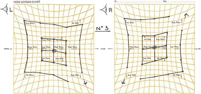
Evidence-Based Supplement
Section titled “Evidence-Based Supplement”Etiology Distribution of Adult Hypertropia
Section titled “Etiology Distribution of Adult Hypertropia”Tamhankar et al. (2011) analyzed 300 consecutive cases of vertical diplopia and found that fourth cranial nerve palsy (trochlear nerve palsy) and thyroid eye disease accounted for more than 50% of causes 1). In addition to these two conditions, post-ophthalmic surgery complications, orbital floor fracture, and myasthenia gravis are major differential diagnoses; thus, a detailed history and ophthalmic examination can identify the cause in most cases.
Limitations of the Parks Three-Step Test Sensitivity
Section titled “Limitations of the Parks Three-Step Test Sensitivity”Manchandia and Demer (2014) reported that among 50 cases with MRI-confirmed superior oblique atrophy, only 35 (70%) met all three criteria of the Parks three-step test 2). Therefore, a negative three-step test does not rule out superior oblique palsy, and a comprehensive assessment of clinical and imaging findings is necessary.
Vertical Gaze Differences in Congenital vs. Acquired Superior Oblique Palsy
Section titled “Vertical Gaze Differences in Congenital vs. Acquired Superior Oblique Palsy”Demer (2022) analyzed 31 cases with MRI-confirmed superior oblique atrophy and showed that the traditional notion that congenital palsy worsens hypertropia in upgaze is not supported in many cases 3). In both congenital and acquired cases, hypertropia is often larger in downgaze, making it difficult to differentiate congenital from acquired based solely on gaze direction comparison.
Differentiation from Skew Deviation
Section titled “Differentiation from Skew Deviation”Wong (2010) proposed skew deviation as a vertical strabismus that is difficult to distinguish from trochlear nerve palsy, and presented the “upright-supine test” in which vertical deviation decreases by 50% or more in the supine position 4). Because it suggests a posterior fossa lesion, neuroimaging is recommended for patients with a positive test.
Masquerading Superior Oblique Palsy
Section titled “Masquerading Superior Oblique Palsy”Demer and Clark (2022) showed that among 83 patients clinically diagnosed with superior oblique palsy, 26 (31%) had no superior oblique atrophy on MRI, indicating that positive three-step test findings can be reproduced by other conditions 5). It is difficult to confirm true superior oblique palsy based solely on clinical diagnosis, and high-resolution MRI evaluation of muscle volume and trochlear pulley position is essential for accurate diagnosis.
References
Section titled “References”- Tamhankar MA, Kim JH, Ying GS, Volpe NJ. Adult hypertropia: a guide to diagnostic evaluation based on review of 300 patients. Eye (Lond). 2011;25(1):91-96. PMID: 21057518.
- Manchandia AM, Demer JL. Sensitivity of the three-step test in diagnosis of superior oblique palsy. J AAPOS. 2014;18(6):567-571. PMID: 25459202.
- Demer JL. Vertical comitance of hypertropia in congenital and acquired superior oblique palsy. J Neuroophthalmol. 2022;42(1):e240-e247. PMID: 34670252.
- Wong AM. Understanding skew deviation and a new clinical test to differentiate it from trochlear nerve palsy. J AAPOS. 2010;14(1):61-67. PMID: 20227626.
- Demer JL, Clark RA. Masquerading superior oblique palsy. Am J Ophthalmol. 2022;242:197-208. PMID: 35618024.