Pediatric vision screening is a system of examinations aimed at early detection of children with amblyopia, strabismus, significant refractive errors, and other eye abnormalities.
Amblyopia and the Significance of Vision Screening
Amblyopia is a visual impairment caused by abnormal visual input during the visual development period, and is classified into the following four types 1).
Refractive amblyopia: A condition in which high bilateral refractive errors prevent clear image formation on the foveal retina.
Anisometropic amblyopia: A condition with a difference in refractive power between the two eyes. It is the most common type of amblyopia.
Strabismic amblyopia: A condition in which the non-fixing eye is chronically suppressed due to misalignment of the eyes.
Form deprivation amblyopia: Visual deprivation due to congenital cataracts or severe ptosis. It is the most severe and treatment-resistant type 1).
The prevalence of amblyopia in Japan is estimated at 0.58% based on a meta-analysis of the referral rate from the 3-year-old health checkup. Internationally, reported prevalence ranges from 0.14% to 4.8%, with epidemiological data from the United States showing 1.5% in African Americans and 2.6% in Hispanics.
Amblyopia can be reversed with appropriate treatment during early childhood, but if left untreated, it can lead to permanent vision loss in one or both eyes and is a leading cause of visual impairment in adults under 40. Even without amblyopia, uncorrected refractive errors can negatively impact learning and school performance.
In Japan, the 3-year-old health checkup is the most important screening opportunity.
Primary screening: Vision test at home (using a visual acuity target of 0.5) and a questionnaire
Secondary screening: Group health checkup at a health center. All children undergo refraction testing, repeat vision testing, and examination by a pediatrician.
Tertiary screening: Children requiring further evaluation receive a detailed examination at an ophthalmology clinic.
Even if missed at the 3-year-old checkup, annual eye exams at nursery schools/kindergartens, school entry health checkups, and annual eye exams at elementary schools can detect problems. However, according to the Japan Ophthalmological Society, approximately 25% of children referred for further evaluation do not visit an ophthalmologist.
In the United States, the U.S. Preventive Services Task Force (USPSTF) recommends at least one amblyopia screening for children aged 3 to 5 years. The American Association for Pediatric Ophthalmology and Strabismus (AAPOS) recommends continued screening every 1 to 2 years after age 5.
QAt what age should vision screening be performed?
A
In Japan, the 3-year-old health checkup is the primary opportunity, but screening is possible from 6 months of age using devices such as infrared video refractometers. The USPSTF recommends screening at ages 3 to 5, and AAPOS recommends continued screening every 1 to 2 years after age 5.
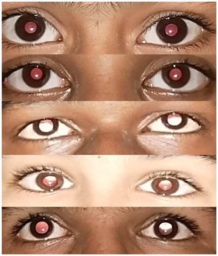
Srivastava RM, et al. Reliability of Smart Phone Photographs for School Eye Screening. Children (Basel). 2022. Figure 5. PMCID: PMC9601144. License: CC BY.
This image shows a comparison of both eyes of a child, highlighting differences in the position and symmetry of the red reflex within the pupils. Differences in reflex patterns are noted for screening refractive errors and anisometropia.
Subjective Symptoms (Signs Noticed by Parents or the Child)
Amblyopia and strabismus often have few subjective symptoms, and the affected person may not notice them. The following items included in the health checkup questionnaire for 3-year-olds can serve as clues for early detection at home.
Abnormal eye alignment: The black part of the eye turns inward, or shifts outward or upward
Abnormal head posture: Tilting the head or looking sideways when viewing objects
Abnormal behavior: Getting close to objects to see, or squinting one eye in bright outdoor light
Abnormal pupil: The center of the black part of the eye appears whitish (white pupil = leukocoria), or the size of the black part differs between the eyes
Clinical Findings (Abnormalities Detected by Screening)
Abnormal red reflex: Shine a light into the pupil using a retinoscope or direct ophthalmoscope and observe the reflection from the fundus. It is normal if both pupils are equally bright and show a symmetrical yellow-orange color. A dim reflection suggests high refractive error, asymmetry suggests anisometropia, and absence of reflection suggests total cataract. This test is essential for early detection of congenital cataract and retinoblastoma.
Abnormal eye alignment: Assessed by the Hirschberg method (observing the position of the corneal light reflex from a penlight held 33 cm in front of the eyes) or the Bruckner method (observing the relationship between pupil brightness in the red reflex and the corneal reflex image). The Bruckner method has fewer false positives and is more suitable.
Significant refractive error: The AAO has established threshold values for refractive errors that pose a risk for amblyopia1). For ages 0–1, myopia ≥ -5.00 D, hyperopia (without strabismus) ≥ +6.00 D, and astigmatism ≥ 3.00 D are risk factors for refractive amblyopia. For anisometropia, myopia ≥ -4.00 D, hyperopia ≥ +2.50 D, and astigmatism ≥ 2.50 D are considered risks.
Abnormal fixation: Eccentric fixation, where the eye fixates using a retinal area other than the fovea, suggests the presence of amblyopia.
Positive aversion response: Determined by the difference in reaction when covering one eye versus the other. If there is strong amblyopia in one eye, covering the non-amblyopic eye may elicit an aversion response such as pushing the hand away.
QHow can I notice vision problems in my child at home?
A
When a child is concentrating on playing with a toy, gently cover one eye at a time and observe any difference in reaction between the eyes. If the child strongly resists covering one side, there may be amblyopia. Also watch for abnormal head posture while watching TV, squinting one eye, or getting extremely close to objects.
Environmental factors: Some reports suggest an association with maternal smoking or alcohol consumption during pregnancy, but epidemiological studies also exist that find no link between smoking and amblyopia.
Children from minority or low-income backgrounds have significantly higher rates of undiagnosed visual impairment. Although 7–20% of school-age children have visual defects, the proportion of undiagnosed and untreated children is even higher among those from socioeconomically disadvantaged environments. The prevalence of vision screening has been declining since 2016, and this trend has continued after the pandemic.
Fixation and pursuit test: Possible from around 2 months of age. Use a penlight or brightly colored toy to check the position and stability of the corneal reflex. If fixation is poor but pursuit is present even with nystagmus, visual acuity is judged to be at least hand motion level.
Aversion reflex: Occlude one eye at a time and observe the difference in reaction between the eyes. It is easier to judge when the child is focused on a toy and you gently cover one eye from above.
Preferential looking (PL) method: Utilizes the tendency of infants to prefer looking at striped patterns over plain ones. Gradually make the stripes finer and estimate visual acuity from the discrimination limit. Useful until around 18 months of age.
Grating acuity card method: Teller Acuity Cards (TAC) or Cardiff acuity test, etc. Based on the same principle as the PL method and can be easily performed in an outpatient setting.
Optokinetic nystagmus (OKN): Rotate a vertical stripe drum to induce nystagmus. Possible from around 2 months of age. Often used as a screening test.
Morizane-style dot card: Have the child point to the eyes in pictures of rabbit or bear faces. Possible from around 2 years of age. Perform at a test distance of 30 cm. Note that this measures the minimum visible threshold, which differs from the usual minimum separable acuity.
Picture optotypes: Use silhouettes of a dog, butterfly, fish, and bird. Used for 2- to 3-year-old children who cannot perform the Landolt C test.
Landolt C ring: Possible from around 3.5 to 4 years of age. This is the standard visual acuity test in Japan. In children, due to the crowding phenomenon (where crowded visual acuity is lower than single optotype acuity until around 8-10 years old), single optotypes should be used until the lower grades of elementary school.
Optotypes used overseas: Snellen, Sloan, HOTV, Lea symbols, etc. Isolated optotypes may overestimate visual acuity due to the crowding phenomenon, so it is recommended to test with a line of optotypes or a single optotype with crowding bars.
Covering with the hand is not recommended because children often peek through finger gaps. Use an adhesive eye patch or an opaque occluder. Pediatric occlusion glasses (with one opaque plastic lens) are also useful.
This is the most basic test that does not require patient cooperation and can be performed from infancy. A light is shone into the pupil using a retinoscope or direct ophthalmoscope, and the color, brightness, and symmetry of the reflex are observed. It is easier to assess in a dark room, but can also be done in a semi-dark or bright room. Abnormalities in the red reflex can lead to early referral for conditions such as congenital cataracts and retinoblastoma.
Used on eyes without dilation, it provides an estimate of refractive error in children. It can be performed on preverbal children and is much faster than visual acuity testing. Main devices include the Grand Seiko binocular autorefractor, Retinomax, and SureSight. However, since most are monocular tests, they do not screen for strabismus.
Because children have strong accommodation, if screening suggests an abnormality, a cycloplegic refraction is essential. Cycloplegic agents include 1% atropine eye drops and 1% cyclopentolate eye drops. For cases of esotropia or amblyopia, it is recommended to test with atropine eye drops at least once.
This method captures corneal reflex images from the pupils to detect strabismus, refractive errors, and anisometropia. Since it tests both eyes simultaneously, unlike autorefraction screening, it can directly screen for manifest strabismus. Changes in the red reflex can also detect anatomical abnormalities such as cataracts, coloboma, and ptosis.
Main devices include iScreen, MTI, plusoptiX, Spot, and Visiscreen. MTI, iScreen, and Visiscreen use visible light flash, while plusoptiX and Spot use infrared light.
In recent years, binocular open-field infrared videorefractometers have been developed, some usable from 6 months of age. They are increasingly used in pediatrics, and the age at which amblyopia is detected tends to be lower.
This records brain waves generated when flash or pattern stimuli (checkerboard or grating) are presented, estimating visual acuity. It reflects the function of the visual pathway from the retina to the occipital visual cortex. VEP acuity tends to be higher than acuity measured by PL or OKN methods, because VEP directly assesses occipital cortical responses. The child must be calm during the test, and it is preferable to perform it at a facility experienced with the procedure.
In the primary screening, a visual acuity test using a 0.5 optotype and a questionnaire are completed at home. The secondary screening is a group checkup at a health center, where all children undergo refraction testing, and if necessary, a repeat visual acuity test, followed by a pediatrician’s examination. If amblyopia or eye disease is suspected (e.g., abnormal refraction test, poor visual acuity, or positive questionnaire items), the child is referred for a detailed examination (complete pediatric eye exam including cycloplegic refraction) at an ophthalmology clinic.
Visual acuity varies depending on the testing method and individual differences, so these values are only guidelines.
QWhat is the difference between a photoscreener and an autorefractor?
A
Most autorefractors are monocular and specialized for estimating refractive errors. In contrast, photoscreeners test both eyes simultaneously, allowing direct screening for manifest strabismus in addition to refractive errors. They also differ in being able to detect anatomical abnormalities such as cataracts and colobomas.
Pediatric vision screening is a testing system; there is no “treatment” for the screening itself. Here, we outline the treatment of major diseases detected by screening.
This is the foundation of amblyopia and strabismus treatment. In Japan, glasses for treating esotropia and amblyopia are covered by insurance up to age 8. Refraction tests must always be performed under cycloplegia. Refractive values in children, especially infants, change significantly, and re-evaluation with cycloplegic agents is necessary during follow-up.
This is the central method of amblyopia treatment. The healthy eye is covered with an eye patch to encourage active use of the amblyopic eye, promoting visual development. There is a risk of occlusion amblyopia (impaired visual development in the healthy eye) due to patching, so regular visual acuity assessment is essential.
Visual sensitivity is highest from 1 to 18 months of age, then gradually declines, but considerable sensitivity remains until around age 8. Generally, the earlier risk factors for amblyopia are identified and treatment is started, the higher the likelihood of developing normal vision.
However, there are reports of cases where treatment started after age 12 improved vision, and adult cases where vision in the amblyopic eye improved due to impairment of the fellow eye, making it difficult to define a clear critical period.
To prevent form deprivation amblyopia, surgery must restore light stimulation by 6–8 weeks of age for unilateral cases and by 10–12 weeks for bilateral cases.
QUp to what age is amblyopia treatment effective?
A
Visual sensitivity is thought to remain considerable until around age 8, but it declines with age. There are reports of vision improvement even after age 12 with treatment, so an absolute upper age limit has not been established. It is well established that earlier treatment is more effective.
Human visual acuity develops through visual experience after birth.
Visual acuity is said to reach 0.1 at 1 year, 0.5 at 2 years, and 1.0 at 3 years.
However, visual acuity is a subjective measurement and difficult to assess in infants; some studies indicate that it takes until an average of 4.5 years to reach 1.0 on actual testing.
Objective measurement methods show earlier improvement in potential visual acuity, with reports of 1.0 equivalent at 1 year.
Visual tracking appears around 1 month after birth and is a check item at the 3-month health examination. Horizontal tracking develops before vertical tracking, and full multidirectional tracking is achieved around 3 months.
Infant visual acuity develops rapidly from birth to 3 years and is nearly complete by 6–8 years.
The developmental process of normal visual acuity in children is shown below.
Age (months/years)
Visual Development Milestones
1 month
Appearance of visual tracking
2 months
Binocular fixation, tracking across midline
3 months after birth
Complete pursuit in all directions. Confirmed at 3-month checkup.
If visual input is blocked during the visual development period, irreversible visual impairment occurs, especially when the blockage occurs earlier, lasts longer, and is more severe. Animal experiments have shown that this involves degeneration and atrophy not only functionally but also organically, extending from the retina to the optic tract and visual pathways.
According to Awaya’s theory, human visual sensitivity is low immediately after birth, becomes very high from 1 month to 18 months of age, then gradually declines, but considerable sensitivity remains until around 8 years of age.
Amblyopia is a functional disorder of the central nervous system resulting from abnormal processing of visual information. It involves not only reduced visual acuity but also impaired contrast sensitivity and accommodation. Subtle functional abnormalities may also be observed in the fellow eye 1).
Strabismic amblyopia: Non-fusible inputs from both eyes compete and suppress each other, and the fixating eye becomes dominant in the visual cortex of the cerebrum. The response to the non-fixating eye chronically decreases, leading to amblyopia1)
Form deprivation amblyopia: Complete or partial obstruction of the visual axis results in a degraded retinal image, impairing visual development. Congenital cataract is the most common cause1)
7. Latest Research and Future Prospects (Investigational Reports)
The blinq™ pediatric vision scanner developed by Rebion uses polarized laser scanning to examine retinal nerve fibers and detect small-angle strabismus and slight foveal misalignment. It is held about 35 cm from the child’s eye and scans both retinas simultaneously in 2.5 seconds.
In a study using the early model Pediatric Vision Scanner, sensitivity of 100% (95% CI, 54%-100%) and specificity of 85% (95% CI, 80%-89%) were reported, with a median measurement time of 28 seconds. The latest model blinq™ has received FDA approval and is funded by the National Eye Institute (NEI). A prospective cross-sectional study of 200 individuals aged 1–20 years showed 100% sensitivity and 91% specificity for detecting amblyopia and strabismus requiring referral.
Smartphone-based deep learning systems have been shown to identify visual impairment in infants due to various causes including anisometropia, strabismus, cataract, and congenital anomalies. In the future, this may greatly improve the efficiency and reach of screening.
The sweep VEP device provided by Diopsys uses sweep VEP to estimate visual acuity or interocular acuity difference and automatically outputs a pass/refer decision.