Membranous conjunctivitis and pseudomembranous conjunctivitis are general terms for conjunctivitis in which a membrane composed of fibrin and inflammatory debris forms on the palpebral conjunctiva.
A pseudomembrane is a gray-white membranous material consisting of fibrin, neutrophils, and exudate, resulting from severe conjunctival inflammation. It does not contain conjunctival epithelial cells and can be easily peeled off with forceps. In contrast, a true membrane is firmly adherent to the conjunctiva because capillaries proliferate into the epithelium; removal causes bleeding and exposes an eroded surface.
The most common causative disease encountered clinically is adenoviral conjunctivitis. In infants and young children, pseudomembrane formation is more likely due to immature epithelial structure.
QWhat is the difference between a true membrane and a pseudomembrane?
A
A pseudomembrane is a coagulation of fibrin and inflammatory cells on the conjunctival surface, without invasion into the epithelium, and can be easily removed with minimal bleeding. A true membrane results from more severe inflammation, with fibrin networks proliferating into the epithelium; removal causes bleeding and leaves an eroded surface. True membranes carry a higher risk of conjunctival scarring.
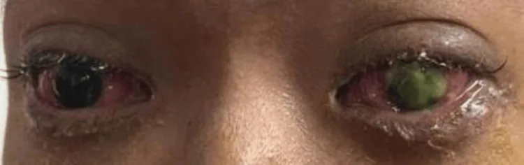
Che Ku Hafiza Che Ku Amran, Qi Zhe Ngoo, Fadil Awis Qarni A Rare Case of Corneal Perforation Secondary to Gonococcal Keratoconjunctivitis 2024 Nov 23 Cureus.; 16(11):e74312 Figure 1. PMCID: PMC11666296. License: CC BY.
Both eyes show marked conjunctival injection and eyelid swelling. In the right eye, a thick yellowish-green membranous deposit is seen on the cornea. This is an ocular surface finding seen in severe membranous or pseudomembranous conjunctivitis.
The main complaints are eye redness, foreign body sensation, tearing, and watery discharge. The pseudomembrane causes strong discomfort due to irritation. Eyelid swelling may make it difficult to open the eyes. Depending on the severity of inflammation, it may be unilateral or bilateral.
A thin yellow membrane is observed on the palpebral conjunctiva and fornix. The membrane may be patchy or cover the entire palpebral conjunctiva. It rarely affects the bulbar conjunctiva.
Fluorescein staining shows the membrane stained bright green, and it may be accompanied by corneal epithelial defects. Conjunctival injection, chemosis, mucopurulent discharge, and preauricular lymphadenopathy are present.
In infants with viral conjunctivitis, follicular formation is weak and pseudomembrane formation is predominant. In adults, the pseudomembrane tends to bleed when removed.
Diagnosis is based on medical history and clinical findings. The membrane is confirmed by slit-lamp microscopy, and fluorescein staining evaluates the extent of the membrane and corneal epithelial damage.
The presence or absence of bleeding when the pseudomembrane is peeled with forceps is used to differentiate between true membrane and pseudomembrane. However, clinical distinction may not be clear 1).
Microscopic examination of conjunctival smear (Diff-Quick) staining is useful for estimating the underlying disease. Viral infections show predominantly mononuclear cells, while bacterial infections show predominantly neutrophils. Note that scraping the pseudomembrane during sample collection may result in neutrophil predominance.
PCR testing is useful for identifying microorganisms such as adenovirus. A positive rapid adenovirus diagnostic kit (immunochromatography) can also confirm the diagnosis.
Evaluation of systemic symptoms helps identify non-infectious causes such as SJS, pemphigoid, and GVHD. In ligneous conjunctivitis, the pseudomembrane is thick, yellowish-white, and hard, and similar membrane formation may occur on the oral mucosa.
The basis of treatment is addressing the underlying disease and controlling conjunctival inflammation.
Management of Pseudomembrane
Pseudomembrane removal: If corneal epithelial damage is present, remove with forceps. Grasp one end of the pseudomembrane widely while minimizing invasion of the conjunctiva.
Debridement of true membrane: This is controversial because removal exposes an eroded surface and increases the risk of scarring 1).
Conservative treatment: Cases have been reported where pseudomembrane resolved spontaneously with only steroid eye drops and artificial tears 1).
Pharmacological Treatment
Steroid eye drops: Instill dexamethasone or fluorometholone 0.1% 3–6 times daily to control inflammation. Use the minimum dose while being cautious of prolonged infection.
Antibacterial eye drops: Used concomitantly to prevent mixed bacterial infection. Important for preventing secondary infection when corneal epithelial defects are present.
Artificial tears: Frequent instillation of preservative-free artificial tears promotes washing out of exudates. Especially actively used in non-infectious causes (SJS, ocular pemphigoid, GVHD).
If symblepharon is present, daily separation of the fornix adhesions with a glass rod is performed. In SJS-related membrane formation, discontinuation of the causative drug and early amniotic membrane transplantation are also considered.
In ligneous conjunctivitis, if tranexamic acid is involved, discontinuation leads to improvement. For other causes, inflammation is reduced with steroids or immunosuppressants.
Re-evaluate within 3–7 days to check healing status and complications.
QShould pseudomembrane always be removed?
A
Removal of pseudomembrane is widely recommended, but recent reports have shown that conservative treatment (only steroid eye drops and artificial tears) can also lead to a favorable course 1). Removal is effective when corneal epithelial disorder is present, but careful judgment is required when true membrane is suspected due to the risk of bleeding and scarring.
Membrane formation results from severe conjunctival inflammation.
Pseudomembrane is a coagulation of fibrinous exudate on the conjunctival epithelial surface. Neutrophils and necrotic epithelial cells are entangled in the fibrin network, and it does not contain blood vessels or lymphatics. It appears translucent and pearl-like, and the epithelium is preserved upon removal.
In true membrane, more intense inflammation causes exudate to penetrate the superficial layer of the epithelium. The fibrin network insinuates between epithelial cells, and a highly vascularized inflammatory membrane forms with capillary ingrowth. The epithelium undergoes coagulative necrosis, and removal strips the epithelium, causing bleeding. Healing occurs as granulation tissue forms under the membrane and epithelium migrates to reconstruct.
In infants, the epithelial structure is immature, so the entire infected epithelium tends to slough off, leading to pseudomembrane formation. In adults, the epithelium is preserved, so bleeding is more likely upon removal.
Pseudomembrane and true membrane form a continuous spectrum, transitioning depending on the degree of inflammation. Histopathologically, a matrix of fibrin, fibronectin, and tenascin contains neutrophils, and macrophages are also seen in older membranes.
Sufficient evidence has not been established for the optimal treatment of adenoviral pseudomembranous conjunctivitis. Debridement of pseudomembrane is generally recommended, but no prospective comparative trials demonstrating its efficacy exist 1).
Good outcomes of adenoviral pseudomembranous conjunctivitis with conservative treatment (only steroid eye drops and artificial tears) have been reported 1). There is also an RCT showing that the combination of povidone-iodine and dexamethasone is effective for early resolution of symptoms. Combination of cyclosporine A and steroid eye drops has also been reported to reduce symptoms.
In the future, comparative studies to verify the efficacy of debridement are needed.
Gilmour KM, Ramaesh K. Case for conservative management of adenoviral pseudomembranous conjunctivitis. BMJ case reports. 2023;16(2). doi:10.1136/bcr-2022-253014. PMID:36810328; PMCID:PMC9945021.