Intracorneal ring segments (ICRS) were introduced in 1987 as synthetic corneal implants for myopia correction. They are placed outside the central optical zone, at approximately two-thirds depth of the corneal stroma.
ICRS function as spacers between corneal lamellae. They shorten the arc length of the central cornea in proportion to the device thickness (arc shortening effect). As a result, the central anterior cornea flattens, and the peripheral area adjacent to the ring insertion is pushed forward.
According to Barraquer’s law, adding tissue to the corneal periphery flattens the center. ICRS utilize this principle. The thicker the device and the smaller its diameter, the greater the refractive correction achieved.
Initially used for myopia correction, ICRS are now positioned as a therapeutic intervention for corneal ectatic diseases such as keratoconus and post-LASIK ectasia, due to limitations in correction range and induced astigmatism.
QCan ICRS cure keratoconus?
A
ICRS is not a curative treatment for keratoconus. It is a surgical alternative aimed at reducing irregular astigmatism and improving vision, thereby at least delaying the need for corneal transplantation. Combined use with corneal cross-linking (CXL) adds the effect of halting progression.
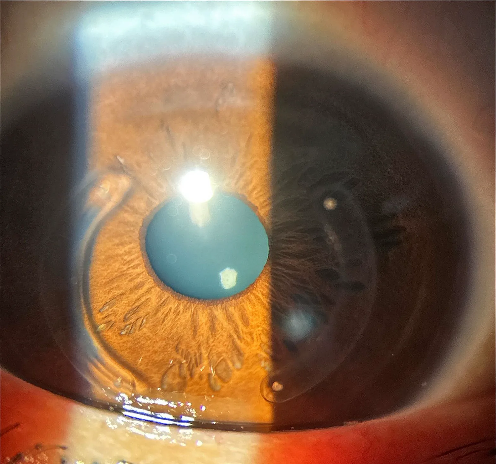
Roberto Albertazzi, Carlos Rocha-de-Lossada, Roger Zaldivar A new technique to implant intracorneal ring-segments from the perilimbal region: one-year prospective pilot study report 2024 Jul 16 BMC Ophthalmol. 2024 Jul 16; 24:288 Figure 4. PMCID: PMC11251366. License: CC BY.
Slit-lamp cross-sectional image showing two arcuate ICRS inserted within the corneal stroma. The stromal structure around the rings, insertion depth, and postoperative corneal shape can be observed.
Corneal ectasia, the condition indicated for ICRS, presents with the following symptoms. The main complaint is decreased visual acuity due to progressive irregular astigmatism. Often, glasses or soft contact lenses do not provide adequate correction.
Keratoconus is characterized by central to paracentral corneal protrusion and thinning. Corneal topography shows a steepening pattern 1). Changes in corneal biomechanics precede morphological changes 1).
Topography after ICRS insertion shows overall corneal flattening, centralization of the corneal apex, maintenance of corneal asphericity, and reduction of surface irregularity.
The etiology of corneal ectasia, for which ICRS is indicated, is multifactorial.
Collagen degradation in the cornea is the essence of thinning 1). Increased matrix metalloproteinases (MMPs) and decreased TIMP are observed 1). Increases in IL-6, TNF-α, and mucosal pemphigoid-9 in tears induce keratocyte apoptosis1).
Eye rubbing is a major risk factor for keratoconus1). It is known to be associated with atopic diseases (hay fever, asthma, eczema, vernal keratoconjunctivitis) 1).
Post-LASIK ectasia can occur when laser refractive surgery is performed on a cornea with unrecognized subclinical keratoconus1). Thinning of the residual stromal bed and weakening of corneal structure are involved.
For the diagnosis of corneal ectasia, combined use of corneal tomography (Scheimpflug imaging or OCT) and corneal biomechanical assessment is recommended 2).
Parameter
Characteristic
TBI (Tomographic Biomechanical Index)
Integrated index of morphology and biomechanics. High diagnostic performance 2)
CBI (Corvis Biomechanical Index)
Indicator of corneal deformation response to air puff 2)
CRF (Corneal Resistance Factor)
Reflects overall corneal stiffness 2)
Since a single index may produce false negatives, comprehensive screening combining corneal tomography and biomechanical evaluation is recommended 2). In keratoconus, biomechanical changes precede morphological changes, making it useful for early detection 1).
Channel creation methods include mechanical dissection and femtosecond laser1). The implantation depth is typically 70–80% of corneal thickness. With femtosecond laser, channels are created at precise depth and diameter based on pachymetry maps 1).
The average change in corneal curvature after ICRS insertion ranges from 2.14 to 9.60 D. Reductions in spherical power, astigmatic power, and spherical equivalent have been reported. It is considered most effective for moderate keratoconus (less than 58.0 D) 1). However, astigmatic changes can be unpredictable 1).
ICRS alone may not halt the progression of keratoconus. Combination with CXL has been shown to be effective for both halting progression and improving visual function1).
Simultaneous ICRS + CXL showed superior results in spherical refractive error and steep-K compared to CXL-first or ICRS-first approaches1). In a report by Chan et al., Intacs + CXL combination was more effective than Intacs alone for improving keratoconus3).
Corneal Allogenic Intrastromal Ring Segments (CAIRS) are an alternative method first reported in 2017, where rings are harvested from donor corneal tissue1). Long-term outcomes of combination with CXL are awaited.
QWhat are the advantages of combining ICRS and CXL?
A
ICRS improves corneal shape and visual acuity, but alone may not halt the progression of keratoconus. CXL increases corneal rigidity through collagen cross-linking, providing a progression-stopping effect. The combination yields synergistic effects of shape improvement and progression arrest. Some reports indicate that simultaneous surgery yields the best results.
The elastic modulus of the cornea is a quantitative measure of its tendency to deform elastically under force. In keratoconus, the elastic modulus is reduced due to pathological changes in the stroma.
The decrease in elastic modulus results from degradation and degeneration of collagen fibers2). This initiates a biomechanical failure cycle. Stress levels increase and redistribute, leading to corneal steepening and thinning2). At the thinning site, local stress further increases, forming a vicious cycle that worsens the protrusion.
ICRS intervenes in this vicious cycle through the following mechanisms.
Placed as spacers between the corneal lamellae, ICRS shorten the arc length. This flattens the central cornea, redistributing curvature and leading to stress redistribution. In some cases, it can interrupt the progression cycle of keratoconus.
The effect of ICRS is closely related to the structural properties of the collagen framework in the corneal stroma. The stroma accounts for 90% of corneal thickness, and its mechanical properties determine the overall biomechanics of the cornea.
QIs the effect of ICRS on corneal biomechanics permanent?
A
The effect of ICRS itself persists as long as the implant remains in the cornea. However, regression of spherical correction may be observed in the medium to long term, and ICRS alone may not completely halt the progression of keratoconus. In case of complications, the ring can be removed, and the cornea generally returns to its original state after removal.
Advances in corneal biomechanical evaluation are attracting attention. New indices such as TBI and CBI complement traditional morphological indices and improve the detection accuracy of early keratoconus2). Integrated assessment of biomechanical indices and corneal tomography has been reported to improve the prediction accuracy of refractive surgery by more than 25% 2).
A meta-analysis on the combination of CXL and ICRS, including 6 studies with 12-month follow-up, showed that simultaneous surgery was superior to CXL-first in spherical refractive error and flat-K, and superior to both CXL-first and ICRS-first in steep-K. 1)
CAIRS is a new approach using allogeneic corneal tissue, and is expected to have advantages in biocompatibility compared to synthetic implants 1). Long-term results including combination with CXL are awaited.
It has also been reported that patients with low corneal stiffness have a 2-3 times higher risk of residual refractive error after KLEx (keratolenticular extraction) 2), highlighting the increasing importance of preoperative biomechanical evaluation.