Ocular mucous membrane pemphigoid (OCP) is a chronic progressive autoimmune blistering disease characterized by scarring of the mucosal epithelium, including the conjunctiva, caused by autoantibodies against adhesion components of the basement membrane. Mucous membrane pemphigoid (MMP) is a group of systemic diseases that can affect multiple mucous membranes such as the oral cavity, ocular surface, nasopharynx, larynx, esophagus, and genitalia. Those with ocular involvement are called ocular pemphigoid or ocular cicatricial pemphigoid. Cases without skin lesions are not uncommon.
In ophthalmology, OCP is considered a representative disease of cicatrizing keratoconjunctival epitheliopathy with corneal epithelial stem cell deficiency, and it follows a staged progression from chronic conjunctivitis to symblepharon and eventually corneal keratinization.
This disease is rare. A surveillance study in the UK reported an annual incidence of cicatrizing conjunctivitis of approximately 1.3 per million population, and the prevalence of OCP is estimated to be about 1 in 10,000 to 50,000 people5). It predominantly affects the elderly in their 60s to 80s, with a female-to-male ratio of about 2:15). The most common mucosal site is the oral cavity (approximately 85%), followed by the eyes (approximately 65%), and about 50% of OCP patients have extraocular involvement such as oral or skin lesions2).
It typically develops insidiously as bilateral chronic conjunctivitis, and the disease duration often extends from several years to over a decade. In the early stages, only nonspecific conjunctivitis symptoms are present, so it is frequently treated as allergic conjunctivitis, chronic conjunctivitis, or dry eye for a long time. Careful history-taking and slit-lamp examination with this disease in mind are extremely important for early diagnosis and improving visual prognosis.
Overview of Mucous Membrane Pemphigoid (MMP)
Target organs: Mucous membranes with stratified squamous epithelium throughout the body, including the oral cavity, ocular surface, nasopharynx, larynx, esophagus, and genitalia.
Skin lesions: Often absent. Erosions and scarring are more prominent than blisters.
Age of onset: 60s to 80s. Female predominance.
Systemic complications: Dysphagia due to esophageal stricture, and laryngeal stenosis can affect life prognosis2).
Features of Ocular Cicatricial Pemphigoid (OCP)
Nature of ocular involvement: Bilateral chronic conjunctivitis with progressive conjunctival scarring.
Extraocular involvement: Approximately 50% of cases have involvement of other sites such as the oral cavity and skin2).
Insidious progression: May be asymptomatic for a long time, and some cases are discovered due to vision loss2).
QAre mucous membrane pemphigoid (MMP) and ocular cicatricial pemphigoid (OCP) the same disease?
A
Mucous membrane pemphigoid (MMP) is a general term for autoimmune blistering diseases that affect multiple mucous membranes, including the oral cavity, eyes, nasopharynx, larynx, esophagus, and genitalia. Among these, cases with ocular involvement are called ocular cicatricial pemphigoid (OCP). OCP may involve only the eyes or be accompanied by extraocular lesions such as oral or skin involvement; approximately 50% of cases have extraocular involvement2). Since skin lesions are often inconspicuous, ophthalmologists are frequently the first to reach the diagnosis.
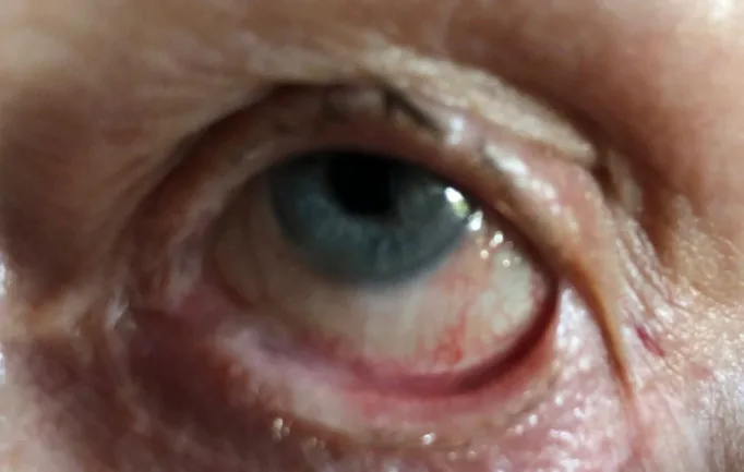
Stan C, Golea A, Gheorghe A, et al. Ocular cicatricial pemphigoid. Rom J Ophthalmol. 2020 Apr-Jun;64(2):226-230. Figure 2. PMCID: PMC7339695. License: CC BY.
In the left eye, symblepharon (adhesion between the eyelid and bulbar conjunctiva) is formed. This shows a specific example of symblepharon described in the section on main symptoms and clinical findings.
In the early stage, chronic conjunctival hyperemia, dryness, foreign body sensation, burning sensation, and tearing are the main complaints. These are resistant to usual dry eye treatment, and their persistence raises suspicion for this disease 1). In advanced cases, ocular movement disorder due to symblepharon, incomplete eyelid closure, corneal irritation from trichiasis, and decreased visual acuity due to corneal opacity or conjunctival epithelial invasion are added.
The progression of this disease is insidious, and it may remain asymptomatic for a long time, with some cases first discovered when visual acuity decreases 2).
The Foster classification (modified Tauber-Foster classification) is widely used for staging. It progresses stepwise from stage I chronic conjunctivitis, to stage II shortening of the inferior fornix, stage III symblepharon and corneal vascularization, and stage IV keratinization of the ocular surface 2).
Stage
Main Findings
Stage I
Chronic conjunctivitis, Rose bengal positive (mucin deficiency), subepithelial conjunctival fibrosis
Stage II
Shortening of the inferior fornix (A–D: progression assessed in 25% increments)
Keratinization of the ocular surface, ankyloblepharon, severe dryness, dermalization of the cornea
In the early stage, scars are observed as linear or patchy white tendon-like tissue on the palpebral conjunctiva. As the disease progresses, shortening of the conjunctival fornix, loss of the palisades of Vogt (POV), conjunctival epithelial invasion into the cornea, lacrimal duct obstruction, loss of conjunctival goblet cells, and severe dry eye due to meibomian gland loss appear. During acute exacerbations, extensive persistent epithelial defects may occur.
Histologically, conjunctival stromal fibrosis, complete loss of goblet cells, and squamous metaplasia are observed 1). Loss of goblet cells means loss of conjunctival mucin secretion function, and the stability of the tear film is significantly impaired. In addition, the lipid layer is also lost due to destruction of the meibomian glands, leading to increased tear evaporation and severe evaporative dry eye.
As systemic mucosal complications, it is necessary to evaluate oral lesions causing desquamative gingivitis and bullous lesions, esophageal lesions causing dysphagia, and laryngeal/tracheal lesions causing hoarseness and dyspnea. Laryngeal stenosis is a life-threatening factor requiring airway management, and urgent collaboration with an otolaryngologist is needed for patients with respiratory symptoms 2). Esophageal stenosis causes dysphagia and malnutrition, and endoscopic evaluation by a gastroenterologist and balloon dilation if necessary are considered. Skin lesions often appear as erythematous or atrophic changes rather than blisters, and tend to occur particularly on the head, face, and neck.
Ocular mucous membrane pemphigoid is caused by an autoimmune mechanism (see Section 6 for details). Risk factors for onset include aging, female sex, and a history of other autoimmune diseases. No clear trigger has been identified, but infection and drug exposure are discussed as possible factors in the breakdown of immune tolerance. An association with HLA-DQB1*0301 has been reported in overseas studies, suggesting a genetic predisposition.
Clinically, it is important to differentiate from other diseases that present with cicatrizing conjunctivitis. The following diseases have different pathologies and treatments, so the cause must be narrowed down at the initial visit.
Autoimmune/Inflammatory Diseases to Differentiate
Stevens-Johnson syndrome/toxic epidermal necrolysis: Acute mucocutaneous disorder due to drug reaction. Differentiated by history of fever and generalized rash.
Graft-versus-host disease (GVHD): Occurs after hematopoietic stem cell transplantation. In severe and progressive adhesion cases, consider OCP comorbidity 4).
Sarcoidosis: Non-caseating granulomatous conjunctivitis that can scar. Reported as a mimic of OCP 3).
Lichen planus/linear IgA bullous dermatosis: Requires dermatological evaluation.
Exogenous Diseases to Differentiate
Drug-induced pseudopemphigoid: Long-term use of antiglaucoma drugs (e.g., latanoprost, timolol, brinzolamide) is a typical example. Improves with drug discontinuation 7).
Chemical injury/thermal burn: Alkali injuries are particularly severe.
Infectious: Trachoma is a major cause in developing countries. In developed countries, severe cases of adenoviral keratoconjunctivitis.
Iatrogenic: History of ophthalmic surgery involving conjunctival incision
Bilateral chronic inflammation of the corneal and conjunctival epithelium slowly progressing to scarring strongly suggests OCP. Especially in elderly women with trichiasis or entropion, carefully evaluate for conjunctival fornix shortening and symblepharon.
Take a detailed history of ocular trauma, ocular infections, and past and current use of eye drops and oral medications. In addition, perform a systemic review of mucosal lesions in the oral cavity, nasopharynx, larynx, esophagus, and skin, and collaborate with dermatology, otorhinolaryngology, gastroenterology, and rheumatology as needed 2).
Impression cytology: Cytology from the conjunctiva confirms loss of goblet cells.
Fluorescein staining: Increased permeability is observed where conjunctival epithelium invades the corneal surface, and together with loss of POV, indicates corneal epithelial stem cell deficiency 8).
The cornerstone of definitive diagnosis is conjunctival biopsy. A specimen is taken from the inferior bulbar conjunctiva, and histopathological examination of formalin-fixed tissue and direct immunofluorescence (DIF) of Michel’s solution-fixed tissue are performed simultaneously 5).
A positive DIF finding in OCP is linear deposition of IgG, IgA, IgM, and complement C3 along the conjunctival epithelial basement membrane zone 5). However, false negatives are not uncommon. Causes include disease quiescence, complete loss of basement membrane in end-stage disease, inappropriate biopsy site, and local variation in immune response. In cases strongly suspected clinically, a negative DIF does not rule out the disease, and multiple biopsies including re-biopsy of the contralateral eye can improve sensitivity 4,5).
Autoantibodies: Anti-BP180 antibodies (anti-type XVII collagen antibodies) and anti-laminin 332 antibodies (anti-laminin 5 antibodies) are detected in many OCP patients. These autoantibodies play a central role in the pathogenesis.
Acute onset due to drug reaction; history of systemic fever and rash
Drug-Induced Pseudo-OCP
Long-term use of epitheliotoxic drugs such as antiglaucoma eye drops. Improvement after discontinuation of the causative drug 7)
Chronic GVHD
History of hematopoietic stem cell transplantation. However, in cases with severe adhesions, attention should be paid to possible coexistence with OCP 4)
Even if ACE/lysozyme levels are normal, sarcoidosis remains in the differential; negative immunofluorescence is useful to exclude OCP 3)
QWhy can OCP not be ruled out even if direct immunofluorescence of conjunctival biopsy is negative?
A
DIF testing is not completely sensitive, and there are multiple factors that can cause false negatives. The main causes include the quiescent phase of the disease, cases where the basement membrane has completely disappeared in the end stage, when the biopsy site is from an area with low activity, and local variation in immune deposition. Cases where results differ between the right and left eyes have also been reported 5). When clinical findings strongly suggest OCP, sensitivity can be improved by repeated testing including re-biopsy from the contralateral eye or another site 4). Detection of serum anti-BP180 antibodies and anti-laminin 332 antibodies is also useful as a diagnostic aid.
The goals of treatment are to halt progressive scarring, prevent and correct corneal and eyelid complications, and relieve symptoms. This disease is a systemic autoimmune disorder, and local therapy alone cannot control its progression. The mainstay of treatment is systemic immunosuppressive therapy, and collaboration with dermatology and rheumatology departments is strongly recommended 2).
Medications are selected stepwise according to disease activity and inflammatory status. In all cases, side effect monitoring and regular blood tests are essential 2,5).
Systemic Therapy: Stepped Selection
Mild to moderate: Dapsone (diaminodiphenyl sulfone) 50–200 mg/day is first-line. Exclusion of G6PD deficiency and monitoring for anemia and methemoglobinemia are mandatory 5).
Dapsone ineffective/moderate: Step up to mycophenolate mofetil 1–3 g/day, azathioprine 1–2 mg/kg/day, or methotrexate 7.5–25 mg/week 2).
Severe/acute exacerbation: Combination of cyclophosphamide 1–2 mg/kg/day and short-term high-dose prednisolone 5).
Glaucoma cases: Switch to preservative-free eye drops as a basic measure. There are also case reports of minimally invasive glaucoma surgery (e.g., XEN gel stent) 6).
End-stage: Cultured mucosal epithelial transplantation for corneal epithelial reconstruction, corneal prosthesis (Boston KPro type 2, OOKP) for severe cases
Many of the drugs are not covered by insurance for this disease, and sufficient explanation and ethical consideration are required before initiation. Note that oral cyclosporine and cyclophosphamide are also not covered by insurance for this disease. Dapsone can induce hemolysis and methemoglobinemia, so glucose-6-phosphate dehydrogenase (G6PD) activity should be measured before administration to rule out deficiency. After starting treatment, regular blood counts, liver function, and renal function should be assessed, and the dose should be gradually increased. Mycophenolate mofetil is relatively well-tolerated and suitable for long-term use, but infection screening should be performed considering the risk of opportunistic infections. Cyclophosphamide is a potent immunosuppressant for severe cases, but it carries risks of bone marrow suppression, hemorrhagic cystitis, and long-term carcinogenicity, so the total dose must be managed. Rituximab, as an anti-CD20 monoclonal antibody, is positioned as salvage therapy for refractory OCP, and assessment of hepatitis B virus reactivation risk before administration is important2).
Local treatment is important as symptomatic therapy adjunctive to systemic therapy. Low-concentration steroid eye drops reduce ocular surface inflammation, and preservative-free artificial tears manage dry eye. Surgical interventions such as punctal occlusion should be carefully considered as they may accelerate scarring.
Surgical treatment should only be performed when inflammation is completely controlled. Surgery under active inflammation can lead to rapid progression of scarring, adhesion, and keratinization2).
Cataract surgery: If necessary, perform after sufficient anti-inflammatory treatment with systemic immunosuppressive therapy starting on the day of surgery. If performed without adequate anti-inflammatory control, adhesion and keratinization may rapidly progress postoperatively.
Corneal epithelial reconstruction: For persistent epithelial defects caused by acute exacerbation, first perform conservative treatment with oral steroids. For refractory cases, options include autologous or allogeneic cultured corneal epithelial sheet transplantation, autologous oral mucosal epithelial transplantation, etc. Autologous oral mucosal epithelial transplantation is approved as advanced medical care in Japan.
Surgery for visual recovery in end-stage: In severe cases with conjunctival invasion and keratinization of the cornea, surgical treatment for visual recovery has a poor prognosis and is usually not performed.
In bilateral progressive diseases such as severe OCP, the indications for autologous limbal stem cell transplantation (CLAu, CLET, SLET) are limited. Many cases are bilateral and lack a healthy donor eye, so allogeneic transplantation or cultured mucosal epithelial transplantation is considered 9). A systematic review of autologous limbal stem cell transplantation, mainly for unilateral chemical trauma, reported an anatomical success rate of 69% and functional success rate of 60% in 1,023 eyes, but it is unlikely to be the first choice in bilateral autoimmune diseases such as OCP 9).
QHow can cataract surgery be safely performed in OCP patients?
A
Surgery in the presence of active inflammation is a major risk factor for rapid progression of symblepharon and corneal keratinization. When planning surgery, systemic immunosuppressive therapy (e.g., steroids, cyclosporine, cyclophosphamide) should be used from the day of surgery to thoroughly suppress inflammation, intraoperative physical damage to the ocular surface should be minimized, and immunosuppression should be continued postoperatively. If surgery is performed with insufficient preoperative inflammation control, there is a high risk of rapid postoperative progression of adhesions and keratinization. Surgical technique selection, systemic management, and collaboration with dermatology and rheumatology are extremely important 2).
OCP is a type II (cytotoxic) autoimmune disease characterized by the production of autoantibodies against hemidesmosome-associated basement membrane components 5).
BP180 (type XVII collagen): A transmembrane protein of hemidesmosomes that connects epidermal/epithelial cells to the basement membrane. Anti-BP180 antibodies are among the most frequently detected autoantibodies in OCP patients.
Laminin 332 (laminin 5): An adhesion protein in the basement membrane that links hemidesmosomes to type IV collagen. Anti-laminin 332 antibodies are also detected in many OCP patients.
Other antibodies against α6β4 integrin and other basement membrane components have also been reported.
When autoantibodies bind to the conjunctival epithelial basement membrane, the complement system is activated and inflammatory cells are recruited. In the acute phase, eosinophils and neutrophils are the main inflammatory cells, while in the chronic phase, lymphocytic infiltration predominates 5). Released cytokines and proteases activate fibroblasts beneath the conjunctival epithelium, promoting excessive production of extracellular matrix such as collagen, leading to conjunctival fibrosis and scarring. Th2-type cytokines such as transforming growth factor-β (TGF-β), interleukin-4 (IL-4), IL-5, and IL-13 are upregulated in the lesion and are considered major drivers of fibrosis. An imbalance between matrix metalloproteinase-9 (MMP-9) and tissue inhibitors (TIMPs) leads to uncontrolled remodeling of conjunctival tissue, which is also a characteristic of the pathology.
Corneal epithelial stem cell exhaustion and corneal keratinization
The progressive stages of conjunctival scarring correspond to the Foster classification. Persistent chronic inflammation leads to subconjunctival fibrosis (stage I), shortening of the conjunctival fornix (stage II), symblepharon (stage III), and ultimately loss of the palisades of Vogt (POV) at the corneal limbus8). Since corneal epithelial stem cells reside in the POV, loss of POV indicates failure of normal corneal epithelial regeneration. Depletion of corneal epithelial stem cells leads to conjunctivalization, where conjunctival epithelium invades the corneal surface, resulting in corneal opacity, vascularization, and eventually skin-like keratinization (stage IV) 8).
Chronic ocular graft-versus-host disease (oGVHD) after hematopoietic stem cell transplantation typically presents with chronic ocular surface inflammation, dry eye, and mild conjunctival fibrosis. However, when severe and rapidly progressive scarring and symblepharon occur, the possibility of OCP should be considered 4). Although oGVHD and OCP have similar clinical features, their treatment strategies differ, making differentiation by conjunctival biopsy and DIF important 4).
QWhat is corneal epithelial stem cell exhaustion in OCP?
A
The corneal limbus contains stem cells responsible for corneal epithelial regeneration, observable as the palisades of Vogt (POV). In OCP, chronic inflammation and scarring extend to the limbus, leading to loss of POV and stem cells. When stem cells are depleted, conjunctival epithelium invades the corneal surface (conjunctivalization), making normal corneal epithelial regeneration impossible. This results in corneal opacity, vascularization, and keratinization, leading to severe visual impairment 8). This condition represents the final common pathway of severe ocular surface diseases seen in ocular mucous membrane pemphigoid, Stevens-Johnson syndrome, and chemical injury.
Case series have shown the efficacy of rituximab and IVIG for refractory OCP, and they are becoming established as treatment options for cases resistant to conventional immunosuppressive therapy2). Rituximab administration eliminates autoantibody-producing B cells, and cases achieving clinical remission have been reported. IVIG has both anti-inflammatory and immunomodulatory effects and is positioned as combination therapy for severe or drug-resistant cases. The potential of new immunomodulators such as JAK inhibitors is also being discussed, and their oral administration, which facilitates outpatient management, is expected to lead to future clinical application.
In the field of corneal epithelial reconstruction, cultured corneal epithelial sheet transplantation and autologous oral mucosal epithelial sheet transplantation have been clinically applied, and their development as advanced medical treatments is progressing. Autologous corneal epithelial sheets are prepared using corneal epithelial cells from the healthy eye, but they cannot be used in OCP with bilateral involvement. Therefore, allogeneic corneal epithelial sheets using corneas provided by eye banks or autologous oral mucosal epithelial sheets prepared from the patient’s own oral mucosal cells are selected. Autologous oral mucosa has no risk of rejection and is expected to provide long-term ocular surface stabilization. Carrier materials such as amniotic membrane, fibrin glue, temperature-responsive culture dishes, and polyvinylidene fluoride (PVDF) membranes have been developed, expanding the options for surgical techniques 8).
For cases with concurrent glaucoma, conventional filtering surgery is difficult due to conjunctival scarring, but successful cases of minimally invasive glaucoma surgery (MIGS) using the XEN gel stent have been reported. After controlling ocular surface inflammation, the stent is inserted via an ab interno approach, and cases achieving intraocular pressure control without eye drops and inflammation remission at one year postoperatively have been demonstrated, attracting attention as a new approach for glaucoma complicated by severe ocular surface disease 6). Future expectations include accumulation of long-term results through multicenter collaborative studies and evaluation of treatment response using biomarkers.
Farrag A, Chan A, Tong L. Cicatricial Conjunctivitis and Concurrent Clinical Features: A Case Study. Clinical medicine insights. Case reports. 2022;15:11795476221100605. doi:10.1177/11795476221100605. PMID:35601266; PMCID:PMC9121463.
Razzak A, Ait Ammar H, Bouazza M, Elbelhadji M. Accidental Discovery of Ocular Cicatricial Pemphigoid. Cureus. 2025;17(1):e77425. doi:10.7759/cureus.77425. PMID:39949452; PMCID:PMC11823481.
Murati Calderon RA, López-Fontanet JJ, Ramirez Marquez E, Tavares Reigada AS, Oliver A. Sarcoidosis: A Mimicker of Ocular Cicatricial Pemphigoid. Cureus. 2025;17(10):e93862. doi:10.7759/cureus.93862. PMID:41194826; PMCID:PMC12584608.
Taketani Y, Dehghani S, Sinha S, Freitag SK, Papaliodis G, Foster S, et al. Concurrence of Ocular Cicatricial Pemphigoid in Chronic Ocular Graft-Versus-Host Disease. Cornea. 2024;43(3):387-390. doi:10.1097/ICO.0000000000003386. PMID:38128104; PMCID:PMC12703942.
Tesorero JCC, Sosuan GMN, Lim Bon Siong R. Ocular Cicatricial Pemphigoid in a Healthy Elderly Male Filipino Patient. Acta Med Philipp. 2025;59(18):117-123.
Hu JCW, Trief D. A narrative review of limbal stem cell deficiency & severe ocular surface disease. Ann Eye Sci. 2023;8:21. doi:10.21037/aes-22-35.
Shanbhag SS, Nikpoor N, Rao Donthineni P, Singh V, Chodosh J, Basu S. Autologous limbal stem cell transplantation: a systematic review of clinical outcomes with different surgical techniques. The British journal of ophthalmology. 2020;104(2):247-253. doi:10.1136/bjophthalmol-2019-314081. PMID:31118185.
Copy the article text and paste it into your preferred AI assistant.
Article copied to clipboard
Open an AI assistant below and paste the copied text into the chat box.