Initial evaluation of ocular trauma is often performed by non-ophthalmologists such as emergency physicians, general internists, and primary care physicians. A systematic approach allows early identification of vision-threatening conditions and optimizes prognosis.
The basic principle of triage is two-tiered: first, prioritize life-threatening conditions (e.g., intracranial injury, airway obstruction), then address vision-threatening conditions. Appropriate initial management before referral to an ophthalmologist directly impacts visual prognosis.
Epidemiological features of ocular trauma are as follows:
According to 2008 US statistics, there were approximately 640,000 emergency department visits for eye injuries (209 per 100,000 population).
44.6% of ocular injuries occur at home, and 44.4% are contusions or corneal abrasions.
The incidence is high in children, peaks in the 20s, and then decreases with age.
Young age and male sex are major risk factors.
The global incidence of open-globe injuries is 3.5 to 4.5 per 100,000 population. 1)
Ocular trauma is broadly classified into mechanical and non-mechanical trauma. Mechanical trauma is further classified into mechanical injuries (open-globe and closed-globe). Checking vital signs (consciousness, respiration, blood pressure, pulse, temperature) is an essential part of the initial assessment.
QHow often is ocular trauma seen in the emergency department?
A
In the United States in 2008, approximately 640,000 emergency department visits (209 per 100,000 population) were due to eye injuries. 44.6% of injuries occurred at home, and contusions or corneal abrasions accounted for 44.4% of all cases.
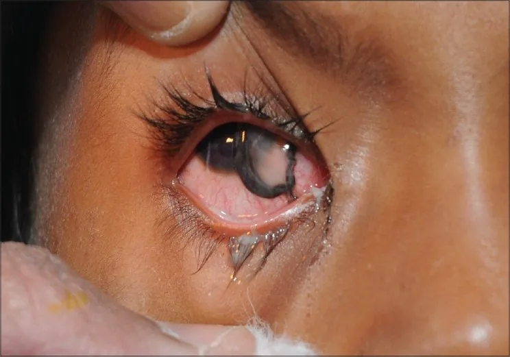
Kumar R, Puttanna M, Sriprakash KS, et al. Firecracker eye injuries during Deepavali festival: A case series. Indian J Ophthalmol. 2010;58(2):157. Figure 5. PMCID: PMC2854452. License: CC BY.
Clinical photograph of a penetrating open-globe injury caused by a firecracker. It is useful for conveying the severity of ocular trauma that should trigger urgent ophthalmic referral.
Orbital fracture: Vertical eye movement restriction (inferior rectus entrapment), diplopia, sensory disturbance in the infraorbital nerve area. Medial rectus entrapment causes horizontal movement restriction. Pseudo-Duane retraction syndrome (globe retraction and palpebral fissure narrowing on attempted abduction in medial wall fracture) may also be observed.
Orbital compartment syndrome: Characteristic findings include proptosis, ophthalmoplegia, periorbital edema, and a firm eyelid.
Other important findings
Shafer sign (tobacco dust): Brown pigment cells in the anterior vitreous. Suggests a retinal tear.
Initial visual acuity less than 5/200: The most important negative prognostic factor in open globe injuries.
The mechanisms of ocular trauma are diverse, and the pathology and prognosis vary greatly depending on the type of injury.
Open Globe Injury
Definition: Full-thickness wound of the cornea or sclera.
Laceration (sharp external force): There are three types: penetrating injury (single wound), perforating injury (entry + exit), and intraocular foreign body (IOFB).
Rupture (blunt external force): Occurs due to a sudden rise in intraocular pressure. It often results in a scleral wound parallel to the limbus, and diagnosis may be delayed because it is covered by conjunctiva and Tenon’s capsule.
Closed Globe Injury
Definition: Injury without a full-thickness wound.
Contusion: Damage to the eyeball and surrounding tissues caused by blunt force. It can cause hyphema, lens subluxation, retinal detachment, etc.
Lamellar laceration: Partial-thickness corneal or scleral laceration that does not extend through the full thickness.
The Birmingham Eye Trauma Terminology (BETT) and the Globe and Adnexal Trauma Terminology are used as classification systems. 1)
The injury site is classified into the following three zones. 1)
Zone
Extent
Characteristics
Zone I
Cornea to limbus
Limited to the cornea
Zone II
Limbus to 5 mm posterior to the sclera
Includes the anterior sclera
Zone III
More than 5 mm posterior to the sclera
Posterior sclera and perioptic area
Main causes of blunt trauma: Sports (baseball, golf, boxing, soccer, etc.), traffic accidents (airbags), falls, fireworks.
Causative agents and mechanisms of chemical trauma: Alkalis saponify fatty acids, causing liquefactive necrosis and penetrating deep into tissues. When they reach the limbal stem cells, healing is severely impaired. Acids cause coagulative necrosis due to protein denaturation, forming a barrier that limits deep penetration. Therefore, alkali injuries are generally more severe.
Risk of orbital fracture in young patients: In children with highly elastic bones, the risk of extraocular muscle entrapment is high, often leading to a surgical emergency.
QWhich is more severe in chemical trauma, alkali or acid?
A
Generally, alkali is more severe. Alkali causes liquefactive necrosis, penetrates deeply, and when it reaches the limbal stem cells, healing is impaired. Acid forms a barrier via coagulative necrosis that limits penetration, but at high concentrations it can also cause severe damage.
4. Diagnosis and Examination Methods (Initial Evaluation)
Visual acuity measurement: Always measure both eyes. Initial visual acuity less than 5/200 is the most important negative prognostic factor in open globe injuries.
rAPD test: Perform the swinging light test. A weak light source like a penlight yields a high detection rate.
Confrontation visual field test: Can easily detect gross visual field defects.
CT scan (axial and coronal views): First-line imaging for orbital fractures, orbital hemorrhage, rectus muscle entrapment, and foreign body detection. MRI is contraindicated if a metallic foreign body is suspected.
X-ray: Useful for detecting intraocular or intraorbital metal. Waters view, orbital projection, and Comberg method are used.
Ultrasound (B-mode): Useful for detecting retinal detachment and choroidal hemorrhage when fundus view is obscured.
POCUS (Point-of-Care Ultrasound): Can diagnose retinal detachment with high sensitivity and specificity when retinal visualization is difficult in closed injuries.
Forced duction test: Used to identify extraocular muscle entrapment. If upward gaze limitation is present, entrapment should be suspected even if not confirmed on CT.
pH testing for chemical injury: Measure pH of the conjunctival fornix using litmus paper. Target pH is 7.0–7.4.
5. Standard Treatment (Initial Management Before Ophthalmologist Visit)
Classification according to urgency and key points of initial management.
Orbital compartment syndrome
Urgency: Ultra-emergency (permanent vision loss within 90 minutes)
Procedure: Emergency decompression via lateral canthotomy and inferior cantholysis. Decision based on clinical diagnosis; immediate procedure required.
Procedure: Avoid maneuvers that increase intraocular pressure. Protect with eye shield (no pressure on the globe). Defer foreign body removal. Administer antiemetics and analgesics, NPO, elevate head 30 degrees. Midazolam can provide sedation without increasing intraocular pressure. Administer broad-spectrum antibiotics and check tetanus immunization.
Procedure: Instruct irrigation with at least 500 mL of running water before arrival. After arrival, continuous irrigation with isotonic saline or lactated Ringer’s solution until pH reaches 7.0–7.4. Use Morgan lens if blepharospasm occurs. Remove residual foreign bodies in the fornix as they may prevent pH stabilization.
Procedure: Elevate head of bed 30–45 degrees, apply eye shield, rest. Avoid NSAIDs and aspirin due to increased bleeding risk from platelet inhibition. Discuss risks and benefits of discontinuing anticoagulation with the internist.
Acute management and surgical indications are as follows.
Acute conservative management: Prohibition of nose blowing (to prevent orbital emphysema). Use steroids to reduce orbital edema. If sinus disease is significant, use antibiotics to prevent orbital cellulitis.
Surgical indications: When any of the following criteria are met.
In Japan, the basic policy is to wait for natural absorption with rest. Hospitalization is recommended for children or when the hyphema level exceeds 1/3 to 1/2 of the anterior chamber.
Medication
Dosage
Atropine ophthalmic solution (1%)
Once daily at bedtime
Rinderon ophthalmic solution (0.1%)
Four times daily
Adona tablets (30 mg)
3 tablets, 3 times a day after meals
Timoptol eye drops (0.5%)*
2 times a day (when intraocular pressure is elevated)
Indications for ophthalmology referral: Full-thickness laceration, orbital fat prolapse, involvement of eyelid margin or lacrimal system, avulsion injury.
Medial canthal lacerations: If canalicular laceration is suspected, do not suture; refer for early surgery.
Superficial lacerations (within 25%): Can be reapproximated with 6-0 silk or plain gut sutures. Eyelid margin lacerations are closed with 6-0 nylon interrupted sutures. Optimal repair within 12–36 hours after injury.
Dog bites: Require copious irrigation, debridement of necrotic tissue, antibiotics covering aerobic and anaerobic bacteria, and post-exposure prophylaxis for rabies and tetanus.
Diagnosis should be made within 24–48 hours after injury. The effectiveness of treatment options is controversial.
Steroid pulse therapy: Prednisone equivalent 1,000 mg for 2–3 days, or high-dose steroids (prednisolone equivalent 80–100 mg) plus hyperosmotic agents (glycerol or D-mannitol 300–500 mL) for 3–7 days.
Contraindications to high-dose steroids: Contraindicated in patients with intracranial hemorrhage.
Optic canal decompression: Indications are controversial; efficacy is unclear except in cases with marked optic canal deformity or bone fragment displacement.
Cases where light perception is lost after injury and does not recover quickly are less responsive to treatment.
Non-open globe injury with good media clarity: Consider scleral buckling surgery.
QWhy is orbital compartment syndrome an emergency?
A
The orbit is a closed space; acute hemorrhage or soft tissue swelling can rapidly increase intraorbital and intraocular pressure. If this exceeds the arterial perfusion pressure of the optic nerve, permanent vision loss can occur within 90 minutes. Emergency decompression via lateral canthotomy and inferior cantholysis is the only effective treatment.
QWhy should aspirin and NSAIDs be avoided in hyphema?
A
Aspirin and NSAIDs inhibit platelet function and increase the risk of rebleeding. In the acute phase of hyphema, these medications must be avoided; if analgesia is needed, acetaminophen is preferred.
Alkalis saponify fatty acids, causing liquefactive necrosis and penetrating deep into the cornea. When they reach the limbal stem cells, epithelial regeneration is impaired. Acids form a barrier through coagulative necrosis due to protein denaturation, self-limiting deep penetration.
Ocular contusion causes anteroposterior compression and equatorial expansion, damaging the “seven rings of trauma.” Blunt force increases intraocular pressure, stretching the limbus, causing aqueous humor to move posteriorly and into the angle, damaging iris and ciliary body vessels and leading to bleeding.
Acute hemorrhage or soft tissue swelling within the confined orbital space rapidly increases orbital pressure and intraocular pressure. When this exceeds the arterial perfusion pressure of the optic nerve, permanent vision loss can occur within 90 minutes.
Two mechanisms combine. The buckling theory involves force transmitted through bone causing wall failure; the hydraulic theory involves force transmitted through the eye, increasing intraocular pressure and causing wall failure. In young patients, higher bone elasticity makes the orbital wall less likely to fracture, so entrapment of extraocular muscles (greenstick fracture type) is more common than open fractures.
Ocular contusion causes compression followed by anteroposterior rebound decompression, leading to vitreous traction on the retina and tear formation. Trauma liquefies the vitreous, and liquefied vitreous accumulates under the retina through the tear, causing detachment. In open globe injuries, direct retinal laceration or secondary traction from incarcerated vitreous gel is the main mechanism. In closed globe injuries, large retinal tears at the vitreous base due to blunt force are characteristic.
Direct traumatic optic neuropathy (rare): a penetrating object directly damages the optic nerve. Indirect traumatic optic neuropathy: blunt trauma causes deformation of the optic canal → shearing of the optic nerve → swelling → compression of the neurovascular bundle → worsening ischemia.
McMaster et al. (2025) conducted a systematic review and meta-analysis of 16 studies involving 10,874 eyes. 1) Primary repair within 24 hours was shown to reduce the risk of endophthalmitis compared to repair after 24 hours. Evidence is particularly strong for penetrating injuries and intraocular foreign body (IOFB) injuries. Regarding visual prognosis, heterogeneity among studies was high, and the evidence level was downgraded to “very low.” For the effect of IOFB removal timing on endophthalmitis rate, 4 studies with 2,216 cases were analyzed, but only one study reported a significant association.
Ocular Trauma Management in Mass Casualty Disasters
Kheir et al. (2021) reported ocular trauma in 39 patients (48 eyes) from the Beirut port explosion. 2) Injuries were predominantly shrapnel-based, and 53.8% required surgical intervention. Final best-corrected visual acuity worse than 20/200 occurred in 14.5% (7 eyes), and all 4 eyes with no light perception required enucleation or evisceration. The study emphasizes the importance of comprehensive ocular trauma management strategies in mass casualty disasters.
David McMaster, James Bapty, Lana Bush, Giuseppe Serra, Theo Kempapidis, Scott F. McClellan, Fasika A. Woreta, Grant A. Justin, et al. Early versus Delayed Timing of Primary Repair after Open-Globe Injury. Ophthalmology. 2025;132(4):431-441. doi:10.1016/j.ophtha.2024.08.030.
Wajiha Jurdi Kheir, Shady T. Awwad, Alaa Bou Ghannam, Ali A. Khalil, Perla Ibrahim, Elza Rachid, Nasrine Anais El Salloukh, Madeleine Yehia, et al. Ophthalmic Injuries After the Port of Beirut Blast—One of Largest Nonnuclear Explosions in History. JAMA Ophthalmol. 2021;139(9):937. doi:10.1001/jamaophthalmol.2021.2742.
Copy the article text and paste it into your preferred AI assistant.
Article copied to clipboard
Open an AI assistant below and paste the copied text into the chat box.