There are three types of surgical procedures involving the eye: enucleation, evisceration, and orbital exenteration. It is important to accurately understand the definitions of each.
Enucleation is a surgical procedure that removes the entire eyeball and its intraocular contents while preserving the extraocular muscles, eyelids, and orbital fat. Histological examination of the removed eye is possible, and in cases of intraocular malignancy, pathological confirmation of extraocular invasion can be obtained.
Evisceration is a surgical technique that removes all intraocular contents (uvea, lens, vitreous, retina) while preserving the scleral shell, extraocular muscle attachments, and orbital adnexa. An orbital implant is placed into the cavity to maintain orbital volume.
Enucleation was first reported as “extirpation” in the 1500s, at which time the conjunctiva and extraocular muscles were not preserved. In the mid-1800s, enucleation without an implant was described in the literature, and the first reports of implant insertion were made in 1886–1887. Dermis-fat grafting (DFG) was introduced in 1978.
Evisceration was first reported by James Bear in 1817 as a treatment after expulsive hemorrhage. In the late 19th century, Noyes developed it as a treatment for intraocular infection, and Mules established orbital implant placement.
In Japan, there is no orbital implant approved by the Ministry of Health, Labour and Welfare, and early approval is desired.
QWhat is the difference between enucleation and evisceration?
A
Enucleation removes the entire eyeball and cuts the optic nerve. Evisceration preserves the sclera and extraocular muscles and removes only the intraocular contents. Evisceration offers better prosthetic eye mobility and cosmetic advantages, but it is contraindicated when malignancy is suspected, and enucleation is chosen. Enucleation also has the advantage of allowing histopathological examination of the removed eye.
Both enucleation and evisceration are “surgeries” rather than “diseases,” so here we describe the clinical conditions that are indications for these procedures.
Uveal melanoma: The most common primary intraocular malignancy in adults. Indicated when eye-sparing therapies have a low success rate. Assessment of optic nerve invasion is essential.
Retinoblastoma: A childhood retinal malignancy caused by RB1 gene mutation. Enucleation is indicated when optic nerve invasion is suspected.
Trauma and Painful Eye
Irreparable ocular trauma: Severe scleral damage or uveal prolapse, or a long interval between injury and presentation.
Painful phthisis bulbi: Indicated only when intraocular tumor has been ruled out.
Absolute glaucoma: End-stage glaucoma refractory to medication and surgery.
Other
Prevention of sympathetic ophthalmia: When severe ocular laceration or rupture is deemed irreparable.
Severe infection: Advanced endophthalmitis with no hope of visual recovery.
Microphthalmos: May be indicated for prosthetic eye fitting.
Indications and Contraindications for Evisceration
The main indications for evisceration are painful blind eye, endophthalmitis, and traumatic eye. Conversely, known or suspected intraocular malignancy is an absolute contraindication.
Main indications:
Endophthalmitis (severe bacterial or fungal intraocular infection)
Penetrating ocular trauma (severe trauma deemed irreparable under surgical microscope early after injury)
Painful blind eye (blind eye with pain refractory to narcotic analgesics)
Painful phthisis bulbi (only when intraocular tumor has been ruled out)
Absolute glaucoma (end-stage refractory to medication and surgery)
Contraindications:
Intraocular malignant tumor (known or suspected): absolute contraindication due to risk of tumor cell dissemination from residual uveal tissue
Phthisis bulbi or microphthalmos (relative contraindication): possible insufficient scleral volume to wrap the implant
Considerations for Primary Enucleation in Acute Trauma
Many surgeons recommend primary closure of open globe injuries first, and consider enucleation only if no light perception persists. Delaying primary repair allows the patient time to weigh the pros and cons after the initial injury.
The classic “14-day rule” (removal within 14 days of injury prevents sympathetic ophthalmia) has been shown to be arbitrary and lacks scientific evidence.
A special indication is self-inflicted eye injury (Oedipism). Approximately 500 cases occur annually, with schizophrenia and chronic depression each accounting for about 50%. It is most common in the 40-50 age group 5), and multidisciplinary collaboration among psychiatry, ophthalmology, and police is important.
QDoes a traumatized eye always require removal?
A
Not necessarily. Many surgeons perform primary closure first and consider removal only if no light perception persists postoperatively. It is important to give the patient time to consider the pros and cons after acute trauma. The classic “14-day rule” (removal within 14 days prevents sympathetic ophthalmia) has been shown to lack scientific evidence.
Ocular trauma is a major cause of enucleation and evisceration. Worldwide, 55 million eye injuries occur annually, about 90% of which are preventable. Blindness from eye trauma is estimated at about 1/100,000 person-years 5).
Incidental intraocular tumors are also a concern in evisceration. Pathological examination of eviscerated contents revealed intraocular malignancy in 1.95% of 13,591 cases. This underscores the necessity of preoperative exclusion of malignancy and submission of specimens for pathology.
Self-inflicted eye injury (Oedipism) is rare, with about 500 cases annually; schizophrenia and chronic depression each account for about 50%, and it is most common in the 40-50 age group 5).
B-mode ultrasonography: Confirms findings characteristic of choroidal malignant melanoma (choroidal excavation, mushroom shape). Used to evaluate calcification seen in retinoblastoma and painful phthisis bulbi.
Contrast-enhanced MRI (or contrast-enhanced CT if MRI is contraindicated): Evaluates the presence, size, and extraocular extension of the tumor. Essential for confirming intraocular malignancy.
CT scan (in trauma cases): In globe rupture, confirms findings such as globe deformity, expulsive hemorrhage, and microphthalmos. In perforating ocular trauma, checks for intraocular foreign bodies5).
Preoperative and intraoperative pathological evaluation
In enucleation for malignant tumors, frozen section pathology is used to confirm the absence of tumor cell infiltration at the optic nerve stump of the removed eye. Confirmation of a negative margin signifies completion of treatment.
In evisceration, the removed intraocular contents must always be submitted for pathological examination. Because intraocular malignancy may be discovered incidentally (1.95% of 13,591 cases), omission is contraindicated.
When the fundus cannot be visualized, B-mode ultrasonography and CT are used to rule out malignancy. If malignancy cannot be excluded, enucleation is recommended.
Anesthesia and incision: Under general anesthesia. A 360-degree conjunctival peritomy is made at the limbus, and the sclera is exposed to near the equator.
Management of extraocular muscles: The four rectus muscles are tagged with 5-0 PGA or other absorbable sutures, then detached from the sclera. The medial and lateral rectus muscles are cut leaving a stump sufficient to grasp the globe. The superior oblique tendon and inferior oblique muscle are cut, and hemostasis of the stumps is ensured.
Subluxation and optic nerve transection: After separating the posterior Tenon’s capsule from the sclera, the globe is gently lifted and rotated to subluxate it. With the enucleation scissors closed, they are inserted posteriorly along the sclera. When the optic nerve is palpated as a cord-like structure with the scissor tips, the scissors are opened slightly and the nerve is cut as far posteriorly as possible in one swift motion. For malignant tumors, attempt to cut the optic nerve longer.
Hemostasis, pathology, and implant insertion: Immediately after transection, digital pressure is applied for hemostasis (usually 3–5 minutes). For malignant tumors, the optic nerve stump is sent for frozen section to check for tumor cell infiltration. A spherical implant is placed within the muscle cone, and the superior and inferior rectus muscles, as well as the medial and lateral rectus muscles, are sutured together over the implant.
Closure: The anterior Tenon’s capsule and conjunctiva are sutured separately. Antibiotic ointment is placed in the conjunctival sac, a conformer is inserted, and a light pressure dressing is applied.
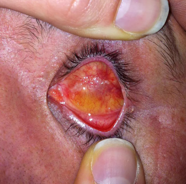
Schmitzer S, Simionescu C, Alexandrescu C, Burcea M. The Anophthalmic Socket - Reconstruction Options. J Med Life. 2014;7(Spec Iss 4):23-29. Figure 1a. PMCID: PMC4962761. License: CC BY.
In the orbit after evisceration, when the eyelids are opened, the outer edge of the scleral shell (conjunctival margin) is preserved, and a methyl methacrylate sphere can be seen placed inside the lumen. This corresponds to scleral shell preservation and implant placement in evisceration, discussed in the section “5. Standard Treatment.”
Evisceration can be performed under either general or local anesthesia. Retrobulbar injection of anesthetic with epinephrine can reduce bleeding. Subconjunctival injection of local anesthetic or preoperative instillation of 10% phenylephrine eye drops may also be used.
Corneal excision: The conjunctiva is incised 360° at the limbus and dissected to the insertion of the rectus muscles. The cornea is incised and excised at the limbus (some methods preserve it).
Evisceration: Curved scissors are inserted between the uvea and sclera to dissect circumferentially. Four radial incisions are made at the limbal stump. The contents are scooped out with a cotton swab and sharp spoon, and residual uveal tissue is removed with a scalpel and gauze. Hemostasis is achieved with bipolar cautery.
Absolute alcohol treatment: This may be used to denature and remove residual uveal tissue and microorganisms. However, some surgeons avoid it due to the risk of excessive irritation and edema. When used, care is taken to keep it within the sclera and avoid contact with the conjunctiva.
Relaxing incisions: Two long relaxing incisions are made in the scleral wall posterior to the equator. This helps prevent accumulation of exudate and blood and facilitates suturing.
Implant placement and closure: The scleral flaps are overlapped and sutured to cover the implant sufficiently. The anterior sclera, Tenon’s capsule, and conjunctiva are closed in layers. After placing a conformer, temporary tarsorrhaphy is performed if necessary.
Pathological diagnosis possible: The entire enucleated eye can be examined histologically. It is the only option for malignant tumors.
Lower risk of sympathetic ophthalmia: Exposure to uveal antigens is completely eliminated (classical view).
Sunken socket is likely to occur: In a US survey, 94% of ocularists responded that “sunken socket and deep superior sulcus are more common after enucleation.”
Features of Evisceration
Slightly better cosmetic outcome: Because the sclera and extraocular muscles are preserved, prosthetic eye mobility is good. In a survey of board-certified ocularists in the US, 82% responded that “evisceration provides the best aesthetic result.”
Lower implant exposure rate: 1.5–21.6% after enucleation vs. 0–3.3% after evisceration6).
Contraindications: Cannot be performed in cases where malignancy is suspected.
Dermis fat graft (DFG) is used for primary or secondary orbital reconstruction 7). DFG consists of dermis (providing rigidity, a suture matrix, and promoting vascularization) and fat (volume filling). The standard anterior dermis diameter is 20–25 mm, and fat diameter is 20–35 mm 7).
Indications for primary DFG: Primary reconstruction in orbits with a history of radiation, severe infection, or multiple surgeries 7).
Indications for secondary DFG: Management of implant exposure, dislocation, volume deficiency, or orbital contraction 7).
In children (especially under 5 years), DFG has the advantage of promoting orbital growth and increasing in size with growth 7). The overall complication rate is 58.8%, but most are mild. Good eyelid position was achieved in 83.3% of primary DFG cases versus 37.5% of secondary DFG cases (p=0.07) 7).
Antibiotics are especially important in cases of endophthalmitis (duration: 10 days to several weeks).
Prescribe analgesics and antiemetics.
Conformer: Essential for preventing adhesion and contraction of the conjunctival sac. It should be worn immediately after surgery and not left out for long periods.
Starting artificial eye use: Begin fabrication of the artificial eye 2–4 weeks after surgery, once pain and inflammation have subsided. Adjustment of the artificial eye should be done by an ocularist 6–8 weeks after surgery. The cost of a custom-made artificial eye is 80,000–100,000 yen (may be eligible for medical expense reimbursement).
Pediatric management: Wear an artificial eye as early as possible to promote eyelid and orbital development (especially important under 5 years of age).
Correction of anophthalmic socket depression: For orbital depression, elevate the socket using autologous tissue (bone, cartilage, dermis-fat) or artificial materials (silicone block, hydroxyapatite).
Ilium: Suitable for orbital bone atrophy
Dermis-fat: Soft and easy to fit with artificial eye; can be re-transplanted if re-atrophy occurs
Hydroxyapatite: Risk of exposure if placed near the surface
Silicone block: Important to insert deeply
When conjunctival sac enlargement is needed: Insert a full-thickness skin graft taken from the groin or lower abdomen, wrapped inside-out around a standard thin artificial eye. The conjunctival sac should be firmly fixed deep to the periosteum of the inferior orbital rim.
QWhen can I start wearing an artificial eye after surgery?
A
The conformer (temporary artificial eye) is worn immediately after surgery. Adjustment of the final artificial eye is done by an ocularist 6–8 weeks after surgery. A custom-made artificial eye is fabricated after the conjunctival sac has stabilized. The general guideline is to start 2–4 weeks after surgery, once pain and inflammation have subsided. Early use of the conformer is important because prolonged disuse can lead to significant contraction of the conjunctival sac.
QIs an implant (orbital implant) always necessary?
A
It is not mandatory, but implantation helps maintain orbital volume and reduces enophthalmos. In Japan, there are no Ministry of Health, Labour and Welfare-approved orbital implants, so they are used off-label. Without an implant, the extraocular muscles are folded into a spherical shape and covered with Tenon’s capsule and conjunctiva.
Rationale for the advantages of enucleation: By removing the entire eyeball and optic nerve, it is possible to histologically evaluate the depth of tumor invasion, residual tumor cells at the optic nerve stump, and the presence or absence of extraocular invasion. Evisceration does not allow for histological evaluation of the entire eyeball.
Risk of sympathetic ophthalmia: There is a theoretical concern that exposure to uveal antigens may trigger an autoimmune reaction (sympathetic ophthalmia) in the contralateral eye. However, a survey of 880 cases found no definitive record of sympathetic ophthalmia after evisceration. The “14-day rule” is considered to lack scientific evidence.
Porous implants promote fibrovascular ingrowth due to their porous structure, improving motility through tissue integration. Fixation with extraocular muscles is also better. Non-porous implants lack tissue ingrowth, which may lead to reduced motility and risk of implant migration.
After enucleation, loss of orbital volume leads to superior sulcus deformity, enophthalmos, and ptosis. This condition is called “post-enucleation socket syndrome.” Inadequate fixation of extraocular muscles to the implant can cause implant migration and worsen symptoms. Appropriate implant size selection and secure suturing of extraocular muscles are key to prevention.
By age 5, orbital volume reaches 80% of adult volume (completed by age 14–15), and from birth to adolescence, eyeball volume triples. Since mechanical stimulation to the bone is essential for orbital growth, appropriate implant selection and long-term follow-up are important in children.
Visual hallucinations called “Charles Bonnet syndrome (CBS)” may occur after enucleation. CBS was traditionally thought to require loss of more than 60% of binocular vision, but it has been shown that it can occur with loss of vision in only one eye.
Forte et al. (2025) reported a 67-year-old woman who developed CBS after enucleation for choroidal melanoma1). Visual hallucinations appeared the day after surgery and persisted for two years. A literature review identified 9 cases of CBS after unilateral vision loss, with a mean age at diagnosis of 69.4 years (range 52–82 years), and hallucinations appeared within hours to 2 days after vision loss in 8/9 cases 1).
It is recommended that all patients undergoing enucleation be informed beforehand about the possibility of CBS and undergo postoperative screening.
QCan hallucinations occur after eye removal?
A
Yes. Visual hallucinations called Charles Bonnet syndrome (CBS) can occur. Hallucinations vary, including moving patterns, colors, and figures, and patients are aware that the hallucinations are not real (insight is preserved). It has been reported that CBS can develop even with only unilateral vision loss 1), so it is important to explain this to patients before surgery.
Traumatic Enucleation and Intracranial Complications
In complete globe avulsion associated with self-inflicted eye injury (Oedipism) in psychiatric patients, optic nerve avulsion can cause subarachnoid hemorrhage (SAH). Because the ophthalmic artery branches from the internal carotid artery at segment C6 and runs through the subarachnoid space, avulsion can lead to SAH and internal carotid artery dissection.
Flippin et al. (2023) reported a case of bilateral self-enucleation in a psychiatric patient 2). Both globes were completely avulsed, and head CTA revealed suprasellar SAH plus intraventricular hemorrhage. In cases with complete globe avulsion and optic nerve avulsion, head CT (preferably CTA) is required to evaluate intracranial hemorrhage.
Excessive daytime sleepiness due to obstructive sleep apnea (OSA) can cause severe ocular trauma and enucleation. Baker et al. (2024) reported a case of right globe avulsion (optic nerve avulsion of about 5 cm) due to sudden sleep onset in an OSA patient 3). A nationwide cohort study in Taiwan (6,915 cases) showed that overall trauma risk in OSA patients was increased by 83.1% compared to non-OSA patients 4).
Reconstruction Using Umbilical Cord Amniotic Membrane (AmnioGuard)
The use of umbilical cord amniotic membrane has been reported as a new reconstruction method for wound dehiscence after evisceration. Umbilical cord amniotic membrane is about 10 times thicker than regular amniotic membrane and is rich in HC-HA/PTX3.
Bunin (2022) performed reconstruction using a donor scleral shell and 2.5×2.0 cm AmnioGuard for suture dehiscence after evisceration of a painful blind eye due to proliferative diabetic retinopathy6). Good cosmetic results were maintained at 8 months postoperatively.
Primary DFG transplantation is considered promising for cases with a history of radiation or complex orbits. In a large-scale analysis of 143 literature reviews plus 34 cases by Jovanovic et al. (2020), the DFG complication rate was 58.8%, but most were reported as mild 7). Primary DFG showed a trend toward a higher rate of achieving good eyelid position compared to secondary DFG (83.3% vs 37.5%, p=0.07) 7).
Forte G, Assaf N, Forte P, Jolly JK. Charles Bonnet syndrome associated with unilateral vision loss: a new diagnostic perspective. Ophthalmic Physiol Opt. 2025;45:681-688. PMID: 40099782.
Flippin JA, Truong E, Kishawi S, Allan A, Ho VP. Traumatic bilateral self-enucleation with subarachnoid hemorrhage. Am Surg. 2023;89(11):4905-4907. PMID: 34459279. doi:10.1177/00031348211041565.
Baker N, Schenck CH, Golden E, Varghese R. A case of accidental self-enucleation caused by obstructive sleep apnea. J Clin Sleep Med. 2024;20(8):1395-1397. PMID: 38752810. doi:10.5664/jcsm.11218.
Cheng AC, Wu GJ, Chung CH, et al. Effect of obstructive sleep apnea on the risk of injuries: a nationwide population-based cohort study. Int J Environ Res Public Health. 2021;18(24):13416. doi:10.3390/ijerph182413416.
Narang U, Maubon L, Shah V, Wagh V. Ocular trauma or oedipism: completing the evisceration. GMS Ophthalmol Cases. 2021;11:Doc13. PMID: 34540525. PMCID: PMC8422941. doi:10.3205/oc000186.
Bunin LS. Reconstruction with umbilical amnion following ocular evisceration: a case study. Am J Ophthalmol Case Rep. 2022;26:101462. PMID: 35265778. PMCID: PMC8899220. doi:10.1016/j.ajoc.2022.101462.
Jovanovic N, Carniciu AL, Russell WW, Jarocki A, Kahana A. Reconstruction of the Orbit and Anophthalmic Socket Using the Dermis Fat Graft: A Major Review. Ophthalmic Plast Reconstr Surg. 2020;36(6):529-539. PMID: 32134765. doi:10.1097/iop.0000000000001610.
Copy the article text and paste it into your preferred AI assistant.
Article copied to clipboard
Open an AI assistant below and paste the copied text into the chat box.