Traumatic subconjunctival hemorrhage is a condition in which a conjunctival blood vessel ruptures due to a blow or other eye trauma, and blood collects in the subconjunctival space (between the bulbar conjunctiva and Tenon’s capsule). It is said to account for 10–20% of all subconjunctival hemorrhages1).
Unlike non-traumatic subconjunctival hemorrhage (idiopathic, hypertension, anticoagulant use, Valsalva maneuvers, etc.), traumatic cases are clinically important because they can be a sign of serious injury inside the eye. Subconjunctival hemorrhage itself is benign, but when it is seen after trauma, it is necessary to systematically rule out hidden ocular injury.
Subconjunctival hemorrhage is found in about 3% of ophthalmology outpatients and reaches 10.1% in people aged 65 years or older2). In Tarlan et al.’s review, idiopathic cases accounted for 30–50% of all subconjunctival hemorrhages, hypertension for 10–30%, and trauma for 10–20%1). Detailed epidemiologic data limited to traumatic cases are scarce, but sports, work, traffic accidents, and accidents at home are the main causes of injury.
If there is no associated intraocular injury, traumatic subconjunctival hemorrhage is absorbed naturally within 1 to 4 weeks and leaves no lasting effects. During the absorption process, the blood changes from red to purple to blue-green to yellow. It does not affect vision. However, the presence or absence of an associated ocular injury determines the prognosis, so careful initial evaluation is essential.
QMy white of the eye turned bright red after an injury. Is that okay?
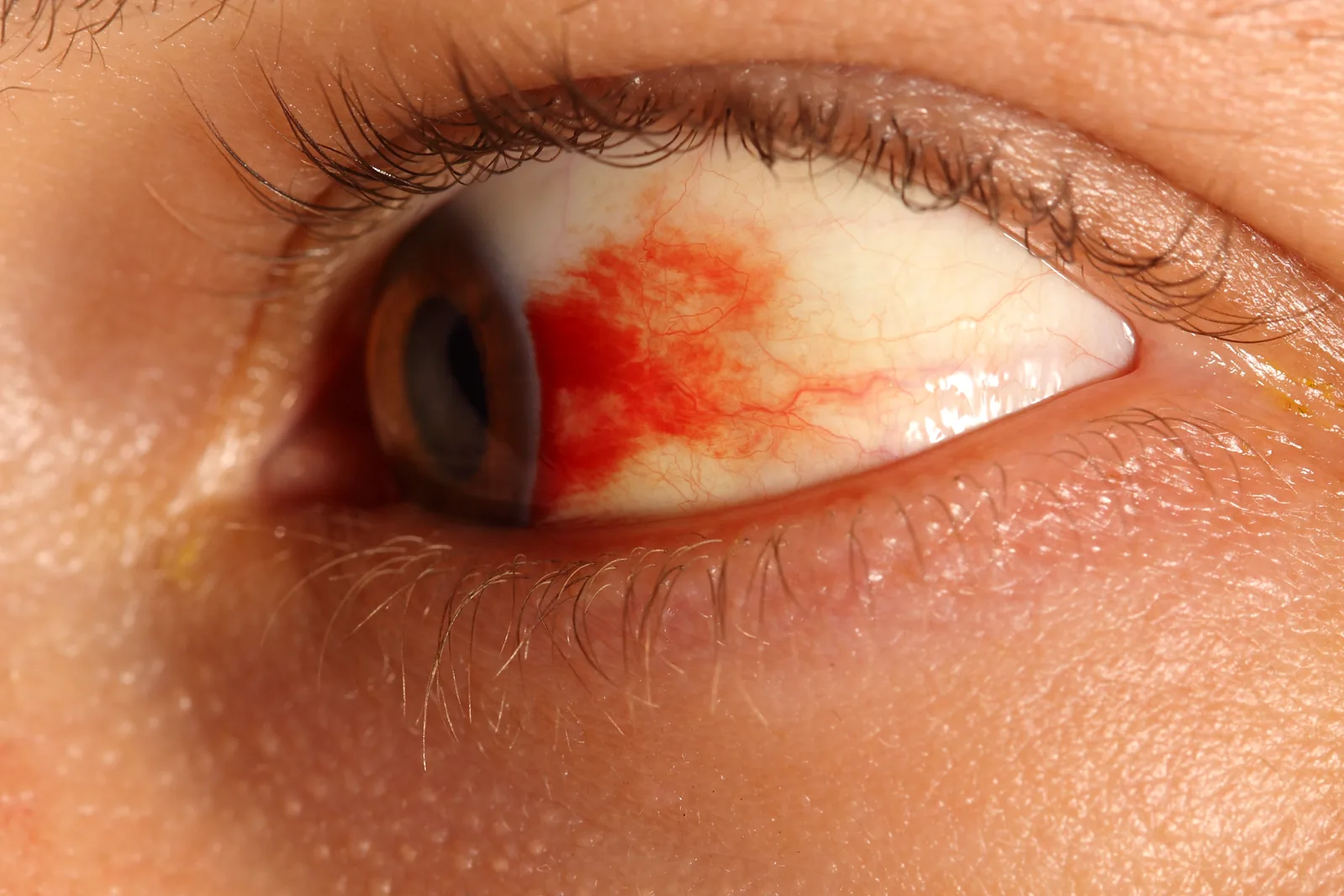
A bright red, sharply demarcated hemorrhagic patch is visible beneath the bulbar conjunctiva, showing blood accumulation under the conjunctiva after trauma. This corresponds to the macroscopic finding of subconjunctival hemorrhage described in the section “Main symptoms and clinical findings”.
Usually asymptomatic: isolated subconjunctival hemorrhage does not cause pain or vision loss. Eye pain after trauma is due to associated injuries (corneal epithelial abrasion, hyphema, etc.).
Noticing the redness: often noticed in the mirror or after someone else points it out.
Foreign body sensation: some people may complain of dry-eye-like symptoms.
Clinical findings (findings the doctor confirms on examination)
Hemorrhagic patch: a bright red or dark red patch is visible beneath the bulbar conjunctiva. It can range from a small, spot-like or stain-like localized area to a broad area spreading across the entire bulbar conjunctiva. As it is absorbed, it changes to pink, orange, and yellow.
Direction of spread of the hemorrhage: over time, it moves downward from the palpebral fissure and spreads.
Marked low eye pressure: The globe feels soft on finger palpation (if open globe injury is suspected, do not use a tonometer).
Pupil deviation, distortion, or fixation: Suggests prolapse of anterior segment tissue.
Anterior chamber hemorrhage and decreased vision: Suggests severe injury inside the eye.
QWhat should I do if the white of the eye is completely red?
A
360-degree subconjunctival hemorrhage may be a sign of a possible occult globe rupture. If it is accompanied by marked low eye pressure, decreased vision, or pupil distortion, the situation is urgent, and orbital CT and ocular exploration may be considered. If these findings are present, you need to see an eye specialist on the same day.
Visual acuity testing and pupillary light reflex check: Check for changes in vision before and after the injury and for abnormal pupils.
Slit-lamp microscopy: Assess the extent and depth of the bleeding and whether there is a conjunctival laceration. A laceration hidden by the bleeding is easy to miss.
Fluorescein staining and Seidel test: Check for conjunctival laceration and corneal epithelial damage, and detect an open globe wound (aqueous leakage).
Intraocular pressure measurement: If open-globe injury (globe rupture or penetrating injury) is suspected, avoid using a contact tonometer and use finger palpation or a noncontact method.
Blood pressure measurement: essential as screening for hypertension.
Blood tests: in patients taking anticoagulants, check INR, PT, APTT, and platelet count.
Orbital CT: in cases of 360-degree hemorrhage or low intraocular pressure, evaluate the continuity of the globe wall, intraocular foreign bodies, and the presence of fractures.
The treatment of traumatic subconjunctival hemorrhage prioritizes treatment of the associated globe injury rather than the subconjunctival hemorrhage itself.
If there is no associated globe injury, let the subconjunctival hemorrhage run its natural course. It is naturally absorbed in 1 to 4 weeks. There is currently no established treatment that speeds absorption.
Medication
Dosage
Indication
Artificial tears
Instill as needed
Symptomatic relief of discomfort
Carbazochrome sodium sulfonate (Adona®) 30 mg
Take orally 3 times a day
Capillary strengthening (if recurrent)
For bothersome symptoms, provide supportive care with artificial tears. If dry eye is present, consider prescribing 3% diquafosol sodium eye drops, 2% rebamipide suspension eye drops, sodium hyaluronate eye drops, and similar treatments. For repeated bleeding, carbazochrome sodium sulfonate (Adona® tablets 30 mg, taken orally 3 times a day) may be used, but the level of evidence is not high.
In patients taking warfarin, confirm by blood test whether the INR is within the therapeutic range (most often 2.0–3.0). Stopping anticoagulants on your own is strictly prohibited because it carries a risk of stroke and cardiogenic embolism. Be sure to discuss the matter with the primary doctor and respond accordingly 3).
QIs there a way to make subconjunctival hemorrhage after trauma heal faster?
A
At present, there is no established treatment that promotes the absorption of subconjunctival hemorrhage. During the resorption process, it changes color from red to purple to blue-green to yellow, which is a normal course. Warm compresses are sometimes recommended empirically, but evidence is limited. If there is a concomitant eye injury (such as conjunctival laceration or hyphema), prioritize treatment of that injury.
QI take blood thinners. If subconjunctival hemorrhage occurs, should I stop the medicine?
A
Stopping anticoagulants on your own is strictly prohibited because it carries a risk of stroke and cardiogenic embolism. Check that the therapeutic range is maintained with tests such as INR, and respond after consulting the primary doctor. The incidence of subconjunctival hemorrhage in patients taking warfarin is 3.7% (1.7% in non-users) 3), and after trauma the bleeding tends to spread more easily, but the decision to stop the medication should be made by a specialist.
6. Pathophysiology and detailed mechanisms of onset
Direct blunt force causes physical injury to the conjunctival vessels, allowing blood to leak into the subconjunctival space. In older adults, the elastic tissue and connective tissue between Tenon’s capsule and the conjunctiva are more fragile, so the same force is more likely to cause bleeding that spreads widely.
Subconjunctival hemorrhage associated with globe rupture
In globe rupture caused by blunt trauma, a sudden rise in intraocular pressure causes a tear at the thinnest part of the eyeball wall, near the corneal limbus and rectus muscle insertions. Intraocular blood and vitreous spill into the subconjunctival space, appearing as widespread subconjunctival hemorrhage. Circumferential subconjunctival hemorrhage is a characteristic finding of this mechanism.
It has been reported that conjunctival lymphatic vessels may be involved in the absorption of subconjunctival hemorrhage. Intraoperative OCT has shown blood within lymphatic vessels with valve-like structures adjacent to the bleeding site, suggesting that lymphatic vessels help clear blood from the subconjunctival space4).
In diabetes, the conjunctival microvessels may become dilated, tortuous, and show changes in blood flow speed, increasing vessel fragility. As a result, even minor trauma can more easily cause subconjunctival hemorrhage.
The drainage mechanism of subconjunctival hemorrhage through conjunctival lymphatic vessels was demonstrated for the first time using intraoperative OCT4). In a case of SCH that occurred during cataract surgery, blood transfer into lymphatic vessels with valve-like structures was confirmed, and marked resolution of SCH within 1 to 2 days after surgery was reported. This finding may also be useful for predicting outcomes after glaucoma filtering surgery.
Standardizing the initial care protocol for traumatic subconjunctival hemorrhage
Standardization of an algorithm to distinguish traumatic subconjunctival hemorrhage from globe rupture is needed. There are also reports that quantitative assessment of the depth and extent of subconjunctival hemorrhage using anterior segment OCT may help improve diagnostic accuracy, but it has not yet become widely used in clinical practice.
Tarlan B, Kiratli H. Subconjunctival hemorrhage: risk factors and potential indicators. Clinical ophthalmology (Auckland, N.Z.). 2013;7:1163-70. doi:10.2147/OPTH.S35062. PMID:23843690; PMCID:PMC3702240.
Mimura T, Usui T, Yamagami S, Funatsu H, Noma H, Honda N, et al. Recent causes of subconjunctival hemorrhage. Ophthalmologica. Journal international d’ophtalmologie. International journal of ophthalmology. Zeitschrift fur Augenheilkunde. 2010;224(3):133-7. doi:10.1159/000236038. PMID:19738393.
Miller KM, Oetting TA, Tweeten JP, Carter K, Lee BS, Lin S, et al. Cataract in the Adult Eye Preferred Practice Pattern. Ophthalmology. 2022;129(1):P1-P126. doi:10.1016/j.ophtha.2021.10.006. PMID:34780842.
Lau AZ, Tang GY, Morgan WH, Chan GZ. Drainage of subconjunctival hemorrhage through conjunctival lymphatic pathways. American journal of ophthalmology case reports. 2025;39:102368. doi:10.1016/j.ajoc.2025.102368. PMID:40686767; PMCID:PMC12272577.
Copy the article text and paste it into your preferred AI assistant.
Article copied to clipboard
Open an AI assistant below and paste the copied text into the chat box.
