Corneal foreign body is a common condition in ophthalmic emergencies. The types of foreign bodies vary widely, including metal fragments such as iron filings, glass shards, plant material (leaves, thorns, wood chips), stone dust, and soil. In ophthalmology clinics in metalworking areas, corneal foreign bodies account for 7.0% of new patients. According to emergency department statistics abroad, corneal foreign bodies are the second most common ocular trauma after corneal abrasion, accounting for 30–40% of all ocular trauma visits 1.
88% of corneal foreign bodies are iron filings. Most foreign bodies entering the palpebral fissure are expelled by blinking, but tiny foreign bodies less than 0.5 mm wide and 0.02 mm thick remain on the cornea. Iron rusts onto the epithelium within 30 minutes and becomes fixed on the cornea, resisting blinking.
Type of foreign body
Frequency
Male-to-female ratio
Iron powder
88%
27:1
Plant fragment
3%
—
Sand/pebble
2%
—
Charcoal/ash
2%
—
Other
5%
7:1
For foreign bodies other than iron filings, the male-to-female ratio is 7:1, which is not as high as for iron filings but still more common in men. The number of foreign bodies is proportional to the width of the palpebral fissure, and they tend to be smaller in the central cornea and larger in the periphery.
The goal of treatment is to remove the foreign body and promote healing of the wound without complications. The condition changes depending on the type of foreign body and the time elapsed. In particular, foreign bodies in the deep cornea or those involving perforation require attention to the risk of infection. Most cases heal with appropriate treatment, but if infectious keratitis develops, it may leave stromal opacity.
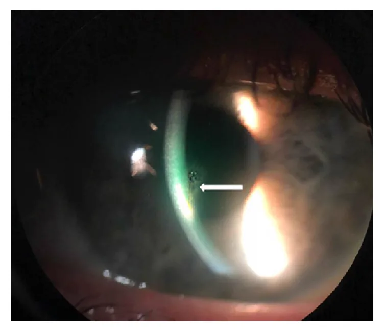
Soon AK, Mather R. Chronic, Stromal Foreign Body of Presumed Human Origin, following Corneal Abrasion. Case Rep Ophthalmol Med. 2019;2019:9607282. Figure 1. PMCID: PMC6444229. License: CC BY.
This is a slit-lamp microscope photograph of a foreign body (arrow) embedded in the central cornea and surrounding stromal opacity. It corresponds to the slit-lamp findings of a corneal foreign body (intrastromal foreign body with corneal infiltration) discussed in the section “Main Symptoms and Clinical Findings.”
During work or outdoor activities, a foreign body enters the eye and causes the following symptoms.
Acute Phase Symptoms
Eye pain: Occurs immediately after the foreign body penetrates the cornea. The severity depends on the size and depth of the foreign body.
Foreign body sensation: The foreign body on the cornea causes irritation with each blink. It persists even after removal until the epithelial defect heals.
Redness: Accompanied by conjunctival injection. In cases of severe inflammation, ciliary injection is also observed.
Tearing: Reflex tearing due to trigeminal nerve stimulation of the cornea.
Symptoms in Prolonged Cases
Decreased vision: Foreign bodies in the pupillary area or rust rings affecting the visual axis can impair vision. Deep foreign bodies may cause irreversible visual dysfunction.
Photophobia: Occurs when complicated by intraocular inflammation or iridocyclitis.
Eye discharge: Present when infectious keratitis is complicated.
The following findings are confirmed by slit-lamp microscopy.
Corneal foreign body: Evaluate the type, location, number, size, and depth of the foreign body. Iron foreign bodies appear brown to black.
Rust ring: A brown annular deposit formed around an iron foreign body. It forms in a cylindrical shape in the epithelial and Bowman layers.
Corneal infiltrate: Inflammatory cell infiltration around the foreign body. When infection is complicated, a stromal abscess forms.
Coats white ring: A small white annular opacity remaining after removal of an iron foreign body. It is due to iron deposition and usually does not require treatment.
Anterior chamber inflammation: Observed when the foreign body reaches deep layers or is left in place for a long time.
Fluorescein staining is essential for confirming epithelial damage. Disruption of the tear film around the foreign body is highlighted by staining, aiding in the detection of transparent or very small foreign bodies. If aqueous humor leakage is present, perforation can be confirmed by Seidel test.
QDoes the rust ring need to be completely removed?
A
It is acceptable if complete removal is not achieved in the initial procedure. After a few days, the corneal tissue melts and the rust ring becomes easier to remove, so removal can be performed in multiple sessions. However, a rust ring extending to the pupillary area requires careful management because the condition of the stroma after removal significantly affects visual function.
Corneal foreign bodies are common in workers in metal, wood, plastic manufacturing, construction, agriculture, and cleaning industries. In daily life, DIY, gardening, and cleaning can also be triggers. Obtaining the circumstances of injury and inferring the type of foreign body helps determine the treatment strategy. A single-center study in Turkey reported that among 100 patients with metallic corneal foreign bodies, 59% were metal industry workers, 65% were injured during metal cutting, and 57% were not wearing protective eyewear at the time of injury despite the availability of safety glasses at the workplace 2.
Iron powder: Rusts within 30 minutes, forming a cylindrical rust ring within 12 hours. After 72 hours, the area around the rust ring dissolves, but infection is unlikely.
Plant fragments (thorns, wood chips): Cause a strong foreign body reaction in the corneal stroma. Over time, swelling and decay progress, allowing bacteria and fungi to invade the cornea, often leading to infectious keratitis.
Chestnut burrs and caterpillar venomous hairs: May cause severe visual impairment due to toxins. Suspect intravitreal invasion of venomous hairs.
Sharp foreign bodies (glass, iron fragments): Can penetrate deep into the cornea and cause perforation.
QWhy are plant-based foreign bodies particularly dangerous?
A
Plant-based foreign bodies cause a strong foreign body reaction in the corneal stroma. Over time, swelling and decay progress, and the foreign body may break or fragment, leading to retained fragments. Additionally, plants often carry bacteria and fungi, increasing the risk of infectious keratitis. Steroid eye drops are contraindicated as they promote fungal infection.
At the initial visit, do not force the eyelid open; use topical anesthetic eye drops to reduce irritation before examination. Handle carefully, considering the possibility of perforating injury.
History: Confirm the circumstances of injury, estimate the type of foreign body, and the time elapsed since injury.
Slit-lamp examination: Confirm the type, location, number, size, and depth of the foreign body. Also assess for anterior chamber inflammation and iridocyclitis.
Fluorescein staining: Assess the extent of epithelial damage. Perform Seidel test to determine the presence of perforation.
Fundus examination: If an intraocular foreign body is suspected, use an auxiliary lens to observe from the posterior pole to the far periphery.
Imaging tests (when deep or intraocular foreign body is suspected)
X-ray examination: Useful for confirming and locating foreign bodies. If an intraocular foreign body is found, confirm its position using the Comberg method.
CT scan: Performed when detection by X-ray is difficult. It can also evaluate changes in the orbit and cranium simultaneously.
Ultrasound examination: Useful when the fundus cannot be visualized due to anterior segment opacity or vitreous hemorrhage. It is also used to search for foreign bodies that cannot be detected by X-ray, such as glass or plastic.
MRI: Can only be performed when it is clear that the foreign body is non-magnetic. It is absolutely contraindicated if a magnetic foreign body is suspected.
Eye irrigation may be effective for powdery or granular foreign bodies on the ocular surface. However, foreign bodies that are embedded or stuck in the cornea cannot be removed by irrigation. An ophthalmologist must thoroughly examine the anterior segment with a slit lamp and select the appropriate treatment.
After topical anesthesia, perform the following procedures under a slit lamp microscope.
Basic techniques for foreign body removal
Removal with a foreign body spud: Use the spud like a scalpel, sharp spoon, or spatula to lift and remove the foreign body. Do not direct the tip perpendicular to the cornea.
27G needle method: Use a disposable needle to lift and remove the foreign body. Foreign bodies other than iron filings are often just resting on the cornea and can be removed by scooping.
Removal with forceps: Grasp the posterior end of the foreign body with jewelers forceps and pull it out. Be careful not to disturb the surrounding tissue.
Removal of Rust Ring
Curettage with foreign body needle: After removing the epithelial rust ring, scrape out the punctate rust remaining in the superficial stroma with the needle tip.
Curettage with drill (Alger brush): Compared to the needle technique, this method results in fewer residual fragments on the first attempt 3. Using low speed and lightly touching the tip can prevent over-scraping.
Staged removal: Even if complete removal is not achieved initially, re-removal is possible after a few days when the corneal tissue has softened.
Removal of Deep Foreign Bodies and Those with Perforation
Perform under an operating microscope with a lid speculum.
Incision + forceps extraction: Make an incision at the entry site of the foreign body and extract it with forceps.
30G needle technique: Insert the needle tip around the foreign body through the incision and lift it out.
Foreign body reaching the anterior chamber: Administer retrobulbar anesthesia to suppress eye movement before removal. There are two approaches: corneal side and anterior chamber side.
Anterior chamber approach: Maintain the anterior chamber with viscoelastic material, pull the foreign body into the anterior chamber with anterior capsule forceps, then remove it through a side port.
Management of perforation wound: Small fresh wounds can be expected to self-seal. Large wounds should be sutured with 10-0 nylon.
After foreign body removal, the wound site has a disrupted epithelial barrier and is susceptible to infection.
Antibiotics: Apply antibiotic eye ointment and administer antibiotic eye drops early postoperatively. A multicenter retrospective study (n=307) reported no significant difference in the incidence of infectious keratitis between picloxidine and tobramycin eye drops (5.3% vs 4.5%, p=0.797), with infectious complications remaining low at approximately 4.9% 4.
Sodium hyaluronate eye drops: Used concomitantly to promote corneal epithelial regeneration and protection.
Steroid eye drops: Generally not used. This is because they promote an environment susceptible to infection and mask signs of infection. Particularly after a vegetative foreign body, they are contraindicated to prevent fungal infection.
Atropine eye ointment: Used as an anti-inflammatory aid when anterior chamber inflammation is severe.
Follow-up: For several days after surgery, careful observation is performed until a robust corneal epithelium regenerates and barrier function is restored.
QWhich is more suitable for rust ring removal: a drill or a foreign body needle?
A
The drill (Alger brush) has the advantage of leaving less residual material initially compared to the foreign body needle. However, it has the disadvantage of potentially over-scraping the stroma. It is important to use a low speed and lightly touch the tip. Especially for rust rings involving the pupillary area, the condition of the stroma after removal affects visual function, so the indication should be carefully considered.
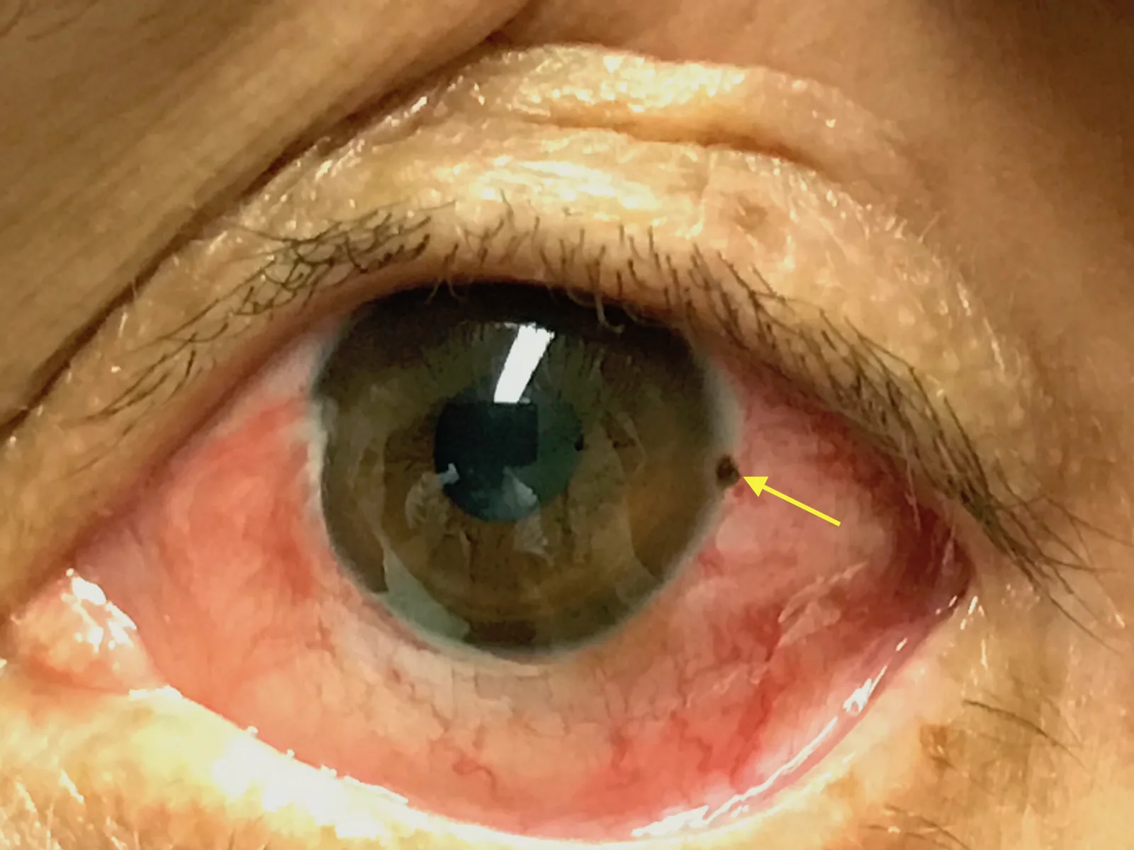
Zakaria J, Peña J. Corneal Rust Ring. JETem. 2018;3(4). DOI: 10.21980/J8X067. Figure 1. License: CC BY 4.0.
A slit-lamp microscope photograph showing a metallic foreign body at the corneal limbus (3 o’clock direction) and a surrounding brown annular rust ring (arrow). This corresponds to the rust ring formation due to an iron foreign body discussed in the section “Pathophysiology and Detailed Mechanism.”
When iron powder adheres to the cornea, oxidation progresses in the presence of moisture and oxygen. It rusts onto the epithelium within 30 minutes, forming a cylindrical rust ring in the epithelial layer within 12 hours. Rust reaching Bowman’s layer spreads in a disc shape proportional to the volume of iron powder, and further forms a cylindrical rust ring in the superficial stroma directly beneath. After 72 hours, the tissue around the rust ring begins to dissolve, but infection usually does not occur at this stage.
A small white annular opacity that remains in the corneal stroma after removal of an iron foreign body. It is an iron deposit that remains as a trace of the foreign body’s presence in the cornea. It is usually asymptomatic and does not require treatment, and may regress over time.
Disruption of the epithelial barrier by a corneal foreign body provides a route for bacterial and fungal invasion. Plant-derived foreign bodies carry a particularly high risk of infection because they introduce microorganisms into the corneal stroma. A case report from Malaysia describes a 61-year-old man who developed Fusarium keratitis after a rambutan fruit struck his eye, causing multiple plant fragments to penetrate deeply near Descemet’s membrane 5. Without appropriate treatment, corneal ulcer can progress to iridocyclitis and panophthalmitis, resulting in significant visual impairment.
For inert foreign bodies such as glass fragments that are difficult to remove, observation may be acceptable if they do not affect vision and cause no chemical or physical irritation. However, follow-up is necessary.
Rebattu B, Baillif S, Ferrete T, et al. Corneal foreign bodies: are antiseptics and antibiotics equally effective? Eye (Lond). 2023 Sep;37(13):2664-2672. doi:10.1038/s41433-022-02380-0. PMID:36639401; PMCID:PMC10482830. ↩
Ozkurt ZG, Yuksel H, Saka G, Guclu H, Evsen S, Balsak S. Metallic corneal foreign bodies: an occupational health hazard. Arq Bras Oftalmol. 2014;77(2):81-83. PMID: 25076469. ↩
Sigurdsson H, Hanna I, Lockwood AJ, Longstaff S. Removal of rust rings, comparing electric drill and hypodermic needle. Eye (Lond). 1987;1(Pt 3):430-432. PMID: 3653447. ↩
Rebattu B, Baillif S, Ferrete T, Risso K, Rabot A, Babeau F, Nahon-Estève S, Martel A. Corneal foreign bodies: are antiseptics and antibiotics equally effective? Eye (Lond). 2023;37(13):2664-2672. PMID: 36639401. ↩
Rosli AH, Abdurrahman MY, Kamal KM. Deeply Embedded Corneal Foreign Bodies With Fungal Keratitis Secondary to Rambutan Fruit Fall. Cureus. 2022;14(2):e22413. PMID: 35371697. ↩
Copy the article text and paste it into your preferred AI assistant.
Article copied to clipboard
Open an AI assistant below and paste the copied text into the chat box.