Cryptophthalmos means “hidden eye” in Latin. It refers to a condition where fused eyelid skin covers the eyeball and orbit, with absence of the palpebral fissure. The forehead skin is continuous with the cheek skin.
The incidence is reported as 0.043 per 10,000 live births and 1.1 per 10,000 stillbirths1). As of 2018, only 55 cases have been reported in the literature. The overall prevalence of eyelid abnormalities is 0.06%, with two-thirds being sporadic and one-third having a genetic predisposition1).
Cryptophthalmos is classified into three types based on morphology.
Complete (typical): Complete occlusion of the orbit. The most severe type, with absence of eyebrows, eyelashes, and glandular structures.
Incomplete (atypical): Rudimentary eyelids remain. A small conjunctival sac is present laterally.
Abortive form (congenital symblepharon): Absence of the upper eyelid. The forehead skin attaches to the upper part of the cornea.
Any type can be unilateral or bilateral, isolated or syndromic. Cryptophthalmos is a condition where the eyeball is covered by fused eyelid skin, and it can occur alone or as part of Fraser syndrome. If the eyelid margin is completely absent, it is diagnosed as ablepharon or cryptophthalmos.
It is very closely associated with Fraser syndrome, and autosomal recessive inheritance due to mutations in FRAS1, GRIP1, and FREM2 genes is common. Cryptophthalmos is observed in 80–93% of Fraser syndrome cases.
QCan a child with cryptophthalmos regain vision?
A
In almost all cases, there is little or no visual function. The purpose of surgery is mainly cosmetic improvement and reconstruction of the ocular region; vision improvement is extremely rare. For details, see the section on “Standard Treatments”.
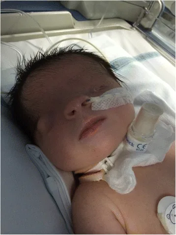
De Bernardo G, Giordano M, Di Toro A, et al. Prenatal diagnosis of Fraser syndrome: a matter of life or death? Ital J Pediatr. 2015 Nov 9;41:86. Figure 1. PMCID: PMC4640198. License: CC BY.
A facial photograph taken shortly after birth shows that the palpebral fissure has not formed and the skin covers the ocular surface. The left eye is microphthalmic, and the right eye also has morphological abnormalities, helping to understand the appearance of cryptophthalmos.
The main clinical findings by type are shown below.
Complete Type
Orbital occlusion: The skin of the forehead and cheek is continuous, with complete absence of the palpebral fissure.
Adnexal absence: Eyebrows, eyelashes, and glandular structures are absent.
Microphthalmia association: The surface skin is fused with the cornea. There is no conjunctival sac, and microphthalmia is very common. Orbital cysts may be present.
Incomplete Type / Partial Type
Incomplete type: Rudimentary eyelids remain. The palpebral fissure length is about one-third of normal. There is a small conjunctival sac laterally. The eyeball is small and almost covered by skin.
Incomplete type: Upper eyelid defect. Forehead skin attached to the upper 75% of the cornea. The covered cornea becomes keratinized and opaque, but the exposed cornea may be transparent.
In a case report, a male infant at 39 weeks gestation presented with bilateral cryptophthalmos, absent eyebrows, disorganized frontal hairline, nasal tip cleft, low-set ears, hypertelorism, and low anorectal malformation1). A mobile orbital cyst of 1×1 cm was palpable in the right eye, and the left eyeball was deeply buried1). CT revealed an abnormal gyral pattern in the occipital region and a small left eyeball1).
Cryptophthalmos is caused by genetic mutations in proteins that form the FRAS/FREM complex.
FRAS/FREM complex: FRAS1, FREM1, and FREM2 proteins maintain adhesion between the basement membrane and epithelium during embryonic development. Mutations cause adhesion defects, leading to failure of eyelid separation1).
Fraser syndrome (FRASRS1/2): Autosomal recessive inheritance due to mutations in FRAS1 or FREM2 genes1).
MOTA syndrome: FREM1 mutation associated with nasal cleft, anorectal malformations, renal agenesis, etc. 1).
Isolated cryptophthalmos: FREM2 mutation can also cause unilateral or bilateral isolated cryptophthalmos 1).
Inheritance pattern: Mostly autosomal recessive. Autosomal dominant inheritance has also been reported.
The mechanisms of congenital eyelid coloboma include incomplete fusion of facial clefts during embryonic development and compression by amniotic bands.
QHow is cryptophthalmos related to Fraser syndrome?
A
Cryptophthalmos is present in 80–93% of Fraser syndrome cases, and mutations in FRAS1 and FREM2 are the main cause. The FRAS/FREM complex is essential for maintaining adhesion between the basement membrane and epithelium during embryonic development, and its mutation leads to failure of eyelid separation 1). However, FREM2 mutations can also cause isolated cryptophthalmos.
Prenatal ultrasound can detect it around 18 weeks of gestation. Findings include absence of the palpebral fissure and continuous skin from the forehead to the cheek. If syndactyly, increased lung echogenicity, and oligohydramnios are present, Fraser syndrome is highly likely.
Visual acuity, strabismus, and fundus examination: Assessment of visual function
Traction test under general anesthesia: Confirmation of potential fibrous bands
CT/MRI: Evaluation of eye morphology and brain. Useful for confirming central nervous system complications such as abnormal occipital gyral patterns 1)
FRAS/FREM panel testing is important for definitive diagnosis 1). Because the phenotypes of Fraser syndrome and MOTA syndrome overlap, differentiation based solely on clinical findings without genetic testing can be difficult.
Differential diagnosis includes congenital eyelid coloboma (in cases of partial defect), cyclopia, and anisophthalmia.
Treatment goals vary by type. In complete and incomplete types, cosmetic reconstruction is the main objective, and the prognosis for visual improvement is extremely limited. In abortive type, management of exposure keratopathy and risk of visual impairment is urgent.
Ocular surface management: Prescription of ophthalmic lubricants and artificial tears (for exposure and dryness). For large defects, use ophthalmic ointment to prevent corneal drying.
Prosthetic eye shell: Option when surgery is contraindicated, impossible, or unsuccessful.
Stage 1: After skin incision of the ocular remnant, a conformer covered with a mucosal graft is inserted to create a conjunctival sac.
Stage 2 (approximately 1 year later): Eyelid reconstruction with posterior lamellar reinforcement and orbital mucosal grafting is performed. If mucosal grafting is ineffective, foreskin may be used as an alternative.
Incomplete and Partial Types
Incomplete type: After conjunctival sac creation (mucosal graft + conformer placement), eyelid reconstruction is performed using eyelid sharing or switch flap technique. There is a risk of recurrence of corneal-eyelid adhesion.
Incomplete type: The main goal is reconstruction of the upper eyelid and superior fornix. One-stage reconstruction using scleral and amniotic membrane grafts is performed.
If the defect in a child is small, it can be repaired by end-to-end suturing. For larger defects, plastic surgery using skin flaps is chosen. In the reported case, exploratory eye examination revealed a rudimentary cystic and adherent eyeball, and no additional surgery was performed1).
QHow is surgery performed for complete cryptophthalmos?
A
It is performed in stages. In the first stage, after a skin incision over the ocular remnant, a conformer covered with a mucosal graft is inserted to create a conjunctival sac. Approximately one year later, in the second stage, eyelid reconstruction with posterior lamellar reinforcement and orbital mucosal grafting is performed. The main goal is cosmetic improvement; visual improvement is extremely rare.
A consensus on the pathophysiology has not yet been established.
Neuroectodermal defect: The neuroectodermal optic vesicle is essential for fetal lens development, and defects in this layer prevent proper development of the cornea, lens, and anterior chamber.
Disruption of eyelid formation: The eyelids cannot form without differentiation of ectoderm and mesoderm. The eyelids appear at 6 weeks of gestation, and the upper and lower eyelids are fused until the 7th month of gestation. Malformation during this period leads to congenital eyelid abnormalities.
Apoptosis defect theory: The combination of syndactyly, laryngeal, and genital abnormalities suggests that a defect in programmed cell death plays an important role.
Dysfunction of the FRAS/FREM complex: Impaired adhesion between the basement membrane and epithelium due to mutations in FRAS1, FREM1, and FREM2 leads to eyelid separation failure 1).
Secondary complications that often accompany microphthalmia include cataracts, lens dislocation, glaucoma, and retinal detachment.
7. Latest Research and Future Prospects (Research-stage Reports)
Mwipopo et al. (2023) reported a case of bilateral cryptophthalmos from Tanzania 1). Genetic testing identified a FREM2 mutation (heterozygous likely pathogenic) along with a CEP85L mutation (lissencephaly 10: LIS10). LIS10 shows heterogeneity ranging from mild intellectual disability to severe phenotypes. This case is the first report from Africa, and it was noted that underreporting in low- and middle-income countries contributes to underestimation of the number of cases.
Since mutations in genes related to the FRAS/FREM complex show overlapping phenotypes in multiple syndromes, clinical diagnosis alone is insufficient, and the role of genetic testing is increasing 1). The need for genetic testing is also growing in low- and middle-income countries 1).
Mwipopo E, Massomo MM, Moshiro R, Manji KP. Bilateral cryptophthalmos with overlapping features of Manitoba oculo-tricho-anal (MOTA) syndrome and Fraser syndrome 2. BMJ Case Rep. 2023;16(7):e252618.
Thomas IT, Frias JL, Felix V, Sanchez de Leon L, Hernandez RA, Jones MC. Isolated and syndromic cryptophthalmos. Am J Med Genet. 1986;25(1):85-98. PMID: 3099574.
Kabra M, Gulati S, Ghosh M, Menon PS. Fraser-cryptophthalmos syndrome. Indian J Pediatr. 2000;67(10):775-8. PMID: 11105430.
Copy the article text and paste it into your preferred AI assistant.
Article copied to clipboard
Open an AI assistant below and paste the copied text into the chat box.