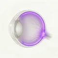
Prodromal Stage
Extraocular symptoms precede: Headache, fever, tinnitus, vertigo, and meningeal irritation signs are predominant.
Ocular symptoms are mild: The stage when mild hyperemia of the optic disc begins to appear.
Vogt-Koyanagi-Harada disease (VKH disease, Harada disease) is defined as an autoimmune disease characterized by bilateral granulomatous panuveitis with or without extraocular symptoms 1). The main lesion is in the choroid, and the autoimmune reaction affects melanocytes in the iris, ciliary body, retina, meninges, inner ear, and skin 1).
In 1906, Vogt and in 1929, Koyanagi independently reported a disease characterized by chronic anterior uveitis, alopecia, vitiligo, and hearing loss. In the same year, Harada reported posterior uveitis with cerebrospinal fluid pleocytosis and exudative retinal detachment. In 1932, Babel combined both and named it “Vogt-Koyanagi-Harada disease.”
Harada disease is more common in people of color, such as Hispanics, Asians, Native Americans, Middle Easterners, and Indians4). It is rare in sub-Saharan Black Africans. In Japan, it accounts for 6.8–9.2% of all uveitis cases. In the United States, it accounts for only 1–4%.
In one study (n=65), 78% of patients were Hispanic, 10% were Asian, and 74% were female4). The average age at onset was 32 years, with the majority in their 20s to 50s4). In Japanese patients, approximately 80% are HLA-DR4 positive, strongly suggesting a genetic predisposition.
It is more common in people of color (Hispanic, Asian, Native American, etc.) in their 20s to 50s, with a tendency for women to be affected more often than men. One study found that 74% were women, and the average age at onset was 32 years 4). In Japan, it accounts for about 7–9% of all uveitis cases.
The symptoms of Harada disease vary depending on the disease stage. Following a systematic course from before onset can aid in diagnosis.
The clinical findings of Vogt-Koyanagi-Harada disease change according to the four disease stages.
Prodromal Stage
Extraocular symptoms precede: Headache, fever, tinnitus, vertigo, and meningeal irritation signs are predominant.
Ocular symptoms are mild: The stage when mild hyperemia of the optic disc begins to appear.
Uveitis stage
Serous retinal detachment: Multiple serous detachments of the posterior pole due to choroidal thickening. In severe cases, it may appear bullous.
Optic disc hyperemia and edema: Inflammatory findings of the posterior pole.
Anterior chamber and vitreous inflammation: As the disease progresses, it presents as panuveitis.
Chronic phase (convalescent stage)
Sunset-glow fundus: A characteristic fundus appearance due to depigmentation of the choroid.
Sugiura sign: Limbal corneal depigmentation. This is the earliest depigmentation finding, appearing about one month after onset.
Vitiligo, poliosis, and alopecia: Systemic skin symptoms become apparent.
Recurrent phase
Granulomatous anterior uveitis: Koeppe nodules, Busacca nodules, mutton-fat keratic precipitates, posterior synechiae.
Iris atrophy and depigmentation: The iris appears thin and atrophic.
Complications: Cataract, secondary glaucoma, choroidal neovascularization4).
It refers to a condition where the fundus appears bright reddish-orange due to depigmentation of choroidal melanocytes. This is a characteristic finding in the chronic phase of Vogt-Koyanagi-Harada disease and often appears within a few months after treatment. It is also included as one of the diagnostic criteria.
The etiology of Vogt-Koyanagi-Harada disease is not fully understood, but an autoimmune reaction against tyrosinase family proteins expressed by melanocytes plays a major role 2). T cell-mediated immune responses (activation of Th1 and Th17 cells) cause inflammation in melanocyte-rich tissues (choroid, meninges, inner ear, skin) 1).
An association with HLA-DR4 subtypes such as HLA-DRB10405 has been established. A systematic review identified HLA-DRB10404, *0405, and *0410 as risk suballeles, and *0401 as a protective suballele 8). Associations with immune response-related genes such as the IL-23 receptor have also been suggested 1).
Infections and vaccinations have been reported to trigger the onset or exacerbation of Vogt-Koyanagi-Harada disease in genetically susceptible individuals.
The revised diagnostic criteria established by the International Vogt-Koyanagi-Harada Disease Committee in 2001 are still used today.
| Type | Criteria |
|---|---|
| Complete type | Meets all criteria 1 to 5 |
| Incomplete type | Meets criteria 1 to 3 and either 4 or 5 |
| Suspected case (ocular-only type) | Meets only criteria 1–3 |
Criterion 1: No history of penetrating ocular trauma or surgery before onset of uveitis Criterion 2: No clinical or laboratory evidence suggesting other diseases Criterion 3: Bilateral ocular lesions (early: serous retinal detachment, diffuse choroiditis; late: sunset glow fundus, Sugiura sign, etc.) Criterion 4: Neurological and auditory findings (meningeal irritation, tinnitus, cerebrospinal fluid pleocytosis) Criterion 5: Skin findings (alopecia, poliosis, vitiligo) — do not appear before onset of ocular disease
The main diseases to be differentiated are listed below.
Ocular syphilis can present with almost the same syndrome as Harada disease, including uveitis, retinal detachment, tinnitus, and headache3). Since administering steroids for syphilis can worsen the condition, serum syphilis tests (RPR, FTA-ABS) and cerebrospinal fluid tests (VDRL) must be performed before starting steroids3).
The treatment goal for Vogt-Koyanagi-Harada disease is rapid suppression of acute inflammation and prevention of transition to the chronic recurrent phase. Early and sufficient steroid therapy is key to not missing the “window of opportunity” for treatment 5).
In fresh cases early after onset, high-dose intravenous steroid therapy (pulse therapy) is common. Methylprednisolone (mPSL) 500–1,000 mg/day or dexamethasone 100 mg/day is infused intravenously over 1–3 hours for 3 days. This is performed after confirming the absence of systemic infection or contraindications. It may also be indicated during pregnancy, but non-fluorinated steroids (mPSL, prednisolone) are preferred over fluorinated steroids (dexamethasone, betamethasone) which have higher placental transfer 9).
After pulse therapy or from the acute phase, oral prednisone/prednisolone is started at 1–1.5 mg/kg/day (maximum 100–200 mg/day)9). After maintaining the initial dose for 2–4 weeks, it is tapered very slowly and discontinued over 6 months or more.
A standard prescription example is shown below.
| Period | Dose |
|---|---|
| 2 days each | 200 mg → 150 mg → 100 mg → 80 mg/day |
| 4 days | 60 mg/day |
| 10 days | 40 mg/day |
| 2 weeks | 30 mg/day |
| Every 4 weeks | 20 mg → 15 mg → 10 mg → 5 mg/day |
| Final 4 weeks | 5 mg/day every other day |
Regarding the relationship between treatment duration and recurrence, it has been reported that patients who received treatment for less than 6 months had a significantly higher recurrence rate of 58.8% compared to those treated for 6 months or more (recurrence rate 11.1%)4).
A systematic review of steroid monotherapy reported that 44% of patients experienced recurrence and 59% developed sunset glow fundus5), indicating that long-term addition of immunosuppressive therapy is often necessary.
In a subanalysis of the FAST Uveitis Trial for non-infectious uveitis (including 93 cases of Vogt-Koyanagi-Harada disease, total 216 cases), the treatment success rates (steroid-sparing inflammatory control at 6 months) were comparable between MTX and MMF. Some analyses indicate that MTX achieved steroid-sparing control in 74% and MMF in 53% at 6 months5).
At the time of recurrence, anterior segment inflammation is often predominant, making local therapy important.
Steroid administration should be tapered over at least 6 months. The recurrence rate when discontinued within less than 6 months is 58.8%, which is significantly higher than the 11.1% rate when continued for 6 months or more 4). When immunosuppressive drugs are added, longer-term continuation is often necessary.
The core pathogenesis of Harada disease is a Th1 and Th17 cell-mediated autoimmune reaction targeting melanocyte-associated antigens (such as tyrosinase, tyrosinase-related protein, and 75 kDa protein) 1). In individuals with genetic susceptibility (e.g., HLA-DRB1*0405), environmental triggers such as viral infection are thought to initiate the process 2).
In the acute phase, granulomatous inflammation extends throughout the full thickness of the choroid. Diffuse lymphocytic infiltration with clusters of epithelioid cells and multinucleated giant cells is observed. Granulomas formed between the retinal pigment epithelium and Bruch’s membrane are known as Dalen-Fuchs nodules, which are relatively specific pathological findings in Vogt-Koyanagi-Harada disease and sympathetic ophthalmia. Immunohistochemically, uveal infiltration consisting of T cells and HLA-DR-positive macrophages is demonstrated, and non-dendritic CD1-positive cells are present near choroidal melanocytes. In the chronic phase, the inflammation transitions to non-granulomatous type.
Regarding the onset of Vogt-Koyanagi-Harada disease after vaccination, proposed mechanisms include molecular mimicry between vaccine peptides and choroidal self-peptides, immune complex deposition due to delayed-type hypersensitivity, and immune reactions to adjuvants (such as aluminum salts)8). In mRNA vaccines, viral proteins may be detected in the blood 1 to 5 days after vaccination7), and it is speculated that this may enhance pre-existing autoimmune activation7).
The FAST trial (NCT01829295) is a large-scale RCT that randomly compared MTX (25 mg orally once weekly) and MMF (1.5 g twice daily) for non-infectious intermediate, posterior, and panuveitis. In a sub-analysis of 93 VKH disease cases (49 in the MTX group, 44 in the MMF group), acute Harada disease showed greater visual improvement and foveal thickness reduction with MMF (both P<.05)5). However, there was no statistically significant difference in treatment success rates between the two groups, and MTX and MMF were considered to have equivalent efficacy as steroid-sparing immunosuppressive agents5).
The use of infliximab, adalimumab, and rituximab for refractory and recurrent cases is accumulating1). However, large-scale RCT data are scarce, and their indication for Harada disease has not been established. Reports indicate that decreased function of CD4⁺CD25⁺ regulatory T cells (Tregs) correlates with disease activity in Harada disease1), and Treg-enhancing therapy is attracting attention as a future research target.
Several groups have successively proposed new diagnostic criteria incorporating optical coherence tomography (OCT) and ICG angiography, stratified by early and late stages5). Standardization of VKH-specific OCT biomarkers (e.g., hyperreflective foci, quantitative choroidal thickness) is still in the research phase.
Reports on VKH management during pregnancy indicate that steroid pulse therapy for VKH disease in the third trimester, followed by a tapering course of oral prednisolone over 1.5 months or more, is safe and effective, with most patients showing no recurrence of uveitis after delivery 9). However, because many immunosuppressive drugs are teratogenic, treatment options during pregnancy are limited, and individualized management by a multidisciplinary team is necessary 9).
Hussain A, Khurana R. Vogt-Koyanagi-Harada Syndrome: A Diagnostic Conundrum. Cureus. 2021;13(12):e20138. doi:10.7759/cureus.20138
Yepez JB, Murati FA, Petitto M, et al. Vogt-Koyanagi-Harada Disease Following COVID-19 Infection. Case Rep Ophthalmol. 2021;12:804-808. doi:10.1159/000518834
Abdelnabi M, Rimu A, Siddiqui S, Mora B, Guerin C. Ocular syphilis mimicking Vogt-Koyanagi-Harada syndrome: a diagnostic dilemma. Proc (Bayl Univ Med Cent). 2023;36(3):380-382. doi:10.1080/08998280.2023.2187184
Tayal A, Daigavane S, Gupta N. Vogt-Koyanagi-Harada Disease: A Narrative Review. Cureus. 2024;16(4):e58867. doi:10.7759/cureus.58867
Acharya NR, Rathinam SR, Thundikandy R, et al. Outcomes in Patients With Vogt-Koyanagi-Harada Disease From the First-Line Antimetabolites for Steroid-Sparing Treatment Uveitis Trial. Am J Ophthalmol. 2024;267:100-111. doi:10.1016/j.ajo.2024.06.004. PMID:38909740.
Kong K, Ding X, Ni Y. Resolution of Harada disease-like uveitis after quadrivalent human papillomavirus vaccination: a case report. Hum Vaccin Immunother. 2022;18(1):e1953349. doi:10.1080/21645515.2021.1953349
De Domingo B, López M, Lopez-Valladares M, et al. Vogt-Koyanagi-Harada Disease Exacerbation Associated with COVID-19 Vaccine. Cells. 2022;11:1012. doi:10.3390/cells11061012
Murtaza F, Pereira A, Mandelcorn MS, Kaplan AJ. Vogt-Koyanagi-Harada disease following influenza vaccination. Am J Ophthalmol Case Rep. 2022;26:101516. doi:10.1016/j.ajoc.2022.101516
Sundararaju U, Subramanian S, Rajakumar HK. Steroid pulse therapy for VKH during pregnancy: a safe and effective option? Orphanet J Rare Dis. 2025;20:366. doi:10.1186/s13023-025-03916-9