Volume of the anterior chamber
Anterior chamber depth: 3.6 mm (Gullstrand model eye)
Anterior chamber volume: 160–200 μL
Aqueous humor turnover time: complete replacement in about 60–70 minutes

This article is a reference that brings together the normal values and threshold values most often used in clinical ophthalmology, organized by anatomical region of the eyeball.
The sources were limited to five Japanese ophthalmology textbooks.
Axial length is the front-to-back length of the eyeball and is the main factor that determines refractive status.
| Time | Axial length |
|---|---|
| Right after birth | 16–18 mm (about 17 mm) |
| 1 year old | About 21 mm |
| 2–5 years | 22.15 mm |
| 5–16 years | 22.71 mm |
| Adult emmetropic eye | about 24 mm |
Axial length increases rapidly in the first year after birth and reaches a plateau around age 6–7 years. In newborns, the anterior segment reaches 70–80% of the adult size, while the posterior segment remains at 50% or less. The axial length of the Gullstrand schematic eye is defined as 24.0 mm.
The normal values by age and the microphthalmia threshold are shown below (measured by ultrasound A-mode, unit: mm).
| Age | Normal (male) | Normal (female) | Microphthalmia (male) | Microphthalmia (female) |
|---|---|---|---|---|
| After birth | 16.85 | 16.60 | 14.70 | 14.44 |
| 2 years old | 20.60 | 20.29 | 17.97 | 17.65 |
| 6–7 years | 22.00 | 21.68 | 19.19 | 18.86 |
| 13 years to adult | 23.40 | 23.06 | 20.42 | 20.06 |
Axial length is about 17 mm in newborns and about 24 mm in adults, a difference of about 7 mm. A simple calculation would predict myopia of more than 15 D, but this is offset by a decrease in corneal refractive power and changes in the lens, keeping the eye emmetropic. This is called emmetropization (emmetropization).
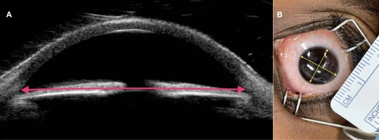
Adult horizontal diameter: 11–12 mm
Adult vertical diameter: 10–11 mm
Newborn horizontal diameter: 9.8 mm (9–10.5 mm)
Newborn vertical diameter: slightly larger, 10.4 mm
Adult corneal radius of curvature: 7.7–8.0 mm
Corneal anterior surface radius of curvature in the Gullstrand model eye: 7.7 mm
Corneal refractive power (Gullstrand model eye): 43.05 D
Newborn corneal refractive power: 47.5–51 D (more steeply curved than in adults)
| Layer / location | Thickness |
|---|---|
| Central cornea (adults) | About 520 μm |
| Total corneal thickness (another reported value) | About 550 μm |
| Epithelium | About 50 μm (10% of total thickness) |
| Stroma | About 500 μm (90% of total thickness) |
| Endothelium | About 5 μm |
| Bowman membrane | About 10 μm |
| Peripheral part (adults) | 0.7–0.9 mm |
The thickness ratio of the epithelium : stroma : endothelium is 0.1 : 1 : 0.01.
Corneal thickness in newborns decreases rapidly from 0.96 mm at birth, to 611 μm the next day, and to 580 μm on day 3, reaching the adult level of 0.5 mm by 6 months after birth.
Corneal endothelial cells do not divide or proliferate in the living body and decrease with age.
| Indicator | Value |
|---|---|
| Corneal endothelial cell density at birth | about 5,000 cells/mm² |
| Corneal endothelial cell density in early childhood | 3,500 cells/mm² |
| Corneal endothelial cell density in older adults | 2,500–3,000 cells/mm² |
| Age-related decrease rate | 0.3–0.7% per year (about 0.6%/year) |
| Bullous keratopathy threshold | 500 cells/mm² or less |
The normal area of endothelial cells is about 300 μm², and the thickness is 4–6 μm. For morphologic evaluation, the normal coefficient of variation (CV value) is about 0.25; 0.35 or higher is considered abnormal. The normal hexagonal cell rate is 70–80%, and 50% or less is considered abnormal.
The turnover of corneal epithelial cells is about one week.
It has not been proven that corneal thickness increases with age. Age-related corneal changes include arcus senilis, vascular ingrowth, and against-the-rule astigmatism.
Corneal endothelial cells maintain the water content of the cornea at a constant level through their pump function and preserve transparency. Because they do not divide in the living eye, they do not recover once they decrease due to aging or surgical trauma. When the count falls to 500 cells/mm² or less, bullous keratopathy develops and the cornea becomes edematous and cloudy.
The sclera makes up about 5/6 of the outer wall of the eyeball. Its thickness varies greatly by location.
| Location | Thickness |
|---|---|
| Around the optic disc (thickest part) | About 1 mm |
| Corneal limbus | 0.8 mm |
| Equatorial region | 0.6 mm |
| Rectus muscle insertion (thinnest part) | 0.3 mm |
In children, the sclera at the equator is 0.45 mm thick, thinner than the 1.09 mm in adults, and in infants and young children it can enlarge easily because of high intraocular pressure (buphthalmos).
The main structures that pass through the sclera are as follows.
Volume of the anterior chamber
Anterior chamber depth: 3.6 mm (Gullstrand model eye)
Anterior chamber volume: 160–200 μL
Aqueous humor turnover time: complete replacement in about 60–70 minutes
Production of aqueous humor
Daytime secretion: about 2.5–3.0 μL/min
Nighttime secretion: about 1.5 μL/min (reduced to about 50% of the awake level)
Active transport: accounts for 80–90% of aqueous humor production
Aqueous humor production decreases by 3.2% every 10 years with age. This is thought to be one reason intraocular pressure in Japanese people decreases with age.
For aqueous humor outflow, the outflow pathway through Schlemm’s canal is the main route and accounts for 90% of total outflow.
Of the eye’s total refractive power of about 58 D, the lens contributes about 15–20 D (19.11 D in the Gullstrand schematic eye). The lens continues to enlarge throughout life.
| Age | Lens diameter |
|---|---|
| At birth | 6.00 mm |
| 2 months | 6.80 mm |
| 3 months | 7.1 mm |
| 6–9 months | 7.66 mm |
| 1 year 9 months | 8.4 mm |
| 2–5 years | 8.5 mm |
| 16 years | 9.3 mm |
Lens diameter correlates most strongly with axial length.
The ciliary muscle consists of three layers: circular muscle (Muller muscle), oblique muscle (radial muscle), and longitudinal muscle (Brucke muscle).
With age, the vitreous becomes more liquefied. At ages 14–18, about 20% is liquefied overall, and at ages 80–90, more than 50% is liquefied. The peak progression from partial posterior vitreous detachment (PVD) to complete posterior vitreous detachment occurs in the 50s to 60s.
Based on the anatomy of the pars plana. To avoid the pars plicata (2 to 2.5 mm from the limbus) and to safely enter anterior to the ora serrata (average 6 mm), the 3.5 to 4.0 mm position is chosen. In phakic eyes, it is placed slightly more posteriorly (4 mm) to avoid contact with the lens.
Foveola
Diameter: 300 to 500 μm
Retinal thickness (histology): 0.13 mm
Retinal thickness (optical coherence tomography): 0.18–0.2 mm
Development stage
Start of macula formation: around 7 months of gestation
Completion of the fovea: around 4 months after birth
Maturation of the macula: almost mature by 15 months after birth, with maturation continuing until around age 5
Kishi pocket appears around age 3, the communicating passage with the Cloquet canal can be seen from age 5, and it is present in 50% of cases by age 11.
The optic nerve is about 50 mm long and is divided into the following four parts.
| Part | Length |
|---|---|
| intraocular | 1 mm |
| intraorbital | 25–30 mm |
| intracanalicular | 4–10 mm (about 6 mm) |
| intracranial | 10 mm |
Each eye has about 100 million rod cells and 6–7 million cone cells. Rods handle scotopic (dim-light) vision, while cones handle photopic vision and color vision. The outer segment of photoreceptors contains 1,000–2,000 disc membranes, and about 10% are phagocytosed and renewed daily by the retinal pigment epithelium.
The muscle length of the four rectus muscles is about 40 mm each, and their insertion distances from the corneal limbus differ (Tillaux’s spiral).
| Extraocular muscles | Tendon length (mm) | Distance from limbus |
|---|---|---|
| Medial rectus (MR) | 3.7 | 5.5 mm |
| Inferior rectus (IR) | 5.5 | 6.5 mm |
| Lateral rectus (LR) | 8.8 | 6.9 mm |
| Superior rectus (SR) | 5.8 | 7.7 mm |
The inferior oblique muscle is 36 mm long (tendon <1 mm), and the superior oblique muscle is 60 mm long (tendon 30 mm).
Tear fluid is a thin fluid layer essential for protecting the ocular surface and maintaining optical quality.
The dimensions of the tear drainage system are as follows.
| Development indicator | Time/value |
|---|---|
| Newborn refractive value (1 month) | Average +3.2 D |
| Refractive value at 3 months of age | Average +3.9 D |
| Refractive value at 1 year of age | Average +1.9 D |
| Peak visual sensitivity | Around 18 months of age (remains until age 8) |
| Completion of normal binocular vision | 2 to 6 months after birth |
| Development of stereopsis | Until around 24 months of age |
| Confirmation of color vision | A few at 4 weeks, all by 12 weeks |
In newborns, hyperopia increases until 3 months after birth (+3.2 D → +3.9 D), then starts to decrease and emmetropization progresses.
Within 4 weeks of birth, most newborns have straight eye alignment, while the others show small-angle exotropia. By 4 months, eye alignment becomes straight and convergence is good. The binocular visual field in infants develops gradually from immediately after birth to 7 weeks, then expands rapidly from 2 months to 6–8 months after birth.
| Tissue/structure | Developmental timing |
|---|---|
| Formation of the anterior chamber angle | Embryonic 10–12 weeks |
| Appearance of Schlemm’s canal | Embryonic 16 weeks |
| Completion of the angle | Around the 8th month of gestation |
| Start of corneal endothelial differentiation | Embryonic 8–10 weeks |
| Completion of the corneal endothelium monolayer | 15–20 weeks of gestation |
| Start of macular formation | Around 7 months of gestation |
| Completion of the fovea | Around 4 months after birth |
| Start of retinal blood vessel development | 14–15 weeks of gestation |
| Development of the extraocular muscles | 4 weeks of gestation |
| Development of the lacrimal gland | 7 weeks of gestation |
| Start of secretion from the lacrimal gland | 3 months of embryonic development |
| Completion of myelination of the optic nerve | Around age 2 |
Myelination of the optic nerve progresses from the brain toward the eyeball and stops at the lamina cribrosa. Development of the lacrimal gland continues through infancy.