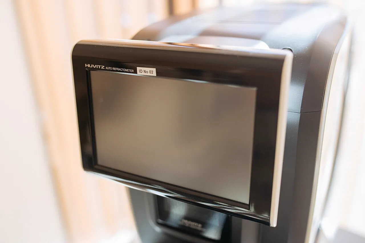
Appearance of a Huvitz autorefractor-keratometer used in ophthalmology clinics. It has a chin rest and forehead rest on the front to hold the patient’s head steady and automatically measure objective refraction values. This corresponds to the objective refraction testing device discussed in the autorefractometer examination section.
Objective refraction testing is an examination that objectively and automatically measures refraction in the patient’s eye. It is one of the most frequently performed tests in ophthalmology clinics, with the aims of shortening the time needed for subjective refraction and providing an objective judgment.
An autorefractor-keratometer is a device that can automatically measure the eye’s refraction (spherical power S, cylindrical power C, axis A) and simultaneously measure the corneal radius of curvature (H/V). The measurement results are used as the starting point for subjective refraction (vision testing), and the final eyeglass prescription value is determined by the subjective examination1).
Refractive errors (myopia, hyperopia, and astigmatism) are diseases, and refractive correction is a medical procedure1). Accurate refraction testing is the basis for proper eyeglass and contact lens prescriptions, and it is directly linked to preventing vision impairment and improving quality of life. In addition, the use of portable autorefractometers in 3-year-old health checkups has greatly increased the early detection rate of amblyopia and severe refractive errors.
Objective refraction tests are broadly divided into two types.
Autorefracto-keratometer: available as fixed (tabletop) and portable (Retinomax® and similar) types
QCan the autorefractor value be used directly as the eyeglass prescription?
A
No. The autorefractor value is the starting point for subjective refraction, and the final eyeglass power is determined by subjective refraction. There is an average difference of about 0.4 D between autorefractor and subjective refraction values, and because it is affected by accommodative influence (instrument myopia), it cannot be used directly for prescribing, especially in children and young people.
Refractive power is expressed in diopters (D). 1 D = the reciprocal of a focal length of 1 m (1/focal length [m])1). The three elements of spherical power (S), cylindrical power (C), and axis (A) determine the eye’s refractive state. Average corneal refractive power (D) can also be calculated from the corneal radius of curvature (the average of H/V).
In adults with accommodative ability, the average difference between objective refraction and subjective refraction is about 0.4 D. Adding +0.50 D spherical power to the autorefractor result brings it closer to the subjective value. If you want to cover the influence of accommodation more broadly, adding +1.50 D to the autorefractor value based on the standard deviation can almost cover it.
Astigmatism value changes sharply with forceful blinking or longer intervals between blinks
QWhy do autoref readings vary?
A
There are four main causes: 1) tear film breakup (dry eye, measurement immediately after blinking, etc.), 2) corneal or lens opacity, 3) abnormalities in corneal shape (irregular astigmatism, keratoconus, etc.), and 4) a mismatch in accommodation timing (if the measurement is not taken immediately after holding back a blink, accommodation can affect the result and cause fluctuations). If the measurements vary, evaluating the tear film condition and using the average of multiple measurements is recommended.
Infrared light (around 850 nm wavelength) is projected onto the fundus, and reflected light from the retina is received by multiple sensors and analyzed. If refractive error is present, the point where the reflected light comes to focus shifts, and from that shift the spherical power (S), cylindrical power (C), and axis (A) are automatically calculated.
In modern devices, two methods are mainly used: the Hartmann-Shack wavefront sensor method and the conventional corneal reflection method.
Hartmann-Shack wavefront sensor method: analyzes the spatial distribution of wavefront aberrations all at once. It can also measure higher-order aberrations.
Corneal reflection method: projects Placido rings onto the anterior corneal surface and measures the corneal radius of curvature at the same time.
At the same time as the measurement, it can measure corneal curvature radius (H and V directions) and calculate the average corneal refractive power (D). This is routinely used in outpatient ophthalmology as the “keratometer” function.
With autorefractors, accommodative involvement (instrument myopia) is unavoidable. The effect is stronger in children and younger people, and even in adults an accommodative effect of about 1 D is not rare. For this reason, autorefractor results need to be compared with subjective refraction.
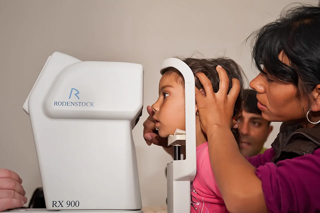
Refractive examination of a pediatric patient using a Rodenstock RX900 autorefractor. The patient rests their face on the chin rest, while the examiner aligns the device and performs the measurement. This corresponds to the steps of head positioning, alignment, and measurement described in the “Examination method and procedure” section.
Autorefractor-keratometer procedure
1. Fall prevention and guidance: Carefully guide the patient so they do not fall when approaching the device.
2. Explanation: Explain clearly to the patient that their refractive value will be measured.
3. Head positioning: Place the chin firmly all the way back and keep the head from tilting.
4. Alignment: Center the device for accurate measurements.
5. Checking the Mayer ring: Adjust the focus firmly so the Mayer ring is clearly visible.
6. Blink prompting and measurement: Ask the patient to blink lightly several times, then measure while keeping the eyes wide open and holding the blink.
Overview of retinoscopy (skiascopy)
Environment: Use divergent light in a dim room.
Examination distance: Fixed at 50 cm (standard for static retinoscopy).
Light and shadow judgment: With movement (same direction) indicates the plus side of refractive power at -2.00 D or more. Against movement (opposite direction) indicates -2.00 D or less.
Neutralization point: Calculate the refractive power from the relation eye refractive power = lens worn + (-2.00 D).
Indications: Especially useful in children, patients with psychogenic visual disturbance, and patients with autism spectrum disorder.
If head position and alignment are not properly fixed, the refractive value may shift toward hyperopia, and astigmatism may increase or the astigmatic axis may change. In particular, the astigmatic axis can change easily because of poor measurement, so it is important to confirm stability at each measurement.
Strong blinking or delayed measurement timing can cause tear film breakup. This leads to large fluctuations in data, increased astigmatism, and changes in the astigmatic axis. Before measurement, have the patient blink gently a few times, then measure immediately after opening the eyelids; this is key to improving accuracy.
By checking the irregularity of the Mayer ring, you can learn whether the tear film has broken up, the state of the cornea, the size of the pupil, and whether the pupil is perfectly round. This can be useful for predicting in advance how the next visual acuity and refraction tests may be affected and for planning the examination.
Portable devices such as Retinomax® are useful in screening settings, for children, and for patients who have difficulty moving around. Performing refraction testing in the second-stage exam of the 3-year-old vision screening has a major effect in reducing missed amblyopia and severe refractive errors.
After objective refraction testing, combine the following subjective refraction tests to determine the final refractive power.
Lens exchange method: adjust spherical and cylindrical power by combining the trial frame and trial lenses
Cross-cylinder method: use a lens with plus and minus powers at right angles to precisely determine the astigmatic axis and power
Two-color (red-green) test: used for fine adjustment of spherical power (to distinguish overcorrection from undercorrection)
A cross-cylinder lens is a lens that has positive power in one meridian and negative power in the perpendicular meridian. In an eye with astigmatism, it can move one focal line to the plus side and the other to the minus side, and it is used to precisely determine the astigmatic axis and power.
If instrument myopia is suspected, or if accommodation must be excluded in children, refraction is performed after instilling a cycloplegic eye drop1). The standard procedure is to measure about 1 hour after instilling Saipurejin® (1% cyclopentolate).
QWhat should be kept in mind during refractive testing in children?
A
There are three main points. ① Instrument myopia (accommodative interference) is more pronounced than in adults, so autorefractor values cannot be used directly as prescription values. ② If marked accommodative interference is suspected, or if amblyopia or strabismus need to be evaluated, cycloplegic refraction with Saipurejin® 1% eye drops is necessary. ③ In the 3-year-old health check, refractive testing with a portable autorefractor contributes greatly to the early detection of amblyopia and anisometropia. Retinoscopy is useful for infants who cannot cooperate with the examination.
It should be performed proactively, especially in esotropia, hyperopia, and infants
When the cylinder power is small and the axis varies a lot
Suspect irregular astigmatism, dry eye, or corneal epithelial damage
Place more weight on the subjective refraction result when deciding the prescription
High refractive error (especially in children)
Confirm with cycloplegic refraction and prescribe appropriate glasses.
Assess whether amblyopia is present, and start amblyopia treatment early if needed.
Recommended frequency of regular refraction testing
During amblyopia treatment: recheck refraction every 3 months
Progressive myopia: recommend refraction testing twice a year
Children in the growth period: regular examinations at least once a year
Full correction (the same power as the autorefractor value) is not always the best eyeglass prescription1). A prescription that takes daily use into account (how much near work is done, wearing time, etc.) leads to more comfortable everyday wear.
Infrared light (around 850 nm) is projected onto the fundus, and the reflected light from the retina is received by a Hartmann-Shack wavefront sensor or a corneal reflection sensor. The sensor analyzes shifts in the distribution of bright spots (wavefront aberrations) to automatically calculate spherical refractive power (S), cylindrical refractive power (C), and axis (A).
Corneal curvature radius is measured by projecting Placido rings (concentric circles of light) onto the front surface of the cornea and calculating the average corneal refractive power and the curvature radius for each meridian from the curvature of the reflected image. This makes it possible to objectively assess the axis and magnitude of corneal astigmatism.
Retinoscopy is a method for calculating refractive power from the movement of the light reflex as reflected light from the retina reaches the examiner’s eye. In static retinoscopy, divergent light is used in a semi-dark room at a test distance of 50 cm. The movement of the light reflex (with or against motion) and the neutral point are confirmed, and the eye’s refractive power is calculated from the lens power needed for neutralization. In dynamic retinoscopy, the examiner changes the distance while looking for the neutral point. Because it can be performed with both eyes open, it has the advantage of being less affected by instrument myopia.
In the power cross notation, a cross-cylinder lens is a lens in which one meridian has positive refractive power and the perpendicular direction has negative refractive power. The examination is performed while focusing on the axis of the minus cylinder lens and the intermediate axis of this lens. In eyes with astigmatism, it can move one focal line to the plus side and the other to the minus side, and is used to determine the astigmatic axis and refine the astigmatic power.
With the spread of digital devices, the prevalence of myopia is rising sharply worldwide1). The importance of refractive correction is increasing, and the role of accurate refraction testing is being reexamined.
Measurement accuracy with the Hartmann-Shack wavefront sensor method has continued to improve, and devices that can evaluate higher-order wavefront aberrations are becoming more widely used. Development of autorefractors equipped with AI (artificial intelligence) is also progressing, and automatic judgment of measurement quality and improved accuracy in irregular corneas are expected.
On the other hand, in eyes after corneal transplantation or refractive surgery (LASIK, PRK, etc.), it is known that large changes in corneal shape reduce the measurement accuracy of the autorefractor. Developing measurement algorithms that can handle such unusual corneal shapes is a challenge. In addition, in high myopia (-10 D or more) and high hyperopia (+10 D or more), the measurement range may be reached, so the results must be interpreted with care.
Arslantürk Eren M, Nalcı Baytaroğlu H, Atilla H. Comparison of Spot Vision Screener and Tabletop Autorefractometer with Retinoscopy in the Pediatric Population. Turk J Ophthalmol. 2024;54(2):56-62. PMID: 38644780.
Demir MS, Muhafiz E. Performance of a photoscreener in detecting accommodation spasm. Clin Exp Optom. 2022;105(8):817-821. PMID: 34751084.
Copy the article text and paste it into your preferred AI assistant.
Article copied to clipboard
Open an AI assistant below and paste the copied text into the chat box.

.jpg)