Nakakura S, et al. Evaluation of rebound tonometer iCare IC200 as compared with IcarePRO and Goldmann applanation tonometer in patients with glaucoma. Eye Vis (Lond). 2021;8:25. Figure 1. PMCID: PMC8247177. License: CC BY 4.0.
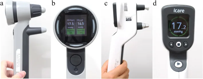
This is a four-panel photograph showing: (a) full view of the IcarePRO body, (b) color display panel of IcarePRO (showing both eye pressure values and reliability indicator), (c) full view of the iCare IC200 body (multi-position model), and (d) color display panel of IC200 (showing 17.2 mmHg). It corresponds to the appearance and display specifications of the iCare series discussed in the section “1. What is the iCare Tonometer (Rebound Tonometer)?”
The iCare® is a rebound (rebound) tonometer, a portable tonometer that can measure intraocular pressure without topical anesthesia. It ejects a small probe toward the cornea and estimates the intraocular pressure from the rebound speed. It correlates well with GAT but often shows slightly higher values. The reliability of the measurements is high, and it is particularly easy to use for measuring intraocular pressure in children 1).
The contact force is extremely small, about 1.0 g, and the measurement time is about 0.1 seconds. Since there is almost no invasion to the cornea, surface anesthesia is unnecessary. The use of disposable probes also reduces the risk of infection between patients.
iCare IC100 (formerly iCare®): Standard portable type. Measurement in the supine position is not possible.
iCare IC200: Normally, when the tip is pointed downward, the probe falls and measurement is not possible, but the IC200 is designed to solve this problem and allows measurement in multiple positions including supine 1).
iCare HOME: A home model for patient self-measurement. Approved by the FDA in 2017 for self-measurement of intraocular pressure.
iCare HOME2: Successor to HOME. Equipped with auto-alignment function, built-in memory, and Bluetooth/cloud data transfer.
There is almost no pain. The probe contact force is about 1.0 g, and the contact time is about 0.1 seconds, making the invasion to the cornea extremely small. Since no topical anesthetic eye drops are needed, there is no irritation from eye drops either. A new disposable probe is used for each patient, ensuring hygiene. It is one of the most widely used tonometers for children and infants, and is useful for screening intraocular pressure abnormalities in children, including congenital glaucoma.
The correlation between iCare and GAT is good, but iCare tends to show slightly higher values than GAT. The difference is generally +0.5 to 2.0 mmHg, and in high intraocular pressure ranges, the variability tends to be larger than with GAT1).
Comparative studies with GAT have generally shown agreement within ±5 mmHg, indicating practical accuracy for screening and follow-up1). However, for detailed evaluation of ocular hypertension or glaucoma, it is advisable to recheck with GAT when abnormal values are found.
GAT is greatly affected by central corneal thickness (CCT), leading to underestimation in thin corneas and overestimation in thick corneas. iCare has been reported to be less affected by CCT than GAT, but caution is needed as underestimation may occur in thin corneas.
iCare performs 6 automatic measurements, excludes the upper and lower limits, and displays the average as the intraocular pressure value. At the same time, a reliability indicator is shown in color.
Green: High reliability; the measurement value can be adopted.
Yellow: Within acceptable range. Consider re-measurement if necessary.
Red: Low reliability of the measurement value. Re-measurement is required.
A magnetic coil launches a lightweight magnetized probe (diameter 1.7–1.8 mm) toward the cornea using an electromagnetic field.
When the probe contacts the cornea, a rebound occurs depending on the hardness (intraocular pressure) of the eye.
The deceleration (departure acceleration) of the probe after rebound is measured by electromagnetic induction.
The higher the intraocular pressure, the shorter the contact time between the probe and cornea, and the greater the departure acceleration.
This physical relationship is converted into intraocular pressure (mmHg) by an analysis algorithm.
The momentary contact (0.1 second) with a minimal force of about 1.0 g is the basis for not requiring topical anesthesia. Because the measurement time is short, the effects of eye movement and pulse wave are minimized.
It has been reported that the influence of central corneal thickness (CCT) is smaller than with GAT, but intraocular pressure may be underestimated in thin corneas. Corneal rigidity and ocular stiffness are also known factors affecting measurements.
Measurement Procedure in the Examination Room (IC100/IC200)
Preparation: Attach a disposable probe to the device. Have the patient fixate on the fixation light in front.
Alignment: Position the device in front of the eye and stabilize it with the forehead rest. Confirm automatic alignment so that the probe is directed at the center of the cornea.
Measurement: Pull the trigger to release the probe. Six consecutive measurements are automatically performed, and the average value and reliability indicator are displayed.
IC200 (multi-position): Can be measured in supine or prone positions. Suitable for bedside or operating room use. Infants and young children are measured while held supine or on a parent’s lap.
Confirmation: If the reliability indicator is red, repeat the measurement. If green or yellow, accept the measurement value.
Self-measurement procedure with iCare HOME/HOME2
Preparation: The patient adjusts the forehead and cheek rests and holds the device steady. Refer to the LED guide light; when horizontal, a green light turns on indicating readiness for measurement.
Measurement: Press the button to automatically perform six consecutive probe releases. The measurement values are stored in the internal memory along with time, date, and eye side information.
Data management: HOME2 enables remote review of measurement data by the physician via Bluetooth/cloud connectivity. Data can be downloaded during clinic visits to evaluate changes over time.
Measurement timing: It is recommended to measure at least three time points: immediately after waking, before bedtime, and in the afternoon. Measuring over multiple days allows assessment of the 24-hour intraocular pressure fluctuation profile.
Patient training: At the initial introduction, simultaneous measurement with GAT is performed in the examination room to confirm the accuracy of self-measurement before starting home use.
QIs there value in measuring intraocular pressure at home?
A
Intraocular pressure measurement in the clinic is typically performed 3–4 times a year, which is insufficient to capture the full picture of IOP fluctuations. IOP varies throughout the day, with peaks sometimes occurring at night or early morning. Self-measurement with iCare HOME provides the following information. First, it can capture the nighttime IOP elevation (3–5 mmHg) that occurs due to the combination of supine posture and circadian rhythm. Second, it allows evaluation of peak IOP in cases such as normal-tension glaucoma where IOP is normal during clinic visits but elevated at other times1). Third, it enables confirmation of the trough effect of eye drops (IOP at the time of least drug effect) and dynamic assessment of treatment efficacy. Additionally, regular self-measurement contributes to improved motivation for treatment (medication adherence).
Intraocular pressure is a dynamic parameter. Even in healthy individuals, fluctuations of 4–5 mmHg per day are observed, and in glaucoma patients, fluctuations can exceed 10 mmHg. There is a growing need for IOP monitoring beyond clinic measurements.
At night, the combination of supine posture and circadian rhythm reproducibly causes an IOP elevation of 3–5 mmHg compared to seated measurements. This nocturnal IOP elevation cannot be captured by clinic measurements performed in the seated position.
Identification of peak IOP: Even if IOP is normal during clinic visits, high IOP may occur at other times. This is especially important in normal-tension glaucoma1)
Dynamic evaluation of treatment effect: Check the trough effect of eye drops (IOP at the time when the drug effect is weakest) and evaluate the comprehensiveness of treatment
Assessment of postural change effects: The magnitude of IOP increase when changing from sitting to supine position is correlated with glaucoma progression risk, and supine measurement with IC200 is useful
Improvement of medication adherence: Self-measurement allows patients to visualize their own IOP fluctuations, increasing motivation to continue treatment
Regular follow-up for pediatric glaucoma: Can be used for screening and follow-up in outpatient settings
The main factors affecting the measurement accuracy of iCare are shown below.
Central corneal thickness (CCT): Although the effect is not as large as with GAT, there are reports of underestimation of IOP in thin corneas. Caution is needed in patients with very thin corneas (e.g., congenital corneal opacity, after corneal resection such as LASIK)
Probe placement (probe angle): Measurement errors occur when the probe is not perpendicular to the center of the cornea. Use the auto-alignment function for accurate positioning
Body position: iCare IC100 and iCare HOME cannot measure in a downward-facing position because the probe falls. Use IC200 for supine or multiple positions
Number of measurements and reliability indicator: Among 6 consecutive measurements, if the reliability indicator is red, always repeat the measurement
Tear film: The influence of tear film thickness and stability is considered smaller than with GAT, but accuracy may decrease in cases of marked corneal dryness or corneal epithelial damage
Corneal rigidity and ocular rigidity: The physical properties of the cornea affect the measurement values. In patients after refractive surgery (e.g., LASIK), the measurements may be biased.
The iCare HOME2 features an automatic alignment function as an improvement over the previous generation HOME, improving the accuracy of patient self-alignment. It can store measurement data long-term in its internal memory, and via Bluetooth/cloud connectivity, physicians can remotely review the data. This opens the way for application in remote glaucoma management through integration with telemedicine.
Development of 24-Hour Continuous Intraocular Pressure Monitoring
A contact lens-type sensor (Triggerfish CLS) has obtained CE marking in Europe, and clinical studies for 24-hour continuous monitoring are underway. However, the limitation in clinical application is that the measured values are in millivolt equivalents (mVeq) and cannot be directly converted to mmHg. The implantable intraocular pressure sensor (EyeMate) has obtained CE marking in Europe as a permanent implantable device inserted into the ciliary sulcus, enabling on-demand and long-term intraocular pressure monitoring.
AI-Based Analysis of Intraocular Pressure Fluctuation Patterns
Research is progressing on using machine learning and AI to analyze the large amount of intraocular pressure time-series data collected by iCare HOME, to predict glaucoma progression risk and evaluate treatment effects in real time. It is expected to be applied to personalized treatment that takes into account individual differences in intraocular pressure fluctuation patterns.
Jorge JM, González-Méijome JM, Queirós A, Fernandes P, Parafita MA. Correlations between corneal biomechanical properties measured with the ocular response analyzer and ICare rebound tonometry. J Glaucoma. 2008;17(6):442-8. PMID: 18794677.
Kim YJ, Moon Y, Kwon AM, Lim HW, Lee WJ. Intraocular Pressure According to Eye Gaze by iCare Rebound Tonometry in Normal Participants and Glaucoma Patients. J Glaucoma. 2021;30(8):643-647. PMID: 33979114.
Copy the article text and paste it into your preferred AI assistant.
Article copied to clipboard
Open an AI assistant below and paste the copied text into the chat box.