Radiation Retinopathy (RR) is a chronic progressive occlusive retinal microvascular disorder that develops when the retina is included in the radiation field during radiotherapy for intraocular tumors, orbital or sinus tumors, intracranial diseases, etc. It occurs after radiation therapy for intraocular tumors such as choroidal malignant melanoma, orbital tumors, head and neck tumors, and brain tumors. It was first reported by Stallard in 1933.
Note that massive accidental exposure can cause necrosis of the retina and optic nerve within a few weeks.
Because retinal vascular endothelial cells with high proliferative capacity show selective sensitivity to radiation, a series of pathological conditions leading to capillary occlusion, ischemia, and neovascularization develop1). Choroidal vascular endothelium is also damaged. Onset often occurs six months or more after irradiation, especially after 2 to 3 years. The overall incidence varies depending on the radiation field.
The incidence by site is shown below.
Irradiated site
Incidence rate
Orbit
85.7%
Paranasal sinuses
45.4%
Nasopharynx
36.4%
Brain
3.1%
A meta-analysis reports that the prevalence of RR after radiotherapy for brain, head, and neck tumors is approximately 6%, and optic neuropathy (ON) is approximately 2% 3). The overall incidence including late-onset cases is reported to be about 17% 4).
QDoes it occur in everyone who receives radiation therapy?
A
The incidence varies greatly depending on the irradiation site, dose, fractionation method, and comorbidities. The overall incidence is reported to be 17%, meaning it does not occur in all patients 4). Early detection through regular fundus examinations is important.
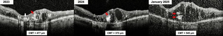
Kayabaşı M, et al. Evolution of the Onion Ring Sign in Radiation Retinopathy. Cureus. 2025. Figure 3. PMCID: PMC12740121. License: CC BY.
OCT images from January 2023, 2024, and 2025 show CMT of 477 μm, 373 μm, and 545 μm, respectively. This corresponds to the macular edema discussed in the section “2. Main symptoms and clinical findings”.
In the early stage, there are often no symptoms, and it may be discovered incidentally during a health checkup. When the lesion affects the macula or optic nerve, the following symptoms appear.
Decreased vision: Caused by macular edema or macular ischemia. Often progresses slowly.
Metamorphopsia (distortion): Caused by disruption of photoreceptor arrangement due to macular edema.
Fundus findings resemble diabetic retinopathy, starting with microaneurysms, retinal hemorrhages, and hard exudates, followed by cotton-wool spots. As it progresses, retinal neovascularization develops, leading to vitreous hemorrhage. Visual acuity decreases due to macular edema and occlusion of perifoveal capillaries. Once it develops, progression is faster than diabetic retinopathy.
The clinical course follows these stages:
Early stage: Microaneurysms, retinal hemorrhages, and hard exudates appear.
Advanced stage: Cotton-wool spots appear. Indicates expansion of ischemic area.
Severe stage: Retinal neovascularization occurs, leading to vitreous hemorrhage.
Neovascular glaucoma (NVG): Refractory glaucoma due to neovascular invasion of the iris and angle. The rate of enucleation due to NVG is reported to be 1–12% 5).
As a late-onset special finding, in a case that developed 17 years later, an onion ring sign due to cholesterol crystals within the cyst cavity was confirmed on OCT, and it is attracting attention as a marker of treatment resistance in the chronic phase 6).
In addition, in a case where RR localized to the superior retina developed 16 months after 30 Gy whole-brain irradiation, the lesion distribution coincided with the 30 Gy isodose line of the radiation field, confirming that even in low-dose areas, the onset pattern corresponds to the radiation field 7).
QWhen does onset usually occur?
A
Onset often occurs six months or more after irradiation, especially 2–3 years later. The median time to diagnosis is reported as 39 months after irradiation 3), but late-onset cases up to 17 years have been reported 4). Long-term regular fundus examinations are necessary after irradiation.
The dose threshold is generally considered to be 35 Gy 4). Onset is more likely with doses exceeding 45 Gy, and the risk is particularly high above 50 Gy 3). However, cases have been reported at 20 Gy, and onset has also been reported after whole-brain irradiation of 30 Gy 7), so caution is needed even at doses below the threshold. Retinal vascular endothelial cells with high proliferative capacity are most susceptible to damage, and choroidal vascular endothelium is also affected.
The latency period after irradiation is six months or more, with a peak at 2–3 years. This is thought to be because endothelial cell damage from radiation accumulates and takes time to exceed the clinical threshold.
Risk factors are listed below.
Risk factor
Description
Total dose
>35 Gy (threshold) 4), high risk above 45 Gy
Fraction dose
High fractionation irradiation
Irradiated area
Orbit or near optic chiasm3)
Diabetes
Worsens microvascular fragility
Concurrent chemotherapy
Increased sensitivity
Irradiation near the optic chiasm has been reported to show a significant correlation with the development of RR (p=0.009)3).
Proliferative RR is reported to occur in 3–25% of all RR cases5). In cases after plaque brachytherapy, progression to proliferative RR is observed at 32 months after irradiation.
QDoes having diabetes increase the risk of radiation retinopathy?
A
Diabetes is an important risk factor for radiation retinopathy. Microvascular fragility due to diabetes acts synergistically with radiation-induced endothelial damage, potentially causing the condition at lower radiation doses. Along with maintaining blood glucose control, more frequent fundus examinations are recommended after radiation therapy.
FA is the basic examination for diagnosis and staging of RR. In the early stage, increased permeability of retinal capillaries is observed, and as the disease progresses, capillaries become occluded. Arterioles also become occluded, leading to extensive enlargement of retinal avascular areas and the development of retinal neovascularization. The Amoaku FA classification (Grades 1–4) is widely used 1).
Grade
Main Findings
1
Microaneurysms and focal capillary dilation
2
Capillary occlusion and extensive vascular abnormalities
OCT is used for quantitative assessment of ME according to the Horgan classification (Grade 1–5), and OCT detection is possible at 4 months after plaque brachytherapy1). OCTA can noninvasively visualize capillary dropout, nonperfusion areas, and changes in the FAZ, and is useful for early detection 1).
RR onset often occurs 6 months or more after irradiation, especially 2–3 years later. The median time to onset is 39 months after irradiation, and careful observation is particularly necessary for >50 Gy irradiation 3). After irradiation, regular fundus examinations and OCT imaging (at least every 6–12 months) are recommended.
For diagnosis, it is important to obtain a history of radiotherapy (for intraocular tumors, orbital tumors, intracranial tumors, or sinus tumors).
Because the fundus findings resemble diabetic retinopathy, differentiation is necessary. Confirming the presence or absence of a history of radiation exposure generally makes differentiation easy.
Diabetic retinopathy: Fundus findings (microaneurysms, hemorrhages, exudates, neovascularization) closely resemble radiation retinopathy. Checking for diabetes and a history of radiation exposure is key to differentiation. Radiation retinopathy is characterized by faster progression than diabetic retinopathy once it develops.
Retinal vein occlusion: Hemorrhage and edema along the occluded vein are predominant, showing a sectoral distribution not seen in radiation retinopathy. Differentiation is easy if there is no history of radiation exposure.
QWhat is the difference from diabetic retinopathy?
A
Fundus findings (microaneurysms, hemorrhages, exudates, neovascularization) are very similar between the two. The most important distinguishing point is the presence or absence of a history of radiation exposure. Additionally, once radiation retinopathy develops, it progresses faster than diabetic retinopathy, and its time course—from six months to several years after irradiation—is characteristic. Management is particularly difficult when both diseases coexist.
Anti-VEGF drugs are currently the first-line treatment for RR. The agents used are bevacizumab (IVB), ranibizumab, and aflibercept1). The use of high-dose ranibizumab 2 mg has also been reported1).
Prophylactic anti-VEGF administration is performed to suppress the onset of RR after radiotherapy. A meta-analysis of 4 studies involving 2109 patients showed the following results2).
The recommended protocol is IVB 1.25–1.5 mg every 4 months for 24 months 2). Prophylactic anti-VEGF therapy over 48 months showed significant improvement in best-corrected visual acuity: 0.54 logMAR (prophylaxis group) vs. 2.00 logMAR (control group) 5).
In a review by Sahoo et al. (2021), the RCT by Schefler and Murray validated the efficacy of anti-VEGF therapy, recommending early intervention for macular edema (within 90 days after irradiation) 1).
A meta-analysis by Victor et al. (2023) of 4 studies involving 2109 patients confirmed that prophylactic IVB significantly reduced ME by 50% and RON by 38% after plaque brachytherapy2).
Laser photocoagulation is performed on the avascular areas of the retina to prevent the development of retinal neovascularization and neovascular glaucoma. Panretinal photocoagulation (PRP) is performed for proliferative RR, with a reported regression rate of 66% 5). Even in post-plaque treatment cases, regression was observed in 64.4% 5). Focal laser is used as an adjunct for ME.
Vitrectomy is performed for vitreous hemorrhage. Vitrectomy is also indicated for tractional retinal detachment. For NVG, filtering surgery or cyclophotocoagulation may be necessary. COMS data reports that 43% of patients have corrected visual acuity of 20/200 or less at 3 years after irradiation 2).
If macular edema is present → anti-VEGF agents (first-line) and steroids (adjunctive)
There is no effective method to stop progression, and the prognosis is often poor.
QHow long should anti-VEGF injections be continued?
A
For prophylactic administration, a protocol of every 4 months for 24 months is recommended 2). For therapeutic administration, the duration varies depending on disease activity. In treatment-resistant chronic cases, more than 72 injections may be required 6).
The central mechanism of radiation-induced retinal damage is selective loss of retinal vascular endothelial cells. Retinal vascular endothelial cells with high proliferative capacity are most susceptible, and choroidal vascular endothelium is also damaged. Endothelial cells are particularly sensitive to radiation, and capillary walls collapse due to DNA damage and apoptosis.
The progression of the pathology follows these stages:
Endothelial cell injury stage: Progresses immediately after irradiation. DNA double-strand breaks and apoptosis of endothelial cells occur, leading to loss of vascular wall integrity.
Capillary occlusion and ischemia stage: Loss of endothelial cells causes capillary occlusion, expanding the retinal ischemic area. Early fluorescein angiography shows increased permeability, but occlusion becomes dominant as it progresses. Arterioles also occlude, leading to extensive enlargement of retinal avascular zones.
VEGF production and angiogenesis phase: VEGF is overproduced in the ischemic retina, inducing proliferation of fragile new blood vessels.
Accumulation of advanced glycation end products (AGEs), pericyte loss, and basement membrane thickening are also thought to contribute to endothelial damage. This mechanism is similar to that of diabetic retinopathy and explains why the risk of RR increases in patients with diabetes.
There is a latent period of six months or more, especially 2–3 years, from irradiation to clinical onset. This reflects the time required for endothelial cell damage to accumulate and for capillary occlusion to become clinically apparent.
7. Latest research and future perspectives (research-stage reports)
The meta-analysis by Victor et al. (2023) is the largest current evidence showing the efficacy of prophylactic anti-VEGF administration, but most of the included studies are observational, and further validation through randomized controlled trials (RCTs) is needed 2). Standardization of optimal dosing intervals, drugs, and treatment duration also remains a future challenge.
Early detection using OCTA (optical coherence tomography angiography)
OCTA can quantitatively assess capillary dropout, FAZ enlargement, and decreased capillary density without the use of contrast agents. It can detect non-perfusion areas from early stages after radiotherapy, and its application to screening and monitoring of RR is advancing 1).
Chronicity markers for treatment-resistant radiation retinopathy
Kayabai et al. (2025) reported a case of a 53-year-old man who developed radiation retinopathy 19 years after radiotherapy for an intraocular tumor 6). The onion ring sign (multilayered deposition of cholesterol crystals within cystoid spaces) observed on OCT is recognized as an imaging marker for chronic, treatment-resistant radiation retinopathy, and the patient required over 72 intravitreal injections over a long-term course.
Next-generation anti-VEGF agents such as brolucizumab and faricimab (angiopoietin/VEGF dual-target) are being investigated for use in radiation retinopathy 5). They are expected as alternative options in cases resistant to existing drugs.
Risk assessment after proton and heavy ion therapy
In addition to conventional X-rays and gamma rays, risk assessment for radiation retinopathy after proton and heavy ion (carbon ion) therapy is underway. Even with highly dose-concentrated particle therapy, retinopathy can occur if the retina is within the irradiation field, making retinal dose evaluation during treatment planning and postoperative monitoring important issues.
Combined Management with Radiation Optic Neuropathy (RON)
RR and radiation optic neuropathy (RON) may occur simultaneously from the same radiation field. The incidence of RON after EBRT is reported to be approximately 2% 3). In cases where RR and RON coexist, visual dysfunction becomes more severe, making regular visual field testing and OCT evaluation of the optic nerve in addition to fundus examination an important research topic.
Sahoo NK, Lim JW, Laude A, et al. Radiation retinopathy—the complex interplay of radiation, vasculature, and clinical outcomes. Clin Ophthalmol. 2021;15:3797-3809.
Victor AA, Mauldin WM, Houston SK, et al. Prophylactic intravitreal bevacizumab and radiation retinopathy after plaque brachytherapy for uveal melanoma: a meta-analysis. Clin Ophthalmol. 2023;17:2997-3009.
Kinaci-Tas B, Wilschut JA, Kilic E, et al. The incidence of radiation-induced optic neuropathy and retinopathy in patients treated with external beam radiation therapy: a systematic review and meta-analysis. Cancers. 2023;15:1999.
Chakraborty K, Jain S, Tripathy K, et al. Delayed onset radiation retinopathy following skull base tumor treatment. Indian J Ophthalmol. 2023;71:303-305.
Mularska W, Nowak-Gospodarowicz I, Golik B, et al. Radiation retinopathy after plaque brachytherapy for uveal melanoma—pathogenesis, diagnosis, and management. J Contemp Brachytherapy. 2023;15:372-382.
Kayabai M, Ilhan S, Celik E, et al. Onion ring sign as a biomarker of chronic treatment-resistant radiation retinopathy. Cureus. 2025;17(11):e97758.
Chan L, Eftekari SC, Nguyen QT, et al. Radiation retinopathy after whole-brain radiotherapy: a case report and literature review. Adv Radiat Oncol. 2021;6:100706.
Copy the article text and paste it into your preferred AI assistant.
Article copied to clipboard
Open an AI assistant below and paste the copied text into the chat box.