Plateau iris syndrome is a type of primary angle closure disease in which the ciliary processes rotate forward, mechanically pushing the iris root, causing narrowing and closure of the angle 1, 2, 3). The central anterior chamber depth is normal or only mildly shallow, while the periphery becomes extremely shallow. This unique morphology of “normal central depth with shallow peripheral anterior chamber” is characteristic of this disease.
The first description of the condition began in 1992 when Pavlin et al. quantitatively demonstrated the abnormal position of the ciliary processes using ultrasound biomicroscopy (UBM) 2). Recent reviews also classify it as a non-pupillary block mechanism in which the anteriorly positioned ciliary body pushes up the iris root 3). Subsequently, the 5th edition of the Japanese Glaucoma Practice Guidelines classified the causes of angle closure into four categories, positioning this disease as an important mechanism following relative pupillary block1).
Additional treatment such as miotics, ALPI, or lens extraction
When accompanied by elevated intraocular pressure and glaucomatous optic neuropathy, it is called plateau irisglaucoma1). Since some cases of PIC correspond to PIS, a mydriatic challenge test is added after LPI to differentiate between the two.
The frequency of plateau iris varies greatly depending on the target population.
In a UBM study from Singapore targeting primary angle closure suspects (PACS), approximately 30% showed anterior positioning of the ciliary processes 5).
It has been reported that about one-third of primary angle closure eyes after LPI have persistent iridotrabecular contact (ITC) 9).
Angle closure itself has a high prevalence in East Asian and Inuit populations, and plateau iris is also frequently encountered in these groups.
In relatively young patients (under 40 years) with acute angle closure attacks, plateau iris should be suspected. While typical primary angle closure is more common in those aged 50 and older, this condition is a typical cause of young-onset disease.
QWhat is the difference between PIC and PIS?
A
PIC (plateau iris configuration) is an anatomical feature where the iris root is positioned anteriorly, and after LPI relieves the pupillary block, the angle opens 1). PIS (plateau iris syndrome) is a condition where iridotrabecular contact (ITC) in two or more quadrants persists after LPI under mydriatic challenge, requiring additional treatment 9). Since some cases of PIC correspond to PIS, a mydriatic challenge test is performed after LPI to differentiate. When plateau iris syndrome is accompanied by glaucomatous optic neuropathy, it is called plateau irisglaucoma1).
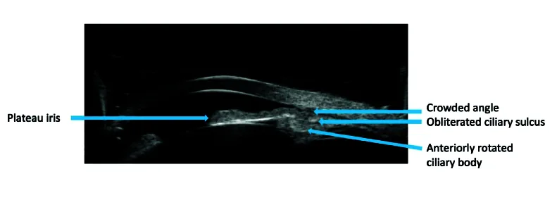
Shamseldin Shalaby W, et al. Contemporary Approach to Narrow Angles. J Ophthalmic Vis Res. 2024. Figure 1. PMCID: PMC11022020. License: CC BY.
Anterior segment UBM shows a narrow angle with the iris root remaining flat and the ciliary body rotated anteriorly. This diagnostic image demonstrates anatomical findings characteristic of plateau iris syndrome.
It often takes the form of chronic angle closure glaucoma, and subjective symptoms are less prominent than objective findings. It is not uncommon for it to be noticed only after progression of elevated intraocular pressure or visual field defects. On the other hand, when the iris root mechanically occludes the angle during mydriasis, it can cause an acute angle closure glaucoma attack, presenting with eye pain, headache, nausea/vomiting, blurred vision, and halos around lights.
Anterior chamber depth: The central depth is normal to slightly shallow. The peripheral depth is extremely shallow. This “normal central depth with shallow peripheral chamber” is a characteristic pattern of plateau iris.
Iris shape: The iris plane is not bulging but flat or mildly anteriorly bowed. The iris root rises steeply forward, forming a trapezoidal peripheral portion.
Gonioscopic findings: The double hump sign is characteristic 9). On compression gonioscopy, the iris appears bimodal. The peripheral hump is due to the iris riding over the ciliary processes, and the central hump is formed by the iris contacting the lens surface.
During acute attack: Corneal edema, ciliary injection, moderate mydriasis with fixed pupil, and marked elevation of intraocular pressure are observed.
Imaging Findings (UBM)
Abnormal position of ciliary processes: The ciliary processes are located more anteriorly than normal and are rotated forward 2, 3, 9). This is the essence of plateau iris.
Kumar’s 4 signs5): On UBM, the diagnosis is made by four findings: (1) iris-trabecular contact (ITC), (2) anterior rotation of the ciliary processes, (3) absence of the ciliary sulcus, and (4) posterior bowing of the iris.
Central anterior chamber depth: Unlike the pupillary block type, the central anterior chamber depth is preserved. The central iris is flat.
AS-OCT: It can quantitatively evaluate angle parameters (AOD, TISA, etc.) non-contact, but visualization of ciliary processes is difficult, and it remains a complementary role to UBM9).
During an acute attack, UBM and gonioscopy may be difficult due to corneal edema. In such cases, first lower intraocular pressure with medication or anterior chamber paracentesis, and perform imaging after the cornea becomes clear.
QWhich is more useful for diagnosing plateau iris, ultrasound biomicroscopy or AS-OCT?
A
UBM is more useful 2, 9). The essence of plateau iris is anterior rotation of the ciliary processes, and UBM is the only clinical examination that can directly visualize the ciliary processes 2). By evaluating Kumar’s 4 signs (ITC, anterior rotation of ciliary processes, absence of ciliary sulcus, posterior bowing of iris), an objective diagnosis can be made 5). AS-OCT can quantitatively evaluate angle parameters (AOD, TISA, etc.) non-contact, but it is difficult to visualize structures behind the iris (ciliary processes) 9). Ideally, both should be used complementarily.
The underlying cause of plateau iris is an anatomical abnormality: the anterior position and forward rotation of the ciliary processes 2, 3). It is thought to result from congenital morphological abnormalities of the peripheral iris and ciliary body. The ciliary processes are located more anteriorly than usual, pushing the iris root forward and causing the angle to close, covering the trabecular meshwork.
During dilation, the tissue of the iris root accumulates in the periphery. In a normal eye, this excess tissue is accommodated in the ciliary sulcus. However, in plateau iris, the ciliary processes occupy the ciliary sulcus, pushing the iris tissue toward the angle and worsening angle closure.
According to the AAO Preferred Practice Pattern, the following are risk factors for primary angle closure disease, including plateau iris9):
Age: Acute angle closure attack at a relatively young age (under 40 years) suggests plateau iris. Typical primary angle closure is more common in those aged 50 and older.
Sex: More common in women.
Axial length: More common in eyes with short axial length and hyperopia. However, some cases do not have as short an axial length as in pupillary block.
Corneal diameter: More common in eyes with small corneas.
Race: Angle closure is more common in East Asians and Inuit populations, and the frequency of plateau iris is also high.
Family history: A family history of angle closure is an important risk factor.
Eyes with plateau iris are at high risk for drug-induced angle closure attacks. The following drugs can trigger attacks through mydriasis or ciliary body edema 9):
Definitive diagnosis of plateau iris requires evaluation after LPI1, 9). After LPI removes the pupillary block component, assess whether residual angle closure is due to plateau iris mechanism.
UBM findings of plateau iris show the following characteristics 2, 5, 9).
Central anterior chamber depth is relatively preserved
Central iris is flat
Iris root is thickened and bends anteriorly
The angle recess is narrowed to a slit-like shape
The ciliary processes are displaced anteriorly and contact the iris root
The ciliary sulcus is obliterated
In 2008, Kumar et al. organized these findings into 4 signs5): (1) iridotrabecular contact (ITC), (2) anteriorly rotated ciliary processes, (3) obliteration of the ciliary sulcus, and (4) posterior bowing of the iris. A diagnosis of plateau iris is made when all four signs are present.
The international diagnostic criteria for PIS (plateau iris syndrome) is the persistence of ITC in ≥2 quadrants upon mydriatic challenge after LPI, with a preserved central anterior chamber depth9). In a study of the Indian population by Mansoori et al., approximately 34% of PACG eyes met these criteria7).
Treatment of plateau iris syndrome is performed stepwise 1, 4, 9). In the 5th edition of the Japanese Glaucoma Treatment Guidelines, treatment for plateau iris mechanism is detailed in Chapter 6 (Laser Surgery) and Chapter 8 (Treatment of Primary Angle Closure Glaucoma, Section 3-B) 1).
First, LPI is performed to eliminate the pupillary block component 1). In plateau iris cases, LPI is recommended to remove the pupillary block component (recommendation level 1C) 1). In plateau iris configuration (PIC), the angle opens after this, and follow-up is done with regular gonioscopy. In plateau iris syndrome (PIS), additional treatment is needed.
Nd:YAG laser alone or argon + YAG combination is standard, and the perforation is enlarged to 100–200 μm, confirmed by pigment dispersion 1). Apraclonidine hydrochloride is instilled before and after the procedure to prevent transient intraocular pressure elevation.
As initial treatment after confirming PIS, low-concentration pilocarpine (0.5–1%) is used 1, 2). Miosis pulls the peripheral iris toward the center, widening the distance between the iris root and the angle, thereby opening the angle (recommendation level 2C) 1). In a 1992 report by Pavlin et al., angle opening after pilocarpine administration was confirmed by UBM2).
However, the effect of miotics is uncertain, and long-term use may cause the following side effects 1):
ALPI is an effective additional treatment for PIS 1, 4, 9). Argon laser is applied to the peripheral iris to photocoagulate and contract the iris stroma, pulling the iris root centrally and widening the angle (recommendation grade 1B) 1).
The laser settings recommended by the Japan Glaucoma Society Guidelines, 5th edition, are as follows 1).
Spot size: 200–500 μm
Power: 200–400 mW
Duration: 0.2–0.5 seconds
Number of burns: Approximately 15 per quadrant, 1–2 rows of coagulation
Treatment area: Full or half circumference of the peripheral iris
Ritch et al. reported long-term outcomes of ALPI for plateau iris syndrome, demonstrating a chronic angle-widening effect 4). Main complications include transient postoperative intraocular pressure elevation, postoperative iritis, pupil distortion, and corneal endothelial damage 1). However, a 2021 Cochrane review noted that RCT evidence for laser peripheral iridoplasty in chronic angle closure is limited 16), and further verification of its efficacy is needed.
For acute primary angle closure attacks, ALPI is recommended as an alternative to first-line pharmacotherapy in the Asia-Pacific Glaucoma Society (APGS) guidelines 14). ALPI can lower intraocular pressure to a safe range (20–30 mmHg) within 15–30 minutes, and is particularly useful in acute attacks with plateau iris14).
Removing the lens deepens the anterior chamber and shifts the ciliary processes posteriorly, thereby widening the angle 1, 9). In cases with coexisting cataract, lens extraction should be actively considered.
The EAGLE study (Azuara-Blanco et al., 2016) 10) was a randomized controlled trial involving 155 patients with primary angle closure disease/primary angle closure glaucoma, showing that early lens extraction was superior to LPI in terms of intraocular pressure control, quality of life, and cost-effectiveness. This result significantly changed the treatment strategy for angle closure disease overall and drew attention to the efficacy of lens extraction for plateau iris syndrome.
However, Tran et al. reported that lens extraction alone leaves iridociliary apposition in some PIS eyes 8). Based on this finding, Francis et al. and Hollander et al. proposed combining lens extraction with endoscopic cyclophotocoagulation (ECP) 11, 15). ECP directly coagulates the ciliary processes, improving their anterior position and providing additional angle widening. Lu et al.’s pilot study showed greater angle deepening in the lens extraction + ECP group, but postoperative intraocular pressure was similar to the lens extraction alone group 12).
Laser Treatment
LPI: Aimed at eliminating the pupillary block component 1). It is also the first step in differentiating PIC and PIS. Settings: Nd:YAG laser with a perforation size of 100–200 μm.
ALPI: Additional treatment for PIS 1, 4). Argon laser: spot size 200–500 μm, power 200–400 mW, duration 0.2–0.5 seconds, approximately 15 shots per quadrant 1). The effect may diminish over time.
During acute attack: APGS guidelines recommend immediate ALPI as an alternative to drug therapy 14). Intraocular pressure decreases to a safe range within 15–30 minutes.
Surgical Treatment
Lens extraction: Deepens the anterior chamber and moves the ciliary processes posteriorly, widening the angle 1, 9). Actively considered in cases with cataract. The EAGLE study demonstrated the efficacy of lens extraction for angle closure disease 10).
Lens extraction + ECP: Directly coagulates the ciliary processes to improve their anterior position 11, 12, 15). An option for severe PIS.
Filtration surgery: Considered when the above treatments are ineffective and glaucoma progresses 1). Trabeculectomy or tube shunt surgery is selected.
If stepwise treatment is ineffective and glaucomatous optic neuropathy progresses, trabeculectomy or tube shunt surgery is selected 1). However, plateau iris syndrome involves structural problems with aqueous humor production and outflow pathways, so caution is needed for complications such as malignant glaucoma after filtration surgery.
QHow long does the effect of ALPI last?
A
The effect of ALPI varies among cases, but it has been reported that the effect may diminish over time 4). This is because the thermal coagulation and contraction effect of the laser on the iris stroma gradually subsides. Long-term results by Ritch et al. have shown the efficacy of ALPI for plateau iris syndrome 4), but a 2021 Cochrane review stated that RCT evidence for iridoplasty in chronic angle closure is limited 16). Regular gonioscopy should be performed to confirm maintenance of angle opening; if the effect is insufficient, additional ALPI or lens extraction should be considered. In the long term, lens extraction provides a more reliable angle-opening effect 1, 10).
The pathology of plateau iris is angle closure due to a non-pupillary block mechanism 2, 3). The mechanisms of angle closure are classified into the following four categories in the Japan Glaucoma Society Guidelines for Glaucoma, 5th edition 1).
Relative pupillary block: The most common mechanism. Aqueous humor outflow obstruction between the iris and lens increases posterior chamber pressure, causing the iris to bulge forward and close the angle.
Plateau iris mechanism: Mechanical pushing of the iris root due to anterior positioning and rotation of the ciliary processes.
Lens-related factors: Angle closure due to a swollen lens or forward movement of the lens.
Retrolenticular factors: Angle closure due to forward pressure from the ciliary body, choroid, or vitreous, such as in malignant glaucoma.
In many cases, these mechanisms are involved in combination. Even in cases diagnosed as plateau iris syndrome, a pupillary block mechanism may coexist, and plateau iris mechanism may be added to pupillary block-type primary angle closure glaucoma. Therefore, strictly speaking, the diagnosis of plateau iris is made “after the pupillary block is relieved by LPI.”
In plateau iris, the ciliary processes are located more anteriorly than usual and are rotated forward 2, 3, 6, 7). This abnormal position of the ciliary processes directly pushes the iris root forward, bringing it into contact with the trabecular meshwork. UBM studies have found this finding in 32–37% of PACG eyes 6, 7).
The mechanism during mydriasis is as follows. In normal eyes, even if iris tissue accumulates in the periphery upon dilation, there is enough space in the ciliary sulcus to accommodate it. However, in plateau iris, because the ciliary processes occupy the ciliary sulcus, the excess iris tissue cannot be accommodated and is pushed toward the angle, causing acute angle closure.
LPI creates a hole through the iris to relieve pupillary block, but it does not affect the underlying cause of the anterior position of the ciliary processes 1). Therefore, in PIS, angle closure persists after LPI. To lower intraocular pressure, it is necessary to pull the iris root with miotics, contract the iris stroma with ALPI, or change the position of the ciliary processes by lens extraction.
Tran et al. reported that iridociliary contact persists in some cases of plateau iris syndrome even after lens extraction 8). This suggests that lens extraction alone may not resolve the fundamental issue of anteriorly positioned ciliary processes. Therefore, a treatment combining lens extraction with endoscopic cyclophotocoagulation (ECP) has been proposed to directly coagulate the ciliary processes 11, 12, 15). By coagulating the anteriorly protruding portion of the ciliary processes with ECP, anatomical normalization is attempted.
QWhy does the angle not open with LPI alone?
A
LPI is a treatment that relieves the pressure difference between the iris and lens (pupillary block) 1). In plateau iris, in addition to pupillary block, mechanical elevation of the iris root due to anterior rotation of the ciliary processes causes angle closure 2, 3). LPI only relieves pupillary block and does not affect the abnormal position of the ciliary processes, so angle closure persists in PIS 1, 9). For residual angle closure, additional treatments such as contraction of the iris stroma with ALPI, traction of the iris root with miotics, or deepening of the anterior chamber and posterior movement of the ciliary processes via lens extraction are required.
The EAGLE study (Effectiveness of early lens extraction for the treatment of primary angle-closure glaucoma) 10) marked a major turning point in the treatment of angle closure. This randomized controlled trial showed that early lens extraction is superior to LPI in terms of intraocular pressure control, quality of life, and cost-effectiveness for primary angle closure disease/primary angle closure glaucoma. The effectiveness of lens extraction for plateau iris syndrome is also being reevaluated; removal of the lens deepens the anterior chamber and improves the positional relationship between the ciliary processes and the iris root, thereby widening the angle 1, 9).
For cases where lens extraction alone does not improve the anterior position of the ciliary processes, combination therapy with endoscopic cyclophotocoagulation (ECP) has been proposed 11, 12, 15). Francis et al. (2016) reported outcomes of lens extraction plus ECP (endocycloplasty: ECPL) in 6 cases of severe plateau iris syndrome, showing angle widening and improved intraocular pressure control 11). Hollander et al. (2017) also reported the usefulness of similar combination therapy 15). A pilot study by Lu et al. (2021) suggested that the angle deepening was greater in the lens extraction plus ECP group, indicating an additional effect of ECP12). However, postoperative intraocular pressure was similar to that in the lens extraction alone group, and long-term outcomes require further investigation.
Treatment strategies for acute primary angle closure attacks are also changing. The APGS (Asia-Pacific Glaucoma Society) guidelines point out the limitations of conventional pharmacotherapy-based approaches and recommend ALPI, anterior chamber paracentesis (ACP), and laser pupilloplasty (LPP) as immediate alternative treatments 14). ALPI can lower intraocular pressure to a safe range within 15 to 30 minutes after administration and is particularly useful in acute attacks with plateau iris14).
In the field of imaging diagnosis, advances in AS-OCT and UBM technology are making mechanism-based classification of angle closure more precise 13). Quantitative assessment of ciliary processes, automated angle evaluation with AI assistance, and wide-field angle imaging using swept-source OCT are being studied. A 2021 Cochrane review showed that RCT evidence for iridoplasty in chronic angle closure remains limited 16), and high-quality comparative trials are needed in the future to standardize treatment for plateau iris syndrome.
Future challenges include long-term comparison of lens extraction alone versus lens extraction combined with ALPI, or lens extraction combined with ECP for PIS; establishment of optimal treatment strategies for young PIS patients; and establishment of individualized treatment based on UBM findings.
Pavlin CJ, Ritch R, Foster FS. Ultrasound biomicroscopy in plateau iris syndrome. Am J Ophthalmol. 1992;113(4):390-395. doi:10.1016/s0002-9394(14)76160-4.
Tabatabaei SM, Fakhraie G, Ansari S, Hamzeh N, Safizadeh M, Beikmarzehei A. Plateau iris: a review. J Curr Ophthalmol. 2023;35(1):11-16. PMID: 37680292. PMCID: PMC10481971. doi:10.4103/joco.joco_319_22.
Ritch R, Tham CC, Lam DS. Long-term success of argon laser peripheral iridoplasty in the management of plateau iris syndrome. Ophthalmology. 2004;111(1):104-8. doi:10.1016/j.ophtha.2003.05.001. PMID:14711720.
Kumar RS, Baskaran M, Chew PT, Friedman DS, Handa S, Lavanya R, et al. Prevalence of plateau iris in primary angle closure suspects an ultrasound biomicroscopy study. Ophthalmology. 2008;115(3):430-4. doi:10.1016/j.ophtha.2007.07.026. PMID:17900691.
Rajesh S. Kumar. Plateau Iris in Asian Subjects With Primary Angle Closure Glaucoma. Arch Ophthalmol. 2009;127(10):1269. doi:10.1001/archophthalmol.2009.241.
Tran HV, Liebmann JM, Ritch R. Iridociliary apposition in plateau iris syndrome persists after cataract extraction. American journal of ophthalmology. 2003;135(1):40-3. doi:10.1016/s0002-9394(02)01842-1. PMID:12504695.
Gedde SJ, Chopra V, Vinod K, Bowden EC, Kolomeyer NN, Challa P, et al.; American Academy of Ophthalmology Preferred Practice Pattern Glaucoma Committee. Primary Angle-Closure Disease Preferred Practice Pattern. Ophthalmology. 2026;133(4):P153-P201. PMID: 41665581. doi:10.1016/j.ophtha.2025.12.030.
Azuara-Blanco A, Burr J, Ramsay C, et al; EAGLE study group. Effectiveness of early lens extraction for the treatment of primary angle-closure glaucoma (EAGLE): a randomised controlled trial. Lancet. 2016;388(10052):1389-1397. doi:10.1016/s0140-6736(16)30956-4.
Francis BA, Pouw A, Jenkins D, Babic K, Vakili G, Tan J, et al. Endoscopic Cycloplasty (ECPL) and Lens Extraction in the Treatment of Severe Plateau Iris Syndrome. Journal of glaucoma. 2016;25(3):e128-33. doi:10.1097/IJG.0000000000000156. PMID:25794042.
Lu M, Chuang AZ, Feldman RM. Comparing the Effect of Lens Extraction With Endocycloplasty to Lens Extraction Alone in Eyes With Plateau Iris Configuration: Pilot Study. Journal of glaucoma. 2021;30(5):436-443. doi:10.1097/IJG.0000000000001793. PMID:33449588.
Pazos M, Traverso CE, Viswanathan A; European Glaucoma Society. European Glaucoma Society - Terminology and guidelines for glaucoma, 6th Edition. Br J Ophthalmol. 2025;109(Suppl 1):1-212. doi:10.1136/bjophthalmol-2025-egsguidelines. PMID:41026937.
Chan PP, Zhang X, Aung T, Chew PTK, Congdon N, Dada T, et al. Controversies, consensuses, and guidelines for acute primary angle closure attack (APACA) by the Asia-Pacific Glaucoma Society (APGS) and the Academy of Asia-Pacific Professors of Ophthalmology (AAPPO). Asia Pac J Ophthalmol (Phila). 2025;14(6):100223. PMID: 40615047. doi:10.1016/j.apjo.2025.100223.
Hollander DA, Pennesi ME, Alvarado JA. Management of plateau iris syndrome with cataract extraction and endoscopic cyclophotocoagulation. Exp Eye Res. 2017;158:190-194. PMID: 27475976. doi:10.1016/j.exer.2016.07.018.
Bayliss JM, Ng WS, Waugh N, Azuara-Blanco A. Laser peripheral iridoplasty for chronic angle closure. Cochrane Database Syst Rev. 2021;3(3):CD006746. PMID: 33755197. doi:10.1002/14651858.CD006746.pub4.
Copy the article text and paste it into your preferred AI assistant.
Article copied to clipboard
Open an AI assistant below and paste the copied text into the chat box.